Nocardia Brain Abscess Mimicking a Metastatic Brain Tumor: A Severe CNS Infection Requiring Aggressive Management
Aleum Lee1, Hee Kyung Kim2
1Department of Radiology, Seoul National University Hospital
2Department of Pathology, Soonchunhyang University Hospital
Nocardiosis is an uncommon Gram-positive bacterial infection caused by aerobic actinomycetes in the genus Nocardia.
Nocardia spp. have the ability to cause localized or systemic suppurative disease in humans and animals. Nocardiosis is typi- cally regarded as an opportunistic infection, but approximately one-third of infected patients are immunocompetent. We report a rare case of pulmonary nocardiosis and a brain abscess caused by Nocardia asteroides in an elderly woman with a history of Crohn’s disease. Radiographic imaging revealed a contrast-enhancing lesion with perilesional parenchymal edema that was preoperatively thought to be a neoplasm. The patient experienced aggressive disease progression simulat- ing a metastatic brain tumor. Early diagnosis of norcadiosis, the absence of underlying disease, and the administration of appropriate antibiotics has a positive impact on prognosis. Familiarity with the magnetic resonance and computed tomog- raphy findings associated with CNS nocardiosis, such as those presented here, is essential for making an early diagnosis.
Index words : Nocardia∙Brain abscess∙Surgical biopsy∙CNS infection
Nocardia brain abscess is a rare central nervous system (CNS) infection that carries a high mortality rate reaching 33% which is considered the highest amongst brain abscesses caused by microorganisms (1, 2). In brain abscess, nocardiosis is a rare intracranial lesion and mostly has been reported in immunocom- promised patients that presents cruel disease progres- sion. All available literature is a single case reports or small case series. An optimal treatment option has not been established. However, early diagnosis and appropriate antimicrobial therapy are very important
factors for a good outcome.
We report a rare case of pulmonary nocardiosis and a brain abscess caused by Nocardia asteroides in an elderly woman with a history of Crohn’s disease. The patient experienced aggressive disease progression, simulating a metastatic brain tumor. She underwent a surgical biopsy of brain mass to differentiation of tumor and infection. After Nocardia Asteroides was seen on Gram’s stain and subsequently identified by culture, appropriate antibiotic therapy was initiated.
A 77-year-old woman presented with dyspnea for 1 week and confused mental state for 2 days. She had Crohn’s disease and diabetes mellitus, and had been treated with long-term mesalazine and prednisone. At admission, her temperature was 37.1�C with 80%
oxygen saturation when breathing ambient air.
Physical and neurological examinations were CASE REPORT
INTRODUCTION
�Received; October 4, 2012�Revised; December 20, 2012
�Accepted; February 19, 2013 Corresponding author : Aleum Lee, M.D.
Department of Radiology, Seoul National University Hospital, 101 Daehak-ro, Jongno-gu, Seoul 110-744, Korea.
Tel. 82-2-3293-3972, Fax. 82-2-747-7418 E-mail : [email protected]
Case Report
unremarkable. Laboratory analyses yielded the follow- ing: leukocytosis (white blood cell [WBC] count, 15300/uL; 96.3% neutrophils, 2.2% lymphocytes), elevated erythrocyte sedimentation rate (75 mm/h in the first hour), and elevated C-reactive protein (CRP) level (11.0). Chest computed tomography (CT) revealed a consolidative lesion with inner necrosis and multiple nodules in the left upper lobe (LUL) of the right lung. Fluid collection in the pleural space and left lower lobe (LLL) collapse were observed, which were suggestive of obstructive pneumonitis (Fig. 1). We
recommended a percutaneous transthoracic needle biopsy (PTNB) to exclude lung cancer.
An initial non-contrast-enhanced CT scan of the brain revealed low attenuation of the right frontal white matter (Fig. 2). Subsequently, a contrast- enhanced axial MR image of the brain revealed a small, ill-defined, T1-low and T2-high signal intensity lesion in the right frontal white matter; a ring-enhanc- ing lesion of approximately 8 mm was observed within this lesion following contrast infusion (Fig. 3). The initial diagnosis was a metastatic intra-axial neoplasm and so a surgical intervention was planned.
First, CT-guided PTNB was performed targeting the localized, round, consolidative lesion in the RLL, and a
Fig. 1. Contrast-enhanced axial chest computed tomography (CT) scan showing a consolidative lesion with multiple nodules and inner necrosis in the left upper lobe (LUL) of the right lung.
Fluid collection was observed in the pleural space along with left lower lobe (LLL) collapse, suggesting obstructive pneumonitis.
Fig. 2. An initial non-contrast-enhanced CT scan of the brain showing low attenuation of the right frontal white matter.
a b c
Fig. 3. Axial T2WI and sagittal T1WI MR image of the brain showing a small, ill-defined T1-low and T2-high signal intensity lesion in the right frontal white matter; a ring-enhancing lesion of approximately 8 mm was observed within the lesion following contrast infusion.
pinkish fresh specimen was obtained that showed some necrosis. The initial pathology report described a necrotizing granulomatous inflammation with fibrosis
and a few branching fungal organisms, which suggested invasive aspergillosis (Fig. 5a). Tests for acid-fast bacilli and Gram stains were negative. Follow-up examination
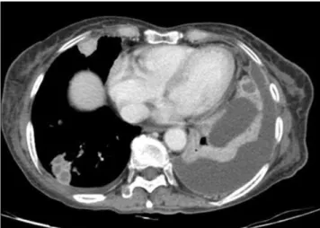
a b
Fig. 4. On follow up examination after two weeks, a T2WI and contrast- enhanced axial MR image of the brain showing the enlarged peripheral enhancing mass with perilesional edema in the right frontal lobe and a newly developed lesion in the right parietal lobe.
a b
c
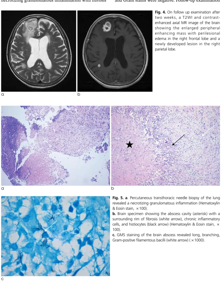
Fig. 5. a. Percutaneous transthoracic needle biopsy of the lung revealed a necrotizing granulomatous inflammation (Hematoxylin
& Eosin stain, 100).
b. Brain specimen showing the abscess cavity (asterisk) with a surrounding rim of fibrosis (white arrow), chronic inflammatory cells, and histiocytes (black arrow) (Hematoxylin & Eosin stain, 100).
c. GMS staining of the brain abscess revealed long, branching, Gram-positive filamentous bacilli (white arrow) ( 1000).
revealed leukocytosis and increased CRP. The patient also reported dyspnea and headache. At that time, a follow-up contrast-enhanced chest CT scan showed aggravation of the nodular consolidation and necrosis, and an enlarged mass. The right pleural effusion was also increased despite percutaneous catheter drainage.
Two weeks after the initial brain MRI, a contrast- enhanced axial MR image of the brain revealed that the peripheral enhancing mass in the right frontal lobe had become enlarged, and a new lesion was detected in the right parietal lobe with focal diffusion restriction (Fig.
4). Under the suspicion of malignancy, we performed a right craniotomy by using a computer-assisted stereotac- tic neuro-navigation system. When the dural incision was made in the right frontal lobe, a mass with internal gray-green pus was found. A dissection plane was created between the lesion and the edematous brain tissue, and the abscess, including the capsule, was completely excised. Microscopic examination of the lesion revealed a thick zone of granulation tissue composed of collagen, fibroblasts, and chronic inflam- matory cells surrounding a central abscess cavity (Fig.
5b). Gomori methenamine silver (GMS) and Gram staining of the abscess revealed long, branching, Gram- positive filamentous rods morphologically similar to Nocardia species (Fig. 5c). GMS staining of the pathologic slide containing the lung tissue biopsy specimen obtained during the preceding PTNB revealed the same branching, filamentous microorganisms.
An early post-operative CT scan confirmed complete removal of the lesion. Except for an erythrocyte sedimentation rate of 44 mm/h, all of the patient’s other laboratory parameters were normal. Treatment with parenteral ceftriaxone and amikacin was initiated post-operatively, and was continued for 6 weeks. The patient was then discharged, asked to continue bactrim administration, and followed-up as an outpatient. At her 6-month follow-up visit, she was able to stand and walk a few steps with assistance and to speak simple words. Her higher mental functions, including memory, attention, language skills, problem solving, and emotional state, were all improving.
Nocardia asteroides was first described in animals by Nocard (1888), and its presence in a human brain
abscess was reported by Eppinger (1890). Nocardia are Gram-positive, partially acid-fast, aerobic, and branching filamentous bacteria. Three species that are human pathogens have been described: Nocardia asteroides, N. brasiliensis, and N. caviae. N asteroides is the most frequently encountered (1).
A Nocardia infection involving the CNS may manifest from meningitis, diffuse cerebral infiltration without localization, granuloma with giant cells, or cerebral abscesses (2, 3).
Nocardia cerebral abscesses are rare and generally occur in immunocompromised patients. They account for only 2% of brain abscesses (2). Mamelak et al.
reported that while the mortality rate among immuno- compromised patients with a Nocardia cerebral abscess is 55%, it is 20% in immunocompetent patients (4). Initially, such abscesses were diagnosed as primarily neoplastic, including high-grade astrocy- tomas, metastasis, or lymphomas (5-7).
In our case, radiographic imaging revealed a contrast- enhancing lesion with perilesional parenchymal edema that was preoperatively diagnosed as a neoplasm such as metastasis or primary brain malignancy. Initially, the mass detected on the chest CT scan was thought to be lung cancer. The distinction between a tumor and an abscess is very important, especially with respect to a potential Nocardia infection. Our patient developed a severe CNS infection in the form of cerebritis, a brain abscess, and ventriculitis. The course of the disease was very serious, and the clinical condition of the patient deteriorated rapidly.
On the image shown, the low signal intensity rim of the lesion on the T2-weight image could be related to the presence of methemoglobin in the wall of the capsule or to the production of free radicals by macrophages (8).
The antibiotic of choice for the treatment of such infections is the synergistic combination of trimetho- prim and sulfamethoxazole (4, 9). The regimen should be maintained for at least 6 weeks and should initially be administered intravenously and then orally for a long period of up to 1 year in immunocompromised patients (4).
In our patient, the initial treatment (based on the Gram staining findings) consisted of broad-spectrum antibiotics (ceftriaxone and metronidazole), and anti- tuberculous and antifungal (fluconazole) medications.
These were switched to trimethoprim/sulfamethoxa- DISCUSSION
zole once the final bacterial culture and sensitivity results were obtained.
However, the optimal management of a Nocardia infection of the brain remains unclear. Mamelak et al.
analyzed 131 patients with this condition along with their own experience of 11 patients (4). They recommended an use of sulfa drugs, empirically when clinically stable extraneural nocardial infection or an immuno-competent patient with a brain abscess less than 2 cm in diameter. If the patient’s condition deterio- rates or if the abscess does not decrease in size within 4 weeks, then the lesion should be aspirated to confirm the diagnosis and decompress the lesion. All abscesses >
2.5 cm in size should be aspirated regardless of the immune status of the patient. If the abscess enlarges after 2 weeks of treatment or if it remains unchanged for 4 weeks despite antibiotic therapy, then a craniotomy should be performed to remove the abscess.
Nocardiosis involving the CNS can be fatal. Early diagnosis and the administration of suitable treatments as well as the performance of appropriate surgical procedures is essential. A Nocardia brain abscess should be considered during differential diagnosis of a cerebral tumor if a contrast-enhancing brain lesion is found in immunocompromised or even immunocom- petent patients. This type of abscess is often misdiag-
nosed as a metastasis or glioblastoma.
References
1. Beaman BL, Beaman L. Nocardia species: host-parasite relation- ships. Clin Microbiol Rev 1994;7:213-264
2. Lee GY. Daniel RT, Brophy BP, Reilly PL. Surgical treatment of nocardial brain abscesses. Neurosurgery 2002;51:668-671;
discussion 671-2.
3. Beaman BL, Burnside J, Edwards B, Causey W. Nocardial infections in the United States, 1972-1974. J Infect Dis 1976;
134:286-289
4. Mamelak AN, Obana WG, Flaherty JF, Rosenblum ML.
Nocardial brain abscess: treatment strategies and factors influencing outcome. Neurosurgery 1994;35:622-631
5. Fleetwood IG, Embil JM, Ross IB. Nocardia asteroides cerebral abscess in immunocompetent hosts: report of three cases and review of surgical recommendations. Surg Neurol 2000;53:605- 610
6. Mogilner A, Jallo GI, Zagzag D, Kelly PJ. Nocardia abscess of the choroid plexus: clinical and pathological case report.
Neurosurgery 1998;43:949-952
7. Roquer J, Pou A, Herraiz J, et al. Primary cerebral abscess due to nocardia presenting as ‘ghost tumor’. Clinical and pathologi- cal study. Eur Neurol 1990;30:254-257
8. Lim JH, Chung TS, Kim HK, Ahn JY, Suh SH. Isolated aspergillosis of the brain in an immunocompetent patient: a case report. JKSMRM 2010;14:64-68
9. Baikie AG, Macdonald CB, Mundy GR. Systemic nocardiosis treated with trimethoprim and sulphamethoxazole. Lancet 1970;2:261
통신저자 : 이아름, (110-744) 서울시 종로구 대학로 101, 서울대학교병원 영상의학과
Tel. (02) 3293-3972 Fax. (02) 747-7418 E-mail: [email protected]
전이성 뇌암으로 오인된 노카디아 뇌농양: 적극적 치료를 요하는 심각한 중추신경계 감염병
1서울대학교병원 영상의학과
2순천향대학교병원 병리과
이 아 름1∙김 희 경2
노카디아병 (nocardiosis)은 actinomycetales 목, nocardia 과에 속하는 호기성 양성 간균에 의한 감염으로 노카디아종은 사람과 동물에서 국소적, 전신적인 화농성 질환을 일으킬 수 있다. 노카디아병은 일반적으로 기회감염 으로 생기지만 감염자의 1/3은 면역기능이 정상이다. 저자들은 고령의 크론병 (Crohn’s disease) 여자 환자에서 노카디아종에 의해 발생한 폐 노카디아병과 뇌농양의 증례를 보고하고자 한다. 이 병변은 영상 검사상 주위 부종을 동 반하며 조영 증강을 보여 수술 전에는 종양으로 생각되었고, 전이성 뇌암처럼 공격적인 병의 진행을 보였다. 노카디아 병의 조기 진단, 기저 질환이 없는 경우, 적절한 항생제 치료가 이루어 졌을 때 예후가 좋다. 저자들은 뇌에 생긴 노카 디아병을 보고하여 자기공명영상과 컴퓨터 단층촬영 소견을 알리고 악성 병변과의 감별에 도움이 되고자 한다.
대한자기공명의과학회지 17:50-54(2013)