ABSTRACT
Purpose: Endoscopic breast surgery for patients with breast cancer was introduced for its superior cosmetic outcomes; it was initially studied in the field of breast-conserving surgery and, more recently, in robotic-assisted nipple-sparing mastectomy (NSM). The main purpose of this study was to investigate the feasibility and safety of endoscopic NSM (E-NSM) in patients with breast cancer by comparing E-NSM and conventional NSM (C-NSM).
Methods: Between May 2017 and October 2020, we retrieved the records of 45 patients who underwent NSM with permanent silicone implants and divided them into the E-NSM group (20 patients) and the C-NSM group (25 patients), depending on the use of the endoscopic device.
We also analyzed demographic information, pathology, operative time, and complications.
Results: No significant differences were observed between the 2 groups based on demographic information, postoperative pathological data, mean length of hospital stay, and total number of complications. The mean preparation time for surgery was comparable between both groups. Compared to the C-NSM group, the E-NSM group had a significantly longer mean operative time and, subsequently, a significantly longer mean total operative time and number of complications.
Conclusion: The results showed that E-NSM was feasible and safe with a more inconspicuous incision in patients with breast cancer.
Keywords: Breast neoplasms; Endoscopic surgical procedure; Mastectomy;
Reconstructive surgery; Robot-assisted surgery
INTRODUCTION
Nipple-sparing mastectomy (NSM) was initially reported in the 1960s by Freeman et al.
[1,2]. It was known to achieve oncological safety comparable to standard mastectomy, with psychological benefits and improved cosmetic results for patients with breast cancer [3,4].
Various types of incisions have been used for NSM, including radial, periareolar, elliptical, and
Original Article
Received: Dec 6, 2020 Revised: Jan 25, 2021 Accepted: Mar 19, 2021 Correspondence to Young Woo Chang
Department of Breast Endocrine Surgery, Korea University Medical Center, 123 Jeokgeum-ro, Danwon-gu, Ansan 15355, Korea.
E-mail: [email protected]
© 2021 Korean Breast Cancer Society This is an Open Access article distributed under the terms of the Creative Commons Attribution Non-Commercial License (https://
creativecommons.org/licenses/by-nc/4.0/) which permits unrestricted non-commercial use, distribution, and reproduction in any medium, provided the original work is properly cited.
ORCID iDs Hye Yoon Lee
https://orcid.org/0000-0001-9077-1412 Young Woo Chang
https://orcid.org/0000-0001-5396-7467 Da Young Yu
https://orcid.org/0000-0001-7931-4034 Tae Yul Lee
https://orcid.org/0000-0002-6511-5453 Duk Woo Kim
https://orcid.org/0000-0003-3382-560X Woo Young Kim
https://orcid.org/0000-0001-6871-6855 Seung Pil Jung
https://orcid.org/0000-0003-3967-2974 Sang Uk Woo
https://orcid.org/0000-0001-6218-4704 Jae Bok Lee
https://orcid.org/0000-0001-7977-3097 Gil Soo Son
https://orcid.org/0000-0001-8684-7875
Hye Yoon Lee 1 , Young Woo Chang 1 , Da Young Yu 1 , Tae Yul Lee 2 , Duk Woo Kim 2 , Woo Young Kim 1 , Seung Pil Jung 1 , Sang Uk Woo 1 , Jae Bok Lee 1 , Gil Soo Son 1
1
Department of Surgery, Korea University College of Medicine, Seoul, Korea
2
Department of Plastic and Reconstructive Surgery, Korea University College of Medicine, Seoul, Korea
Comparison of Single Incision Endoscopic Nipple-Sparing
Mastectomy and Conventional Nipple-
Sparing Mastectomy for Breast Cancer
Based on Initial Experience
Funding
This work was supported by grants from the Korea University Ansan Hospital Grant (K1811041) and Korea Breast Cancer Foundation (Q1809151).
Conflict of Interest
The authors declare that they have no competing interests.
Author Contributions
Conceptualization: Chang YW, Lee HY;
Data collection: Chang YW, Lee HY, Yu DY, Kim DW; Formal analysis: Chang YW, Lee HY; Investigation: Chang YW, Lee HY;
Methodology: Chang YW, Lee HY, Yu DY, Lee TY, Kim DW; Supervision: Chang YW, Lee HY, Jung SP, Woo SU, Lee JB, Son GS; Validation:
Chang YW, Lee HY, Jung SP, Woo SU, Lee JB, Son GS; Writing - original draft: Chang YW, Lee HY; Writing - review & editing: Chang YW, Lee HY.
inframammary incisions [5,6]; however, further studies on the approach of NSM have been extensively conducted to explore ways to create a more inconspicuous and efficient incision.
With the development of minimally invasive devices, laparoscopic or endoscopic surgeries have been developed in many areas. Endoscopic breast surgery for patients with breast cancer was also introduced for its superior cosmetic effects. It was initially studied in the field of breast-conserving surgery [7-9] and, more recently, in robotic-assisted NSM [10-14].
To the best of our knowledge, few studies have compared single incision endoscopic and conventional methods in patients with breast cancer undergoing NSM with immediate reconstruction using a permanent silicone implant. Thus, the main purpose of this study was to investigate the feasibility and safety of endoscopic NSM (E-NSM) in patients with breast cancer by comparing E-NSM and conventional NSM (C-NSM) in terms of pathology, operative time, and outcome.
METHODS
Study population
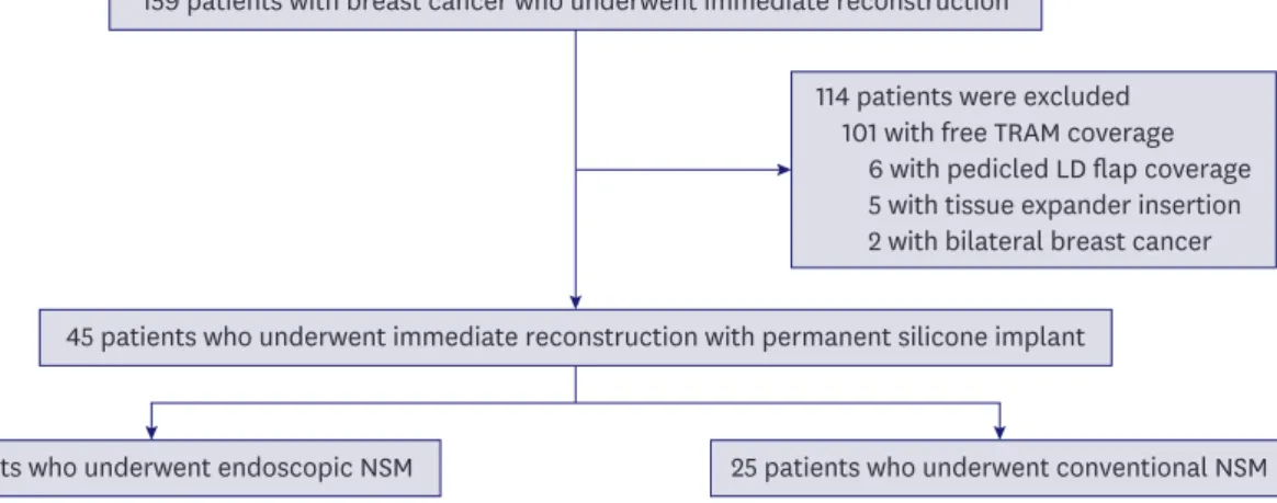
We retrieved the records of 159 consecutive patients with breast cancer who underwent skin- sparing mastectomy with immediate reconstruction between May 2017 and October 2020.
Among these, 101 patients who underwent free transverse rectus abdominus myocutaneous (TRAM) coverage, 6 patients who underwent pedicled latissimus dorsi flap coverage, and 5 patients who underwent tissue expander insertion were excluded. Two patients with bilateral breast cancer were also excluded from the analysis in order to focus only on unilateral breast cancer. The finalized pool of eligible participants comprised 45 patients who underwent NSM with permanent silicone implants, who were divided into 2 groups depending on the use of endoscopic devices. There were 20 patients in the E-NSM group and 25 patients in the C-NSM group (Figure 1).
This retrospective study was approved by the Institutional Review Board of Korea University Medical Center, Ansan (approval number: 2020AS0322). A waiver of informed consent was requested and was approved.
159 patients with breast cancer who underwent immediate reconstruction
20 patients who underwent endoscopic NSM
45 patients who underwent immediate reconstruction with permanent silicone implant
25 patients who underwent conventional NSM 114 patients were excluded
101 with free TRAM coverage 6 with pedicled LD flap coverage 5 with tissue expander insertion 2 with bilateral breast cancer
Figure 1. Flow chart of the study population.
TRAM = transverse rectus abdominis muscle; LD = latissimus dorsi; E-NSM = endoscopic nipple-sparing mastectomy; C-NSM = conventional nipple-sparing mastectomy.
Surgical procedures of endoscopic NSM
Patients were placed in a supine position with both upper limbs on arm boards abducted at 90° (Figure 2). An incision of 5 cm was made along the anterior axillary line starting from the inferior mammary fold. The sentinel lymph node, which was dyed with a technetium-99m- labeled nanocolloid, was excised through this incision. A workspace for the insertion of a Glove Port (Nelis Corporation, Bucheon, Korea) was created within a radius of 3 cm from the incision, and the subcutaneous tissue flaps were raised. Approximately 250 mL of tumescent solution was infiltrated into the subcutaneous fat layer of the breast with a Veress needle through the workspace, and blunt dissection was performed using a straight tunneler before the insertion of the Glove Port.
The Glove Port was inserted into the incision site. Upon lifting the lateral breast to approach the retromammary space, carbon dioxide (CO
2) gas was insufflated, and the pressure was maintained at approximately 6 mmHg. The retromammary space was dissected with caution to avoid dissecting the interpectoral space, using an energy device and an endoscopic grasper guided by a flexible endoscope (ENDOEYE FLEX 10 mm, LTF-S190-10; Olympus Corporation, Tokyo, Japan). The breast was elevated laterally to the edge of the latissimus dorsi muscle, inferiorly to the thoracoabdominal aponeurosis, superiorly to the level of the clavicle, and medially to the edge of the sternum.
After dissecting the retromammary fat plane, the Glove Port was relocated to approach the subcutaneous space, which had already been dissected bluntly with a straight tunneler.
Structures remaining after blunt dissection were cut off using an energy device, and the duct beneath the nipple was cut off using endoscopic scissors. The subcutaneous flap was completely dissected along the boundaries of the breast, and the entire breast was removed (Figure 3).
Axillary lymph node dissection (ALND) was performed in 2 patients in the E-NSM group who had metastatic sentinel lymph nodes; 1 patient underwent ALND using endoscopic devices and the other, directly through the anterior axillary incision.
Subsequently, the inferior origin of the pectoralis major was released, and an acellular dermal matrix sling was made for immediate reconstruction with a silicone implant. Surgery was performed after control of bleeding and drain insertion.
Figure 2. The patient's position for endoscopic nipple-sparing mastectomy.
Data analysis
We analyzed demographic information and pathology obtained from medical records, including invasive tumor size, positive resection margin, status of the axillary lymph nodes, and tumor node metastasis classification according to the 8th American Joint Committee on Cancer [15]. Breast sagging was classified as follows based on the Regnault ptosis classification: grade 1, the nipple at the level of the inframammary fold; grade 2, the nipple below the level of the inframammary fold, but above the lower breast contour; grade 3, the nipple below the level of the inframammary fold and at the lower breast contour;
pseudoptosis, the nipple above the level of the inframammary fold, but the breast hypoplastic and hanging below the fold [16].
The preparation time for the surgery was calculated (in minutes) from the induction of general anesthesia to the initial skin incision. Total operative time was defined from the initial skin incision to the closure of the wound, and it was divided into the time of NSM and of reconstruction based on the end of bleeding control after breast removal in all cases.
Complications were also analyzed. Cases requiring debridement and evacuation of a hematoma were considered as postoperative skin necrosis and hematoma, respectively.
Postoperative implant infections requiring implant change were also identified.
Statistical analysis
All data were analyzed using IBM SPSS Statistics ver. 25 (IBM Corp., Armonk, USA).
Continuous data are presented as means with standard deviations, and categorical data are
A B C
D E F
Figure 3. Illustrations of the flap dissections. (A) Images showing lifting of the lateral breast to approach the retromammary area (arrow, the lateral breast).
(B) Schematic images showing the retromammary space dissection (yellow, glandular tissue; red, muscles in the axilla; blue, Glove Port; green, flexible
endoscope). (C) Images showing the retromammary space dissection guided by a scope. (D) Images showing pulling of the lateral breast downward to approach
the subcutaneous layer (arrow, the lateral breast). (E) Schematic images showing the subcutaneous flap dissection. (F) Images showing the subcutaneous flap
dissection guided by a scope.
presented as numbers with percentages. The t-test was used to compare continuous variables, and categorical variables were compared using the χ
2or Fisher's exact test to analyze the significance of differences. Differences were considered statistically significant at p < 0.05.
RESULTS
Clinical pathological characteristics
The study population included 20 patients in the E-NSM group and 25 in the C-NSM group.
There were no significant differences between both groups in terms of mean age at operation (47.2 ± 9.5 years in E-NSM vs. 44.6 ± 9.6 years in C-NSM, p = 0.38) and body mass index (BMI) (24.1 ± 3.8 in E-NSM vs. 22.3 ± 3.6 in C-NSM, p = 0.11). The mean tumor extent and multicentricity based on preoperative radiological findings and the proportion of breast ptosis did not significantly differ between the 2 groups (p = 0.97).
Based on postoperative pathological data, no significant differences were observed in the mean size of the invasive tumor (0.92 ± 1.3 cm in E-NSM vs. 1.39 ± 1.2 cm in C-NSM, p = 0.21), the number of positive resection margin cases, and the mean number of metastatic (p = 0.93) and harvested (p = 0.26) lymph nodes. Tumor, node, metastasis staging did not significantly differ between the 2 groups (Table 1).
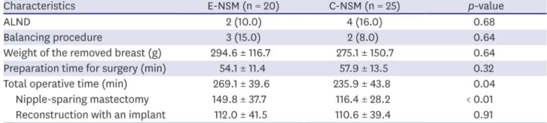
Operative details
ALND was performed in 2 patients in the E-NSM group and 4 patients in the C-NSM group (10.0% in E-NSM vs. 16.0% in C-NSM, p = 0.68), and contralateral balancing augmentation was performed in 3 patients in the E-NSM group and 2 patients in the C-NSM group (15.0%
in E-NSM vs. 8.0% in C-NSM, p = 0.64). The weights of the removed breast tissues were comparable between the 2 groups (294.6 ± 116.7 g in E-NSM vs. 275.1 ± 150.7 g in C-NSM, p Table 1. Clinical and pathological characteristics of breast cancer patients who underwent nipple-sparing mastectomy with immediate reconstruction with permanent silicone implants using endoscopic or conventional methods
Characteristics E-NSM (n = 20) C-NSM (n = 25) p-value
Age at operation (yr) 47.2 ± 9.5 44.6 ± 9.6 0.38
Body mass index 24.1 ± 3.8 22.3 ± 3.6 0.11
Tumor extent * (cm) 4.1 ± 1.9 3.9 ± 2.1 0.68
Multicentricity 12 (60.0) 14 (56.0) 0.79
Breast ptosis 0.97
Normal 10 (50.0) 13 (52.0)
Grade 1 6 (30.0) 6 (24.0)
Grade 2 1 (5.0) 2 (8.0)
Grade 3 1 (5.0) 2 (8.0)
Pseudoptosis 2 (10.0) 2 (8.0)
Invasive size (cm) 0.92 ± 1.3 1.39 ± 1.2 0.21
Positive resection margin 0 (0.0) 0 (0.0)
Metastatic LNs 0.45 ± 1.2 0.48 ± 1.1 0.93
Harvested LNs 4.40 ± 5.7 6.84 ± 8.1 0.26
TNM stage 0.25
0 10 (50.0) 6 (24.0)
IA 3 (15.0) 11 (44.0)
IIA 5 (25.0) 5 (20.0)
IIB 1 (5.0) 2 (8.0)
IIIA 1 (5.0) 1 (4.0)
Values are presented as mean ± standard deviation or number (%).
E-NSM = endoscopic nipple-sparing mastectomy; C-NSM = conventional nipple-sparing mastectomy; LN = lymph node; TNM = tumor, node, metastasis.
* Based on preoperative radiological findings.
= 0.64). Regarding operative time, the mean preparation time for surgery was comparable between the 2 groups. However, the E-NSM group had a significantly longer mean total operative time (269.1 ± 39.6 minutes in E-NSM vs. 235.9 ± 43.8 minutes in C-NSM, p = 0.04) because the mean operative time for NSM was significantly longer in the E-NSM group (149.8
± 37.7 minutes in E-NSM vs. 116.4 ± 28.2 minutes in C-NSM, p < 0.01). The mean time for reconstruction with the implant was comparable between the 2 groups (Table 2).
Outcome and morbidity details
The mean length of hospital stay showed no significant difference (14.4 ± 4.5 days in E-NSM vs. 12.0 ± 6.2 days in C-NSM, p = 0.16) between the groups. Regarding complications, in the C-NSM group, 2 (8.0%) patients experienced postoperative skin necrosis requiring debridement, and 2 (8.0%) developed hematomas. There was no skin necrosis or hematoma in the E-NSM group. Postoperative implant infection requiring an implant change was found in one patient (5.0%) in the E-NSM group. There was no significant difference in the total number of complications between the groups (Table 3). However, it is difficult to say that the statistical difference in surgical complications is absolute due to the small number of patients with events.
DISCUSSION
Comparing the positive resection margin rate between both groups helped to assess the feasibility of E-NSM, as a positive resection margin is a known risk factor for local recurrence in patients with breast cancer [17,18]. There were no cases of positive resection margins, including frozen and permanent pathologic findings, in both groups, demonstrating that the E-NSM method allowed for the complete and proper removal of the glandular and cancer tissue.
The results showed that the operative time for E-NSM was significantly longer than that for C-NSM. Despite a thorough preparation through previous studies on E-NSM, it took time to adapt to the new surgical view, endoscopic device, and position of the endoscope. The Table 2. Operative details of E-NSM and C-NSM groups
Characteristics E-NSM (n = 20) C-NSM (n = 25) p-value
ALND 2 (10.0) 4 (16.0) 0.68
Balancing procedure 3 (15.0) 2 (8.0) 0.64
Weight of the removed breast (g) 294.6 ± 116.7 275.1 ± 150.7 0.64
Preparation time for surgery (min) 54.1 ± 11.4 57.9 ± 13.5 0.32
Total operative time (min) 269.1 ± 39.6 235.9 ± 43.8 0.04
Nipple-sparing mastectomy 149.8 ± 37.7 116.4 ± 28.2 < 0.01
Reconstruction with an implant 112.0 ± 41.5 110.6 ± 39.4 0.91
Values are presented as mean ± standard deviation or number (%).
E-NSM = endoscopic nipple-sparing mastectomy; C-NSM = conventional nipple-sparing mastectomy; ALND = axillary lymph node dissection.
Table 3. Outcome and morbidity details of E-NSM and C-NSM groups
Characteristics E-NSM (n = 20) C-NSM (n = 25) p-value
Length of hospital stay (day) 14.4 ± 4.5 12.0 ± 6.2 0.16
Complications 1 (5.0) 4 (16.0) 0.36
Skin necrosis 0 (0.0) 2 (8.0)
Hematoma 0 (0.0) 2 (8.0)
Infection 1 (5.0) 0 (0.0)
Values are presented as mean ± standard deviation or number (%).
E-NSM = endoscopic nipple-sparing mastectomy; C-NSM = conventional nipple-sparing mastectomy.
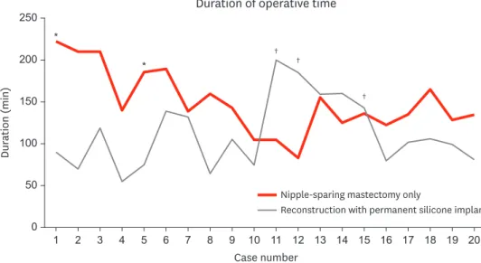
study by Hung et al. [19] on the learning curve of endoscopic total mastectomies showed that the inflection point occurred after 15 to 17 cases. In our cases, the operative time for NSM decreased sharply after approximately 10 cases (Figure 4). Blunt dissection of the subcutaneous flap using a straight tunneler and dissection of the subcutaneous flap after dissection of the retromammary flap reduced the operative time [20]. The preparation time for E-NSM was comparable to that for C-NSM. This was because, unlike in robotic- assisted NSM, which has recently been studied quite extensively, no further preparation for the patient's position was needed in E-NSM. In addition, as E-NSM was performed on the operating field, the preoperative injection of indigo carmine to check the borders of the breast through the scope was not needed [21,22].
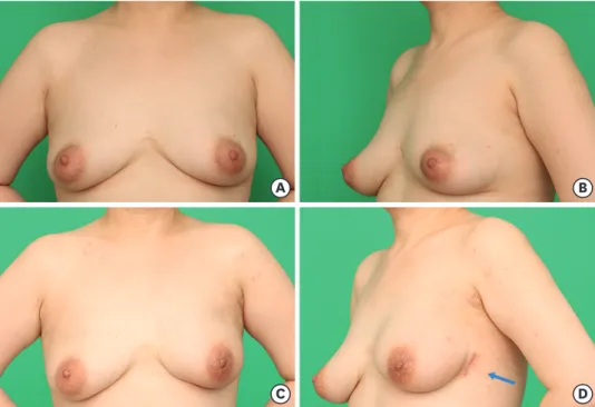
Only one incision, 5 cm along the anterior axillary line, was used in all patients in the E-NSM group. The incision was made starting on the midaxillary line; however, this made subcutaneous dissection more difficult because of the leverage effect of the pectoralis major muscle. Later, we used the anterior axillary line, approximately 2 cm above the midaxillary line ventrally; this allowed us to overcome the leverage effect without differences in cosmetic outcomes. The conventional method of NSM uses various incisions to remove the whole breast effectively and to hide the incision; however, conspicuous wounds from the front view are unavoidable. In contrast, the surgical wound was completely invisible and covered by the undergarment of the patients who underwent E-NSM, which improved patient satisfaction (Figure 5).
Studies on endoscopic mastectomy using CO
2insufflation reported that the pressure was maintained at 8–10 mmHg without any issues related to CO
2[11,23]. In our cases, a constant CO
2pressure of 10 mmHg was used in the first patient who underwent E-NSM, and this patient experienced high end-tidal and arterial partial pressure of CO
2after bleeding while undergoing dissection of the retromammary fat plane. These levels were corrected to normal levels by lowering the CO
2pressure to 6 mmHg. After the event, a decreased CO
2pressure of 6 mmHg was used in all cases thereafter, and no complications occurred due to CO
2. There were also no interruptions in the surgical view due to decreased pressure.
Case number 0
Dur ation (min)
200 250
150
100
50
20 19 18 17 16 15 14 13 12 11 10 9 8 7 6 5 4 3 2 1
Duration of operative time
†
†
†