A papillary thyroid carcinoma is the most common neoplasm in the thyroid gland and accounts for approxi- mately 70% of all thyroid carcinomas (1). Metastatic cer-
vical lymphadenopathy is a common finding in patients with papillary thyroid carcinoma, and its detection is crucial for planning the surgical management of these patients (2-4). Many investigators have noted the sono- graphic differentiation between normal and abnormal lymph nodes (5-7). Although criteria for the diagnosis of abnormal nodes are well established, overlap between the criteria for metastasis and various other pathological conditions still exists.
Although many studies have described the ultrasono- graphic or MR imaging appearance of metastatic lymph nodes from papillary thyroid carcinoma (5-10), many
Diagnostic Value of CT for the Detection of Cervical Lymph Node Metastases in Papillary Thyroid Carcinoma
1Kyu Ri Son, M.D., Dong Gyu Na, M.D.2, Kee-Hyun Chang, M.D.2
1Department of Radiology, SMG-SNU Boramae Medical Center, Seoul, Republic of Korea.
2Department of Radiology, Seoul National University College of Medicine, Institute of Radiation Medicine, Seoul National University Medical Research Center, and Clinical Research Institute, Seoul National University Hospital, Seoul, Republic of Korea.
Received September 15, 2008 ; Accepted March 4, 2009
Address reprint requests to : Kyu Ri Son, M.D., Department of Radiology, SMG-SNU Boramae Medical Center, 39, Boramae Road, Dongjak-ku, Seoul, 156-707, Korea
Tel. 82-2-870-2544 Fax. 82-2-747-7418 E-mail: [email protected]
Purpose: To determine the diagnostic accuracy of CT for the detection of cervical lymph node metastases in patients with papillary thyroid carcinoma (PTC).
Materials and Methods: Two hundred twelve consecutive patients with surgically proven PTC were included in this study. CT images were retrospectively evaluated to determine the presence of a node metastasis using morphologic CT criteria (at least one of the following: strong nodal enhancement without hilar vessel enhancement, heterogeneous enhancement, calcification, and cystic change). The diagnostic accura- cy of CT for the diagnosis of a metastatic lymph node was assessed using a level-by- level analysis.
Results: The accuracy of the CT finding for strong nodal enhancement was greater than the other morphologic CT criteria (81.6% and 74.5-78.5%, respectively). The sen- sitivity, specificity, and accuracy were 64.4%, 91.4%, and 84.3% by the morphologic CT criteria, and were 34.6%, 93.9%, and 78.2% by size criteria, respectively.
Conclusion: The morphologic CT criteria are more accurate than the size criteria in the detection of cervical lymph node metastases in patients with papillary thyroid car- cinoma; and, strong nodal enhancement on a CT scan is the most important factor for its diagnostic accuracy.
Index words :Thyroid neoplasms Papillary carcinoma
Head and neck neoplasms, metastases Tomography, X-ray computed
practitioners are reluctant to use a contrast-enhanced CT scan as a diagnostic tool due to a concern about de- layed subsequent radioiodine therapy. Recently, several investigators have reported that a CT scan increases the sensitivity of detecting metastatic lymph nodes from a papillary thyroid carcinoma (11, 12). The criteria of the CT findings for metastatic lymph nodes of papillary thy- roid carcinomas; however, have not yet been estab- lished. In this study, we evaluated the usefulness of CT for the detection of cervical lymph node metastases in patients with papillary thyroid carcinomas, and assessed the optimal criteria for the detection of a nodal metasta- sis on CT images.
Materials and Methods
Patients
We retrospectively reviewed 212 patients with a papil- lary thyroid carcinoma that also underwent a neck dis- section and a thyroidectomy. The subjects included 182 women and 30 men, ranging in age from 21 to 78 years (average age, 46 years). A contrast-enhanced neck CT was performed in all patients as a preoperative evalua- tion. The surgeons dissected all visible or palpable lymph nodes on a level-by-level basis according to the preoperative findings of clinical palpation and the CT findings.
CT Imaging
CT examinations were performed using a series of multi-detector row CT scanners. The CT scanners in- cluded a four-detector row CT scanner (MX 8000, Philips Medical Systems, Cleveland, OH U.S.A. (n = 68), an eight-detector row CT scanner (Light Speed Ultra, GE Medical Systems, Milwaukee, WI U.S.A.) (n
= 46), a 16-detector row CT scanner (Sensation 16, Siemens, Erlangen, Germany) (n = 53), and a 64-detec- tor row CT scanner (Brilliance 64, Philips Medical Systems) (n = 45). The respective scanning parameters used for the 4-, 8-, 16- and 64-multidetector row CT scanners were respectively as follows: the detector con- figurations were 4 × 2.5, 8 × 1.25, 16 × 0.75, and 64
× 0.625 mm; slice thicknesses, 3.2, 2.5, 3 and 3 mm; re- construction intervals 3, 2.5, 3 and 2 mm; table speeds, 12.5, 13.5, 24, and 46 mm/rotation. The time interval be- tween the CT examination and neck dissection ranged from one to sixty days (mean, 28 days).
Unenhanced images were first obtained in a cranio- caudal direction between an aorticopulmonary window
level and the skull base. After acquiring unenhanced im- ages, 90 ml of an iodinated contrast agent (Ultravist 370, Schering, Berlin, Germany) was injected at 3 ml/s using an automated injector and a scan delay time of 45 s. We additionally injected 45 ml of normal saline at 3 ml/s for saline flushing immediately after injection of the con- trast agent to avoid artifacts induced by stagnated con- trast agents within the subclavian or innominate vein, which may obscure the lymph nodes of the low central neck and supraclavicular fossa.
Image Analysis
The resected nodes were classified according to the nodal classification scheme of the American Joint Committee on Cancer level system (13). Accordingly, all cervical lymph nodes were divided into central (level VI) and lateral (Levels I-V) groups on each of the sides.
Upon CT examination, the cervical lymph nodes were assessed on a level-by-level basis by two radiologists (D.K.N. and K.R.S.) with consensus. We could not reli- ably identify whether a node was positive or negative, as the nodes were too small to distinguish from other nodes of similar size. Therefore, the largest node was considered the representative of each nodal compart- ment and was used for CT and pathological correlation.
The CT images were retrospectively evaluated to de- termine the presence of node metastasis using the size, morphological CT, and combined criteria. We measured the short transverse diameter of the largest lymph node
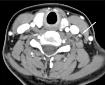
Fig. 1. A 38-year-old woman with papillary thyroid cancer in the left lobe of the thyroid gland. A CT demonstrates a small metastatic lymph node (arrow) with focal strong enhancement as adjacent neck vessels in left level IV. A pathologic specimen revealed metastatic papillary carcinoma in the lymph node.
at each neck level to determine the optimal size criteria.
Morphological CT criteria for metastatic lymph nodes included strong nodal enhancement, cystic change, cal- cification, and heterogenous enhancement (Fig. 1-4).
We considered nodes as being malignant based on mor- phological CT criteria if they had at least one out of these four findings. A lymph node with strong nodal en- hancement was defined as a node showing higher en- hancement than the adjacent sternocleidomastoid mus- cle without hilar vessel enhancement. A lymph node with heterogenous enhancement was defined as a node showing inhomonogenous enhancement without focal cystic change. A lymph node with strong nodal en-
hancement and hilar vessel enhancement was regarded as a reactive lymph node. We considered nodes as being malignant using the combined criteria if they were con- sidered malignant by either size or morphological crite- ria.
Statistical Analysis
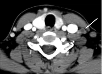
The size of malignant and benign nodes was com- pared using the t-test. The optimal cutoff value for the short transverse diameter in terms of differentiating metastatic from benign lymph nodes was calculated us- ing the receiver operating characteristic curve analysis in the central and lateral neck compartment. The sensi- tivity, specificity, and accuracy for the detection of lymph node metastases were calculated on a level-by- level basis for all neck levels or the central and lateral nodal compartment using each of the three previously mentioned criteria (size, morphological criteria and combined criteria). The three criteria were compared using the McNemar test. A univariate analysis was per- formed to identify the parameters that were used to dif- ferentiate malignant and benign nodes. A multivariate analysis was performed using the multiple logistic re- gression to determine if these parameters were useful in distinguishing malignant from benign lymph nodes if they are independent of each other. The odds ratios and 95% confidence intervals were determined. For all sta- tistical analyses, a p-value less than 0.05 was considered to indicate a significant difference. The data were ana- Fig. 2. A 47-year-old man woman with papillary thyroid can-
cer in the left lobe of the thyroid gland. A CT demonstrates a small lymph node (arrow) with a focal cystic change in left lev- el IV revealed to be a metastatic lymph node.
Fig. 3. A 55-year-old man with papillary thyroid cancer in the left lobe of the thyroid gland. A CT demonstrates small hetero- geneously enhancing lymph nodes (arrows) in both level IVs.
A pathologic specimen revealed metastatic papillary carcino- ma in the lymph node.
Fig. 4. A 50-year-old woman with papillary thyroid cancer in the right lobe of the thyroid gland. A CT demonstrates a lym- phadenopathy (arrow) in the left level VI, which is regarded to be positive when applying size criteria, and negative when ap- plying morphologic criteria. The formal report suggested possi- ble lymph node metastasis, but pathologic exam revealed a re- active lymphadenopathy.
lyzed using the SPSS statistical software (version 10.0;
SPSS, Chicago, IL, U.S.A.).
Results
A central neck dissection was performed at 198 levels (right and left central neck levels). Of the 198 dissected central neck levels, 48 (24%) were proven pathologically to have metastatic nodes. Lateral neck dissection was performed at 183 levels [level I (n = 6), level II (n = 47), level III (n = 71), level IV (n = 44) and level V (n = 15)].
Metastatic lymph nodes were found at 53 (29%) lateral neck levels [level I (n = 2), level II (n = 9), level III (n = 22), level IV (n = 17) and level V (n = 1)]. Overall, metastatic lymph nodes were shown to be present after surgery at 101 levels (27%) of the 381 dissected neck lev-
els in 212 patients on a level-by-level analysis.
Malignant nodes were larger than benign nodes in the central and lateral neck compartments (Table 1). A short transverse diameter of 5 mm and 8 mm were deter- mined as the cutoff values for the discrimination of metastatic nodes from benign nodes at the central neck and lateral neck levels by ROC analysis, respectively.
Table 2 shows the sensitivity, specificity, positive pre- dictive value (PPV), negative predictive value (NPV), and accuracy of four morphological CT findings for the detection of metastatic cervical lymph nodes on all neck levels. The morphological CT criteria revealed a sensi- tivity of 64% and a specificity of 91% for the diagnosis of metastatic nodes. The accuracy of the CT finding for strong nodal enhancement was higher than that of the other morphologic CT criteria (81.6% and 74.5-78.5%, respectively).
The detection of nodal metastasis, sensitivity, speci- ficity, and accuracy when the short transverse diameter of 5 mm for the central neck levels and 8 mm for the lat- eral neck levels was used as the threshold were 34.6%, 93.9% and 78.2%, respectively (Table 3). When the mor- phological characteristics were analyzed, sensitivity, specificity, and accuracy were found to be 64.4%, 91.4%, and 84.3%, respectively (Table 3). The morpho- Table 1. The Size Distribution of Lymph Nodes in the Central
and Lateral Neck
Malignant Benign
Nodes(*mm) Nodes (*mm) p value Central group 5.73 ± 2.63 3.95 ± 1.38 0.00003 Lateral group 6.78 ± 3.56 5.07 ± 1.97 0.0008 Mean ± standard deviation
The size (*mm) refers to the transverse minimal diameter
Table 2. Comparison of the Diagnostic Accuracy of Morphologic CT Findings at All Levels of the Neck
CT Findings Diagnostic Values
Sensitivity (%) Specificity (%) PPV (%) NPV (%) Accuracy (%) Strong enhancement 43 (43/101) 96 (268/280) 78 (43/55) 82 (268/326) 81.6 (311/381)
Cystic change 23 (23/101) 99 (276/280) 85 (23/27) 78 (276/354) 78.5 (299/381)
Calcification 6 (6/101) 99 (278/280) 75 (6/8)00 75 (278/373) 74.5 (284/381)
Heterogenous enhancement 20 (20/101) 96 (269/280) 65 (20/31) 77 (269/350) 75.9 (289/381)
*Morphologic criteria 64 (65/101) 91 (256/280) 73 (65/89) 88 (256/292) 84.3 (321/381) PPV, positive predictive value, NPV, negative predictive value, * Morphologic criteria means at least one of four morphologic CT findings
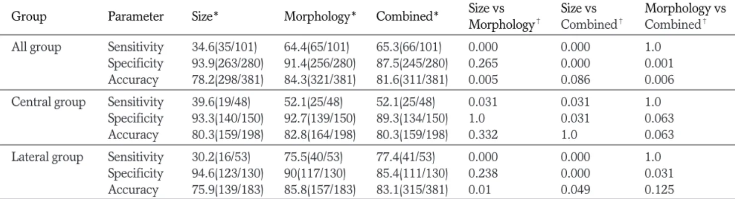
Table 3. Diagnostic Accuracy of CT For Nodal Staging Using the Sensitivity, Specificity, and Accuracy on a Level-by-Level Basis
Group Parameter Size* Morphology* Combined* Size vs Size vs Morphology vs
Morphology� Combined� Combined� All group Sensitivity 34.6(35/101) 64.4(65/101) 65.3(66/101) 0.000 0.000 1.0
Specificity 93.9(263/280) 91.4(256/280) 87.5(245/280) 0.265 0.000 0.001 Accuracy 78.2(298/381) 84.3(321/381) 81.6(311/381) 0.005 0.086 0.006 Central group Sensitivity 39.6(19/48) 52.1(25/48) 52.1(25/48) 0.031 0.031 1.0
Specificity 93.3(140/150) 92.7(139/150) 89.3(134/150) 1.0 0.031 0.063 Accuracy 80.3(159/198) 82.8(164/198) 80.3(159/198) 0.332 1.0 0.063 Lateral group Sensitivity 30.2(16/53) 75.5(40/53) 77.4(41/53) 0.000 0.000 1.0
Specificity 94.6(123/130) 90(117/130) 85.4(111/130) 0.238 0.000 0.031 Accuracy 75.9(139/183) 85.8(157/183) 83.1(315/381) 0.01 0.049 0.125
* Data are in percentage. The numbers in parentheses are raw data.
�p-values were calculated using the McNemar test.
logical criteria showed better sensitivity and accuracy than the size criteria without any significant decrease in specificity.
A univariate analysis and multiple logistic regression analysis demonstrated that the four morphologic CT findings and size were significant parameters for depict- ing malignant nodes (Table 4).
Discussion
The frequency of regional node metastases in patients with papillary carcinomas is reported to vary from 39%
to 90% (2-4, 10), depending on the surgical method un- dertaken and the population studied. In this study, mor- phological findings on CT images were more important for the detection of cervical lymph node metastases in patients with papillary thyroid carcinoma. Compared with the size criteria alone, the morphological criteria showed a higher sensitivity of 52.1% at the central neck level and 75.5% at the lateral neck level. A multivariate analysis demonstrated that the four morphological crite- ria and the size criteria were found to be significant pre- dictors for the identification of nodal metastases; how- ever, strong nodal enhancement (odds ratio, 10.0) and calcification (odds ratio, 6.9) had higher odds ratios.
Most of primary thyroid cancers appeared as relative- ly low attenuated lesions on CT scans because of the high level of vascularity in normal thyroid tissue (13- 15). Although the contrast enhancement pattern for pap- illary thyroid carcinomas has been rarely reported, the presence of strong enhancement within a lymph node strongly suggests the presence of nodal metastasis from
papillary thyroid carcinoma (9), which indicates that the papillary thyroid cancer itself has hypervasularity.
Strong enhancement of lymph nodes showed relatively high sensitivity (43%) among the morphological CT find- ings, and a high odds ratio (10.0) by multivariate analy- sis. In clinical practice, lymph nodes with strong en- hancement should be considered as malignant, even if the node is small in size. Unlike metastatic nodes, reac- tive lymphadenopathy occasionally demonstrates hilar vessel enhancement or central enhancement rather than peripheral enhancement (6, 7).
The various cystic nature of a metastatic papillary thy- roid carcinoma has also been demonstrated on ultra- sonography and MRI (10, 16). Cystic changes have been reported to be present in 70% of metastatic lymph nodes on ultrasonography (16) and in 33% of metastatic lymph nodes by MRI (10). Cystic changes consist of a small solitary cystic area, multiple peripheral cystic areas, or almost complete replacement of the node by cystic for- mation. These cystic changes represent liquefaction necrosis, and appear to be characteristic of a metastatic papillary thyroid carcinoma (16). In our study, 23% of the metastatic lymph nodes showed cystic changes on the CT images.
Microcalcification in the papillary thyroid carcinoma is a common ultrasonographic finding and is well known, histologically, as a psammomatous body (17- 19). Microcalcification had been reported to be seen in approximately 60% of papillary thyroid carcinomas and metastatic lymph nodes by ultrasonography (8, 18, 19).
The peripheral punctuate calcification in the cervical nodes is important for the diagnosis of metastatic papil- Table 4. CT Findings Discriminating Malignant from Benign Cervical Lymph Nodes in Patients with Papillary Thyroid Carcinoma
Pathology p Value at Multiple Logistic Regression Variable
Benign Malignant Univariate
p value Odds
Confidence interval
Analysis Ratio
Strong absent 268 58 0.000 0.000 10.0 4.4 22.9
enhancement present 012 43
Cystic change absent 276 78 0.000 0.033 03.8 1.1 12.7
present 004 23
Calcification absent 278 95 0.005 0.041 06.9 1.1 43.6
present 002 06
Heterogenous absent 269 81 0.000 0.000 06.6 2.7 15.8
enhancement present 011 20
size ≤5 mm (central group) or 263 66 0.000 0.020 02.6 1.2 05.7
≤8 mm (lateral group)
>5 mm (central group) or 017 35
>8 mm (lateral group)
lary carcinomas of the thyroid (8); however, microcalci- fication can barely be detected on CT. In our study, only 6% of metastatic nodes showed calcification on CT im- ages.
As the size of lymph nodes varies according to the var- ious levels in the neck, and because small metastatic de- posits inside lymph do not always cause enlargement of a lymph node, it is very difficult to define an optimal size criteria for determining malignancy (5, 20, 21). Any chosen size criterion is a compromise between high sen- sitivity and specificity. We suggested that the optimal size criteria (8 mm or more for the minimum transverse diameter in the lateral group and 5 mm or more in the central group) was slightly smaller than the minimum transverse diameter (10 mm) for a metastasis of a squa- mous cell carcinoma (5, 20-22).
The different anatomic levels in the neck may affect the performance of diagnostic imaging for identifying metastatic lymph nodes. Several CT and sonography studies have confirmed that enlarged nodes at different levels require different size criteria to predict whether the nodes are metastatic (20-22). Therefore, our results suggest specific size criteria for central or lateral neck node metastases in papillary thyroid carcinomas as well as the presence of smaller lymph node metastases com- pared to other head and neck malignancies.
The diagnosis of metastatic nodes using CT also de- pends on size determination and assessment of changes in the internal architecture. Within this context, Curtin et al. (23) studied the effect of size criteria and internal architectural changes of nodes for the diagnostic accura- cy for metastatic nodes. These researchers showed that the addition of information on the internal architecture of a node resulted in a substantial improvement in the diagnostic performance of CT using a long axis length (maximum axial diameter). In this study, a multivariate analysis demonstrated that the four morphologic criteria and size criteria as significant parameters for diagnosing a nodal metastasis; however, strong nodal enhancement (odds ratio, 10.0) had the highest odds ratio among those factors.
CT requires the use of intravenous iodinated contrast agents to opacify normal vascular structures and delin- eate abnormal enhancement in patients with papillary thyroid carcinoma who may require a subsequent ra- dioiodine ablation. The iodine load may alter radioactive iodine uptake for 6 weeks after its administration.
Although the use of an iodinated contrast agent for CT has the potential to delay or reduce the effectiveness of
subsequent radioiodine therapy, in our current practice (2-3 months after thyroidectomy), undergoing a CT ex- amination seems to have no significant effect on the sub- sequent therapy.
There are several limitations in this study. First, we analyzed lymph node metastases based on the various neck levels; however, we did not perform a node-by- node analysis. We could not have full assurance that a possible metastatic lymph node demonstrated on CT completely matched the pathological results. Thus, the accuracy of the CT criteria could be over- or underesti- mated. However, the preoperative CT evaluation for lymph node metastases in papillary thyroid carcinomas provided an easy surgical approach by providing presur- gical anatomical information. The use of the preopera- tive CT evaluation may be helpful for the establishment of a therapeutic plan as an assessment for mediastinal and cervical lymph node metastases. Second, we mea- sured the short transverse diameter of the largest lymph node at each neck level on an axial CT scan. We did not measure the short transverse diameter of lymph node on the sagittal and coronal image. Thus, node size can be overestimated according to the axis of lymph node.
In conclusion, we found that morphologic CT criteria are more accurate than the size criteria in the detection of cervical lymph node metastases in patients with pap- illary thyroid carcinoma; and, strong nodal enhance- ment on CT scan is the most important predictive diag- nostic factor.
References
1. Hundahl SA, Fleming ID, Fremgen AM, Menck HR. A national cancer data base report on 53,856 cases of thyroid carcinoma treat- ed in the United States, 1985-1995. Cancer 1998;83:2638-2648 2. Mazzoferri EL, Kloos RT. Current approaches to primary therapy
for papillary and follicular thyroid cancer. J Clin Endocrinol Metab 2001;86:1447-1463
3. Kouvaraki MA, Shapiro SE, Fornage BD, Edeiken-Monro BS, Sherman SI, Vassilopoulou-Sellin R, et al. Role of preoperative ul- trasonography in the surgical management of patients with thyroid cancer. Surgery 2003;134:946-955
4. Stulak JM, Grant CS, Farley DR, Thompson GB, van Heerden JA, Hay ID, et al. Value of preoperative ultrasonography in the surgi- cal management of initial and reoperative papillary thyroid cancer.
Arch Surg 2006;141:489-496
5. van den Brekel MW, Castelijns JA, Snow GB. The size of lymph nodes in the neck on sonograms as a radiologic criterion for metas- tasis: how reliable is it? AJNR Am J Neuroradiol 1998;19:695-700 6. Na DG, Lim HK, Byun HS, Kim HD, Ko YH, Baek JH. Differential
diagnosis of cervical lymphadeonpathy: usefulness of color Doppler sonography. AJR Am J Reontgenol 1997;168:1311-1316 7. Tschammler A, Ott G, Schang T, Seelbach-Goebel B, Schwager K,
Hahn D. Lymphadenopathy: differentiation of benign from malig-
nant disease-color Doppler US assessment of intranodal angioar- chitecture. Radiology 1998;208:117-123
8. Ahuja AT, Chow L, Chick W, King W, Metreweli C. Metastatic cervical nodes in papillary carcinoma of the thyroid: ultrasound and histological correlation. Clin Radiol 1995;50:229-231
9. Som PM, Brandwein M, Lidov M, Lawson W, Biller HF. The var- ied presentations of papillary thyroid carcinoma cervical nodal dis- ease: CT and MR findings. AJNR Am J Neuroradiol 1994;15:1123- 1128
10. Takashima S, Sone S, Takayama F, Wang Q, Kobayashi T, Horii A, et al. Papillary thyroid carcinoma: MR diagnosis of lymph node metastasis. AJNR Am J Neuroradiol 1998;19:509-513
11. Kim E, Park JS, Son KR, Kim JH, Jeon SJ, Na DG. Preoperative di- agnosis of cervical metastatic lymph nodes in papillary thyroid car- cinoma: comparison of ultrasound, computed tomography, and combined ultrasound with computed tomography. Thyroid 2008;18:411-418
12. Ahn JE, Lee JH, Yi JS, Shong YK, Hong SJ, Lee DH, et al.
Diagnostic accuracy of CT and ultrasonography for evaluating metastatic cervical lymph nodes in patients with thyroid cancer.
World J Surg 2008;32:1552-1558
13. Weber AL, Randolph G, Aksoy FG. The thyroid and parathyroid glands: CT and MR imaging and correlation with pathology and clinical findings. Radiol Clin N Am 2000;38:1105-1129
14. Takashima S, Morimoto S, Ikezoe J, Takai S, Kobayashi T, Koyama H, et al. CT evaluation of anaplastic thyroid carcinoma. AJNR Am J Neuroradiol 1990;11:361-367
15. Kim HC, Han MH, Kim KH, Jae HJ, Lee SH, Kim SS, et al.
Primary thyroid lymphoma: CT findings. Eur J Radiol 2003;46:233-
239
16. Kessler A, Rappaport Y, Blank A, Marmor S, Weiss J, Graif M.
Cystic appearance of cervical lymph nodes is characteristic of metastatic papillary thyroid carcinoma. J Clin Ultrasound 2003;31:
21-25
17. Frates MC, Benson CB, Charboneau JW, Cibas ES, Clark OH, Coleman BG, et al. Management of thyroid nodules detected at US: society of radiologists in ultrasound consensus conference statement. Radiology 2005;237:794-800
18. Kim EK, Park CS, Chung WY, Oh KK, Kim DI, Lee JT, et al. New sonographic criteria for recommending fine-needle aspiration biopsy of nonpalpable solid nodules of the thyroid. AJR Am J Reontgenol 2002;178:687-691
19. Jeh SK, Jung SL, Kim BS, Lee YS. Evaluating the degree of confor- mity of papillary carcinoma and follicular carcinoma to the report- ed ultrasonographic findings of malignant thyroid tumor. Korean J Radiol 2007;8:192-197
20. van den Brekel MW, Stel HV, Castelijns JA, Nauta JJ, van der Waal I, Valk J, et al. Cevical lymph node metastasis: assessment of radiologic criteria. Radiology 1990;177:379-384
21. Don DM, Anzai Y, Lufkin RB, Fu Y, Calcaterra TC. Evaluation of cervical lymph node metastases in squamous cell carcinoma of the head and neck. Laryngoscope 1995;105:669-674
22. Friedman M, Roberts N, Kirshenbaum GL, Colombo J. Nodal size of metastatic squamous cell carcinoma of the neck. Laryngoscope 1993;103:854-856
23. Curtin HD, Ishwaran H, Mancuso AA, Dalley RW, Caudry DJ, McNeil BJ. Comparison of CT and MR imaging in staging of neck metastases. Radiology 1998;207:123-130
대한영상의학회지 2009;60:383-389
유두상 갑상선암의 경부림프절 전이의 진단을위한 CT의 진단능1
1서울시립보라매병원 영상의학과
2서울대학교 의과대학 방사선의학연구소, 임상의학연구소, 서울대병원 영상의학과 손규리∙나동규2∙장기현2
목적: 유두상 갑상선암 환자에서 경부림프절 전이의 진단을 위한 CT의 진단의 정확성을 평가하고자 한다
대상과 방법: 수술로 유두상 갑상선암으로 증명된 총 212명의 연속된 환자를 대상으로 한다. 조영증강 CT를 수술 전 검사로 모든 환자에서 시행하였다. 형태적 CT 기준(다음 중 적어도 하나; 혈관의 조영증강 없이 강한 조영 증강, 불규칙한 조영증강, 석회화와 낭성변화)에 의해 림프절 전이를 후향적으로 분석하였다. 수준별 분석으로 림프절 전 이에 대한 CT의 진단의 정확성을 결정하였다.
결과: 강한 조영증강의 정확도가 다른 기준보다 높았다(81.6%와 74.5-78.5%). 형태적 CT 기준의 민감도, 특이도, 정확도는 각각 64.4%, 91.4%, 84.3%이었고, 림프절 크기 기준으로는 각각 34.6%, 93.9%, 78.2%이었다.
결론: 유두상 갑상선암의 경부 림프절 전이를 예측하는 데 형태적 CT 기준이 림프절 크기보다 더 정확하며, 강한 조 영증강이 가장 중요한 인자이다.