Address reprint requests to Sang Hee Im, MD, PhD
Department of Rehabilitation Medicine, Myongji Hospital, Kwandong University College of Medicine 697-24, Hwajung-dong, Deokyang-gu, Goyang, Gyeonggi, South Korea,
TEL: 82-31-810-5406, FAX: 82-31-969-0500, E-mail: [email protected] 투고일: 2012년 10월 7일, 수정일: 2012년 11월 21일, 게재확정일: 2012년 11월 21일
This work was supported by the research grant of the Jeju National University in 2009.
수근관 증후군에서 스테로이드 주사 후 초음파 소견의 변화
제주대학교 의학전문대학원 재활의학교실✽,
제주대학교 의학전문대학원 내과학교실✽✽, 관동의대 명지병원 재활의학과
한은영✽・김진석✽✽・이상철・김용균・임상희
– Abstract –
Ultrasonographic Findings of Carpal Tunnel Syndrome after Local Steroid Injection: A Preliminary Study
Eun Young Han, M.D., M.S. * , Jinseok Kim, M.D. ** , Sang Chul Lee, M.D., Ph.D., Yong Kyun Kim, M.D., Ph.D., Sang Hee Im, M.D., Ph.D.
Department of Rehabilitation Medicine, Jeju National University School of Medicine
*Department of Rheumatology, Jeju National University School of Medicine
**Department of Rehabilitation Medicine, Myongji Hospital, Kwandong University College of Medicine
목목적적:: 수근관 증후군에서 스테로이드 주사 치료 후 초음파 검사를 통한 정중신경의 구조적 변화를 관 찰하고, 초음파 검사를 통해 주사 효과를 예측할 수 있는지 알아보고자 하였다.
방
방법법:: 임상증상, 전기진단학적 검사 또는 초음파 검사에서 수근관 증후군으로 진단된 15수를 대상으로 하였다. 모든 환자는 40 mg의 triamcinolone acetonide 주사 치료를 받았으며, 주사 전과 주사 후 1주, 4주, 12주에 주사 효과를 평가하였다. 임상적 평가 지표는 시각상사척도(visual analogue scale, VAS)를 사용 하였고, 초음파 검사를 통한 정중신경의 단면적(cross-sectional area, CSA), 전기진단학적 검사를 통한 정중운동 및 감각신경의 신경전도검사를 시행하였다. Repeated ANOVA test를 이용하여 12주까지 시간에 따른 변화를 비교하였고, 주사치료 전과 각 시기별 임상증상, 정중 신경의 단면적, 전기진단학적 차이를 비교하였다. Spearman’s rank test를 이용하여 주사 치료 전 정중신경의 단면적과 전기진단학적 소견이 임상증상의 변화 정도와 연관성이 있는지 알아보았다.
결
결과과:: 주사 전과 비교하였을 때, 정중신경의 단면적 및 전기 진단학적 소견 모두가 12주까지 시간에 따른 호전을 보였으며(p<0.05), 시각상사척도와 전기진단학적 검사상 정중 감각 신경의 생리학적 호전 은 주사 후 1주부터, 정중 운동 신경의 생리학적 호전은 주사 후 4주부터, 초음파 검사상 정중신경의 단면적의 변화는 주사 후 12주부터 관찰되었다. 주사 전 정중신경의 단면적이 넓을수록, 정중운동신경 진폭이 작을수록 주사 후 시각상사척도의 변화 정도가 크게 관찰되었다(p<0.05).
결
결론론:: 수근관 증후군에서 전기 진단학적 소견의 변화뿐 아니라 초음파 검사를 이용한 단면적 측정은 주사치료의 반응을 추적 관찰하고, 효과를 예측하는 유용한 방법으로 생각된다.
Key Words: Carpal tunnel syndrome, Median nerve, Steroid, Ultrasonography
INTRODUCTION
Carpal tunnel syndrome (CTS) is a very common mononeuropathy that causes various symptoms such as pain, numbness, sensory change and weakness in the hands.
It is caused by the compression of the median nerve at the wrist and is commonly related with a repetitive hand strain injury. Ischemic compression, perineural edema, or fibrosis is observed in the carpal tunnel syndrome.
Fluid accumulation around the nerve contributes to increased pressure and subsequently, neuropathy.
1,2Among the several treatment options for CTS, local steroid injection not only provides symptomatic relief but also improves nerve conduction parameters.
3-8Dammers et al. concluded that a single injection of steroids close to the carpal tunnel might result in a long-term improvement and should be considered before surgical decompression.
9Most of the responders maintained their response until 12 months after the injection without any additional therapy. However, the mechanism of action in the steroid injection of the CTS is not clearly understood yet. In addition, although one study recently reported significant changes in the median nerve cross- sectional area (CSA), mobility, and vascularity after a steroid injection,
10most of the other studies primarily focused on the clinical or neurophysiologic change after steroid injections.
3-8The purpose of current study is to evaluate the struc- tural change of the median nerve after local steroid injec- tion using ultrasonography, and to identify the correla- tion between the clinical improvement (change of VAS score) and the baseline ultrasonographic findings of median nerve in CTS. The knowledge of sequential change of the median nerve, following an injection, will help us to understand the pathophysiology of CTS. Also, by revealing the usefulness of ultrasonography in man- aging CTS, it may play a role as the evidence in predict- ing and evaluating the treatment effectiveness in other peripheral neuropathies, as well as in CTS.
METHODS
In our prospective study, 10 individuals with 15 affect- ed wrists with CTS were enrolled. We obtained the approval from the institutional review board and human subjects review committee at our hospital before conducting the study. Written informed consent was obtained from all participants after they were briefed
on the purpose of the study and the examination proce- dures.
Inclusion criteria contained clinical symptoms such as pain, numbness, or sensory change along the median nerve distribution. CTS confirmed by electrodiagnostic study using AANEM guidelines
11or by ultrasonographic examination
12were also included. Subjects with cervical radiculopathy, diabetes mellitus, thyroid disease, peripheral polyneuropathy, or history of previous surgical or injection treatment for CTS were excluded. Patients who refused to participate in the current study were also excluded.
Each subject underwent an electrodiagnostic study and median nerve ultrasonography at baseline (prior to steroid injection), at 1 week, 4 weeks, and 12 weeks after injection. Simultaneously, clinical symptoms were eval- uated using a visual analogue scale (VAS).
Electrodiagnostic Examination
The electrodiagnostic study was performed by one physiatrist using Synergy
Ⓡ(Oxford Medelec, Wies- baden, Germany). Initial electrodiagnostic study included motor and antidromic sensory nerve conduction tests of median and ulnar nerves, as well as needle elec- tromyography. As follow-up studies, only median motor and antidromic sensory nerve conduction tests were completed. Studies were performed with the subjects in a supine position. Hand temperature was maintained higher than 32℃
Ultrasonographic Examination
An ultrasonography was performed by one physiatrist and 1 assistant who are blinded to the electrodiagnostic findings. The patients were placed in a supine position with a fully supinated forearm on the examination table.
We used an ultrasound machine (L12-5/38 mm, HDI 5000; Philips Healthcare, Bothell, WA) with a 7 to 12 MHz linear array probe. Ultrasonographic analysis was conducted without additional weight when pressure was applied on the skin surface by the probe. A transverse view of the affected median nerve was obtained at the distal wrist crease. The CSA of median nerve was mea- sured using the continuous tracing method of nerve cir- cumference. The average value of 3 measurements was recorded.
– 81 –
Injection procedures
On the first visit, all patients were injected with a com- bination of 1 ml of lidocaine and 40 mg of triamcinolone acetonide under the ultrasound guidance. Using a sterile technique, injection was done with a 23-guage needle.
The penetration point of the needle was just ulnar to the palmaris longus tendon at a 30-degree angle and 1 cm proximal to the distal palmar crease.
Statistical analysis
Statistical analysis was performed with SPSS version 12.0 software for Windows (SPSS Inc, Chicago, IL).
Statistical analyses of all parameters in clinical, electro- diagnostic, and ultrasonographic findings after injection across time-points were performed using repeated- measures ANOVAs. The correlation between clinical improvement (change of VAS score) and baseline ultrasonographic and electrodiagnostic parameters was identified by the Spearman’s rank test. p <0.05 was considered statistically significant. All data are presented as mean ± standard deviation.
RESULTS
Out of the participants, 9 participants were women and 1 participant was a man. 5 patients showed a bilateral wrist involvement. The mean age of patients was 50.5 years (range 45-59). The mean duration of symptoms was 9.5 months (range 0.5-36).
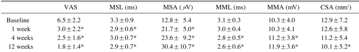
Initial VAS score was 6.5 (range 2-10). The VAS score has been significantly reduced since 1 week after injection (Table 1). Thirteen wrists showed symptomatic improvement at 1 week and all wrists showed improve- ment at 4 weeks after injection. This clinical improvement was still observed at 12 weeks after injection treatment.
Among electrophysiological parameters, median sensory latency and median sensory amplitude showed significant differences, compared with baseline results since 1 week after injection treatment. However, median motor latency and median motor amplitude had shown significant differences since 4 weeks after injection treat- ment (Table 1).
At baseline study, ultrasonography revealed enlarged median nerve, demonstrated as mean CSA, 12.9
mm2. The CSAs of median nerve showed the tendency of serial reduction since 1 week after injection treatment, however statistical significance was observed at 12 weeks after injection treatment (Table 1).
The clinical improvement (change of VAS score) was significantly correlated with baseline CSA (Table 2).
Among electrodiagnostic parameters, baseline median motor amplitude was negatively correlated with change of VAS score (Table 2).
No complications or adverse effects were observed during and after injection treatment.
DISCUSSION
CTS is usually diagnosed on the basis of characteristic symptoms and electrodiagnostic studies. Although
Table 1. Sequential Changes of Clinical, Ultrasonographic and Electrodiagnostic Parameters Analyzed by Repeated-Measures Analysis of Variance
VAS MSL (ms) MSA (μV) MML (ms) MMA (mV) CSA (mm2)
Baseline 6.5±2.2* 3.3±0.9c 12.8±*5.4* 3.1±0.3* 10.3±4.0* 12.9±7.2*
1 week 3.0±2.2* 2.9±0.6* 21.7±*5.0* 3.0±0.4* 10.3±4.1* 12.6±5.8*
4 weeks 2.5±1.6* 3.0±0.7* 23.6±*9.2* 2.8±0.5* 11.2±3.8* 11.2±5.4*
12 weeks 1.8±1.4* 2.9±0.7* 30.4±10.7* 2.6±0.6* 11.9±3.6* 10.1±5.2*
VAS, visual analogue scale; CSA, cross-sectional area; MML, median motor latency; MMA, median motor amplitude; MSL, median sensory latency; MSA, median sensory amplitude
*p <0.05
Table 2. Correlation between Changes of VAS and Initial Ultrasonographic and Electrodiagnostic Parameters
R p-value
CSA (mm2) 0.518 0.048*
MML (ms) 0.426 0.114
MMA (mV) -0.549 0.034*
MSL (ms) 0.419 0.120
MSA (μV) -0.304 0.271
VAS, visual analogue scale; CSA, cross-sectional area; MML, median motor latency; MMA, median motor amplitude; MSL, median sensory latency; MSA, median sensory amplitude
*p <0.05
– 83 –
electrodiagnostic study is highly specific,
13it does not provide spatial information. Moreover, false negative results can be seen in 10~20% of the cases.
14Ever since the introduction of ultrasonography to the musculoskeletal field, the ultrasonographic findings of CTS are well reported. Ultrasonography is useful in the diagnosis of CTS. The increased CSA of the median nerve, more than 10.5
mm2provides a diagnostic sensitivity of 89% and specificity of 94.7%.
15However, there are not enough reports regarding a structural change, after local steroid injection, conjoined with clinical improvement.
In our current study, we evaluated a sequential struc- tural change of the median nerve after local steroid injec- tion using ultrasonography in CTS. All of the 15 wrists with CTS, which received ultrasound-guided local steroid injection, demonstrated clinical improvement.
Initially enlarged median nerve showed serial reduction tendency in CSA and this became significant at 12 weeks after injection. The initial CSA of the median nerve correlated with the clinical improvement (r=0.518). Therefore, we could confirm that ultrasonog- raphy is a useful tool to predict the prognosis of CTS after injection treatment, as well as to diagnose a CTS.
However, as significant morphological change of edema- tous median nerve was observed after clinical and elec- trophysiological improvement, the follow-up evaluations of median nerve using ultrasonography might be helpful at least several weeks after a local steroid injection.
Increased pressure in the narrow carpal tunnel induces the compression and ischemic change of the median nerve. Moreover, the neural edema
16deteriorates clinical symptoms as well as the nerve injury. Through an ultra- sonographic evaluation of the median nerve in the trans- verse plane, we confirmed an increased CSA of the median nerve, probably resulting from local inflamma- tion and edema. Steroids are known to lower the local leukocyte and inflammatory modulator response, and also reduce neural and perineural edema. And this has been proposed as hypothesis for a structural change of the median nerve after steroid injection in the CTS.
Actually, we observed a continuous reduction of the CSA of the median nerve, which was consistent with a recent report showing significant improvement of ultrasonographic parameters, such as CSA, mobility, and vascularity, after a local steroid injection in the CTS.
10These results help us to understand the action mechanism of local steroid injection in the view of macro-structural change. In other words, the reduction in CSA of median nerve by steroid injection might lower
the pressure within carpal tunnel and might release the compressed median nerve from adjacent structures, followed by eventual improvement of clinical symptom.
Another interesting finding of current study is that the clinical improvement was significantly correlated with baseline CSA (Table 2). In other words, the amount of median nerve swelling in CTS could be one of prognostic factor after injection therapy. Previously, Mondelli et al.
reported presurgical values of CSA at the tunnel inlet could predict the normalization of its postsurgical value, normalization of the clinical severity scale.
17Naranjo et al.
also reported with CSA at the tunnel inlet of median nerve was the predictor of success after 3 months of surgical release.
18However, subjects enrolled in previous studies with surgical treatment might have more severe degree of CTS than those of current study. Disease duration of CTS could also effect on the injection outcome. For example, an injection treatment conducted to acute edema of median nerve might result in better response regardless of ultrasonographic CSA findings. Therefore, before more discussion on the feasibility of ultrasonography, we need further studies including more CTS patients with various severities and disease durations. Nevertheless, current report has the implication because it is the first one to reveal the possible usefulness of ultrasonography as a predicting tool of clinical outcome after injection treatment in CTS.
However, this study has some limitations. This is a preliminary study with small sample size and it included only those with a mild to a moderate degree of the carpal tunnel syndrome. We could not recruit a control group, either. Secondly, our study was conducted for a short period such that we observed a structural change of the median nerve only for 12 weeks after the injection treat- ment. Further research, including a control group with larger numbers of patients and long term follow-up should be conducted to obtain more informative results to understand the pathophysiology of CTS and to reveal feasibility of ultrasonography in evaluation and prediction of treatment effect in CTS.
CONCLUSIONS
The ultrasonographic evaluation of the median nerve
after a local steroid injection in CTS revealed significant
structural changes, concurrent with clinical and electro-
physiological improvement. The initial amount of medi-
an nerve swelling was correlated with symptomatic
improvement after injection therapy. These results might
help us to understand the action mechanism of steroid injection in CTS and to predict the effectiveness of injec- tion treatment in CTS as well as in other peripheral neu- ropathies.
REFERENCES
1. Osamura N, Zhao C, Zobitz ME, An KN, Amadio PC:
Permeability of the subsynovial connective tissue in the human carpal tunnel: a cadaver study. Clin Biomech (Bris- tol, Avon) 2007: 22: 524-528.
2. Tuncali D, Barutcu AY, Terzioglu A, Aslan G: Carpal tun- nel syndrome: comparison of intraoperative structural changes with clinical and electrodiagnostic severity.
British journal of plastic surgery 2005: 58: 1136-1142.
3. Agarwal V, Singh R, Sachdev A, Wiclaff, Shekhar S, Goel D: A prospective study of the long-term efficacy of local methyl prednisolone acetate injection in the management of mild carpal tunnel syndrome. Rheumatology (Oxford) 2005: 44: 647-650.
4. Ayhan-Ardic FF, Erdem HR: Long-term clinical and elec- trophysiological results of local steroid injection in patients with carpal tunnel syndrome. Funct Neurol 2000:
15: 157-165.
5. Gelberman RH, Aronson D, Weisman MH: Carpal-tunnel syndrome. Results of a prospective trial of steroid injec- tion and splinting. J Bone Joint Surg Am 1980: 62: 1181- 1184.
6. Giannini F, Passero S, Cioni R, Paradiso C, Battistini N, Giordano N, et al: Electrophysiologic evaluation of local steroid injection in carpal tunnel syndrome. Arch Phys Med Rehabil 1991: 72: 738-742.
7. Girlanda P, Dattola R, Venuto C, Mangiapane R, Nicolosi C, Messina C: Local steroid treatment in idiopathic carpal tunnel syndrome: short- and long-term efficacy. J Neurol 1993: 240: 187-190.
8. Hagebeuk EE, de Weerd AW: Clinical and electrophysio- logical follow-up after local steroid injection in the carpal tunnel syndrome. Clin Neurophysiol 2004: 115: 1464-
1468.
9. Dammers JW, Veering MM, Vermeulen M: Injection with methylprednisolone proximal to the carpal tunnel: ran- domised double blind trial. BMJ 1999: 319: 884-886.
10. Cartwright MS, White DL, Demar S, Wiesler ER, Sarliki- otis T, Chloros GD, et al: Median nerve changes following steroid injection for carpal tunnel syndrome. Muscle Nerve 2011: 44: 25-29.
11. Stevens JC: AAEM minimonograph #26: the electrodiag- nosis of carpal tunnel syndrome. American Association of Electrodiagnostic Medicine. Muscle Nerve 1997: 20:
1477-1486.
12. Kang S, Kwon HK, Kim KH, Yun HS: Ultrasonography of Median Nerve and Electrophysiologic Severity in Carpal Tunnel Syndrome. Annals of Rehabilitation Medi- cine 2012: 36: 72-79.
13. Nathan PA, Keniston RC, Meadows KD, Lockwood RS:
Predictive value of nerve conduction measurements at the carpal tunnel. Muscle Nerve 1993: 16: 1377-1382.
14. Duncan I, Sullivan P, Lomas F: Sonography in the diagno- sis of carpal tunnel syndrome. AJR Am J Roentgenol 1999: 173: 681-684.
15. Yesildag A, Kutluhan S, Sengul N, Koyuncuoglu HR, Oyar O, Guler K, et al: The role of ultrasonographic mea- surements of the median nerve in the diagnosis of carpal tunnel syndrome. Clin Radiol 2004: 59: 910-915.
16. Omer GE, Jr.: Median nerve compression at the wrist.
Hand Clin 1992: 8: 317-324.
17. Mondelli M, Filippou G, Aretini A, Frediani B, Reale F:
Ultrasonography before and after surgery in carpal tunnel syndrome and relationship with clinical and electrophysio- logical findings. A new outcome predictor? Scand J Rheumatol 2008: 37: 219-224.
18. Naranjo A, Ojeda S, Arana V, Baeta P, Fernandez-Pala- cios J, Garcia-Duque O, et al: Usefulness of clinical find- ings, nerve conduction studies and ultrasonography to pre- dict response to surgical release in idiopathic carpal tunnel syndrome. Clin Exp Rheumatol 2009: 27: 786-793.