Hereditary Hemorrhagic Telangiectasia
Jeong-Seung Kwon, D.D.S.,M.S.D.,Ph.D., Hyung-Joon Ahn, D.D.S.,M.S.D.,Ph.D., Jong-Hoon Choi, D.D.S.,M.S.D.,Ph.D.
Department of Orofacial Pain & Oral Medicine, Yonsei Dental Hospital, Yonsei University College of Dentistry, Seoul, Korea
Hereditary hemorrhagic telangiectasia is a rare autosomal dorminant disease that features abnormal and fragile vascular dilations of terminal vessels in skin and mucous membranes, as well as arteriovenous malformations of internal organs, particularly lungs, brain, and liver. Often patients have not been diagnosed with HHT for a long time, and undiagnosed HHT patients unnecessarily develop serious complications such as severe life-threatening hemorrhage, stroke or brain abscess. Therefore, early detection and appropriate screening is very important. Early detection of HHT allows the appropriate screening for the presence of silent disease such as AVMs in the lungs, liver, or brain, and preventive treatment in the patient and their affected family members. Dentists should be familiar with HHT because the telangiectases on skin and oral mucosa are often the most dramatic and most easily identified component of HHT.
Recently, we experienced a case of HHT. We present the case with a review of the literature.
Key words: Telangiectasia, arteriovenous malformation, hemorrhage
1)ⅠⅠ. INTRODUCTION
Hereditary hemorrhagic telangiectasia (HHT), known as Osler-Weber-Rendu syndrome or Rendu- Osler-Weber syndrome, is a rare autosomal dorminant disease. It features abnormal and fragile vascular dilations of terminal vessels in skin and mucous membranes, as well as arteriovenous malformations (AVM) of internal organs, parti- cularly lungs, brain, and liver.1) The telangiectases are small dilated blood vessels near the surface of
Corresponding author : Jong-Hoon Choi Professor, Department of Orofacial Pain & Oral Medicine, Yonsei Dental Hospital, Yonsei University College of Dentistry, 50 Yonsei-ro, Seodaemun-gu, Seoul 120-752, Korea
Tel: 82-2-2228-3113, 82-2-2228-8880 Fax: 82-2-393-5673
E-mail: [email protected] Received: 2012-08-11 Accepted: 2012-09-10
the skin or mucous membranes, and arteriovenous malformations are much larger than telangiectases.
Vascular malformations in HHT lack capillaries and consist of direct connections between arteries and veins.2)
Often patients have not been diagnosed with HHT for a long time, and undiagnosed HHT patients unnecessarily develop stroke or life- threatening hemorrhage.3) It is now considered to be more common than previously thought, and the associated brain and pulmonary lesions are sources of substantial morbidity and mortality.2) Therefore, early detection and appropriate screening is very important. Recently, we experienced a case of HHT.
We present the case with a review of the literature.
Ⅱ. CASE
A 41-year-old female visited to the department of orofacial pain and oral medicine, Yonsei Dental Hostpital with the complaint of the red spots
developed on tongue, lip and body. According to the patients, single red spot was developed on tongue before 1 year, and the number of spots increased by degrees on tongue, and spread to other parts. The lesions were not accompanied by pain and other symptoms. Occasionally, bleeding from red spot was caused by aggressive tongue cleansing. She visited an otolaryngologist, and heard that it may be attributed to leukemia. 6 months prior, laboratory test was preformed for regular health check up, but it was unremarkable. 4 months prior, laboratory test was done in oriental medical hospital, and it was also within normal limit. She was given herb medicine for a month, but the lesion did not change. Past medical history was unremarkable, and she received no medications, except the occasional use of gastrointestinal medication.
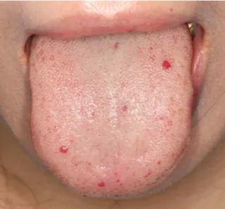
Clinical examination revealed multiple small round red macules or papules on dorsal surface of tongue (Fig. 1), hard palate (Fig. 2), upper lip (Fig.
3), and hand (Fig. 4). Those blanched on pressure.
Laboratory studies showed mildly decreased WBC (3,700/μL) and RBC (3,880,000/μL) count, and slightly elevated cholesterol level. prothrombin time (PT), activated partial thromboplastin time (aPTT), and Factor VIII were within normal limit, and von
Willebrand factor antigen was not detected.
When she was asked about epistaxis history and family history about similar conditions on the second visit, she reported her mother had recurrent
Fig. 2. Telangiectases on hard palate.
Fig. 3. Telangiectasia on lip.
Fig. 4. Telangiectasia on finger.
epistaxis and stroke history, and had been diagnosed with telangiectasia before 10 years in department of neurosurgery. She had also epistaxis history that was mild. She also reported that brain magnetic resonance imaging was performed on regular health check up before 10 years, and revealed no abnormalities.
Ⅲ. DISCUSSION
Consensus diagnostic criteria known as the Curaçao criteria were published in 2000, and recently validated.4,5) Diagnosis of HHT is based on 4 criteria: 1) a history of spontaneous and recurrent epistaxis; 2) the presence of multiple telangiectases;
3) visceral lesions (gastrointestinal telangiectasia, and pulmonary, hepatic, cerebral or spinal AVMs);
and 4) family history (a first-degree relative with HHT according to these criteria). The diagnosis of definite HHT is made if three or more criteria are present; possible or suspected HHT if two are present, and unlikely HHT if only one is present.
The most important sign is the appearance of telangiectases at characteristic sites (lips, oral cavity, hands, and nasal septum). If patient has an isolated visceral vascular lesion, but not these, HHT is very unlikely.6) But, most patients with pulmonary arteriovenous malformations (PAVMs) have HHT.7) It is impossible to obtain a definite diagnosis of HHT without a more specific visceral feature or a family history. If an individual from an HHT family has a visceral AVM, the diagnosis of HHT is essentially confirmed.5) Manifestations of HHT usually increased over a lifetime. However, those can be subtle even well into adulthood.1,6)
Small AVMs are called telangiectases. Telan- giectases appear on skin (lips, face, fingers, trunk, arms, nail beds), and nasal, oral (tongue, palate, buccal mucosa), gastrointestinal, genitourinary, and conjunctival mucosa. They appear as pink to red, pinpoint to pinhead-size macules or papules, or occasionally as larger, even raised purple lesions.
1,2,6) Telangiectases are distinguished from petechiae
by diascopy: a test for ability to blanch by applying
pressure with a finger or glass slide and observing color changes. This blanching indicates that the red color is due to blood contained within blood vessels.8)
The most common sign is epistaxis caused by spontaneous bleeding from telangiectases of the nasal mucosa, which occurs in the vast majority of affected persons, but not in all.9)Bleeding from oral lesions is also common, and may be difficult to control. Chronic hemorrhage may cause iron deficiency anemia.
Telangiectases of the skin typically present later in life than epistaxis. By the age of 40, most affected persons have multiple telangiectases.
There may be bleeding from cutaneous telangiectases, but it is rarely clinically important.
Telangiectases can cause cosmetic problem.2) PAVMs consist of direct connections between a branch of a pulmonary artery and vein through a thin-walled aneurysm.2) PAVMs are often asymptomatic, but those may result in direct right-to-left shunts, and may produce profound dyspnea, fatigue, cyanosis, or polycythemia. Often, however, their initial manifestations are brain abscess and stroke due to right-to-left shunting that facilitates the passage of septic and bland emboli into the cerebral circulation.10)
Neurologic symptoms, including migraine headache, brain abscess, transient ischemic attack, stroke, seizure, and intracerebral and subarachnoid hemorrhage, are common in patients with HHT.2) Brain abscess, transient ischemic attack, and ischemic stroke occur exclusively in patients with PAVMs who have right-to-left shunting.10)
Generally, genetic testing is not required for patients who were diagnosed with definite clinical HHT by the Curaçao criteria. Genetic testing is helpful when the diagnosis of HHT cannot be excluded clinically. It is especially most useful in children and young adults, who have no epistaxis or telangiectases, but PAVMs or CVMs.3) Identifi- cation of the specific HHT mutation in an HHT family allows diagnosis among those relatives (often children and young adults) who do not meet
clinical diagnostic criteria.3) The recent international guidelines recommended gene testing for adults and children with possible HHT.5)
Mutations in two genes account for the majority of cases of HHT: endoglin (ENG, resulting in HHT type 1) on chromosome 9, and activin A receptor type II-like 1 gene (ACVRL1, resulting in HHT type 2), or activin receptor-like kinase 1 (ALK 1), on chromosome 12.1,3,8) Mutation in these genes causes a disturbance in the transforming growth factor β (TGF-β) signaling pathway, which is important in vascular development and repair.1,6), and appear to result in the inability of a blood vessel to mature appropriately.5) Patients with HHT1 tend to more pulmonary involvement, whereas those with HHT2 have a later onset of telangiectasias and a greater degree of hepatic involvement.8)
Mutations in the SMAD4 gene can cause a rare syndrome which combines juvenile polyposis and HHT. Most HHT patients with SMAD4 mutation have juvenile polyposis that carries a risk of GI malignancy.11) Family and medical history should include questions about the occurrence of GI polyps and cancer when HHT is suspected.6)
Von Willebrand disease (vWD) can cause diagnostic confusion. Like HHT, vWD is inherited as an autosomal dominant trait. It also frequently causes epistaxis, and can be associated with mucocutaneous and gastrointestinal telangiectasia.
If there is no personal or family history of visceral AVMs, and no known HHT mutation, vWD should be ruled out.5) Telangiectasia may also be resulted from radiation therapy.1) CREST syndrome might be considered in a differential diagnosis. It includes calcinosis cutis, Raynaud's phenomenon, esophageal dysfunction, sclerodactyly, and telangiectasia.
Serologic studies for anticentromere autoantibodies often help to distinguish between the two conditions because these antibodies typically would be present only in CREST syndrome.8) The telangiectases by increased estrogen levels in the blood or chronic liver disease are characterized by a central red spot and small vessels radiating
outward like a spider's web. Most telangiectases in HHT are punctuate or macular.6)
Antibiotic prophylaxis prior to dental and surgical procedures is recommended for all patients with HHT and PAVMs, based on the endocarditis paradigm.3,6,12,13) Antibiotic prophylaxis is in accor- dance with the American Heart Association protocol with dental procedures with risk for bacteremia. This may reduce the risk of bacteremia that may cause the brain abscess, associated with right to left shunting in patients with PAVMs.14) In HHT patients, anticoagulants such as warfarin and nonsteroidal anti-inflammatory agents such as aspirin and ibuprofen that interfere with platelet function should be avoided unless required for treatment of other medical conditions.6)
HHT is rare condition, but it can cause serious complications such as severe hemorrhage, stroke or brain abscess. Early detection of HHT allows the appropriate screening for the presence of silent disease such as AVMs in the lungs, liver, or brain, and preventive treatment in the patient and their affected family members. Dentists should be familiar with HHT, because the oral lesions are often the most dramatic and most easily identified component of HHT.8)
Ⅳ. REFERENCE
1. Regezi JA, Sciubba JJ, Jordan RCK. Oral pathology:
clinical pathologic correlations. 6th ed., St. Louis, 2012, Elsevier. p. 117.
2. Guttmacher AE, Marchuk DA, White RI. Hereditary hemorrhagic telangiectasia. The New England journal of medicine 1995;333(14):918-924.
3. Faughnan ME, Palda VA, Garcia Tsao G et al.
International guidelines for the diagnosis and mana- gement of hereditary haemorrhagic telangiectasia.
Journal of medical genetics 2011;48(2):73-87.
4. Shovlin CL, Guttmacher AE, Buscarini E et al.
Diagnostic criteria for hereditary hemorrhagic telan- giectasia (Rendu-Osler-Weber syndrome). American journal of medical genetics 2000;91(1):66-67.
5. Shovlin CL. Hereditary haemorrhagic telangiectasia:
pathophysiology, diagnosis and treatment. Blood reviews 2010;24(6):203-219.
6. McDonald J, Bayrak Toydemir P, Pyeritz RE.
Hereditary hemorrhagic telangiectasia: an overview of diagnosis, management, and pathogenesis.
Genetics in medicine 2011;13(7):607-616.
7. Curie A, Lesca G, Cottin Vet al.Long-term follow- up in 12 children with pulmonary arteriovenous malformations: confirmation of hereditary hemorrhagic telangiectasia in all cases. The journal of pediatrics 2007;151(3):299-306.
8. Neville BW, Damm DD, Allen CM, Bouquot JE. Oral and maxillofacial pathology. 3rd ed., St. Louis, 2009, Saunders Co., pp. 754-755.
9. Aassar OS, Friedman CM, White RI. The natural history of epistaxis in hereditary hemorrhagic telan- giectasia. The Laryngoscope 1991;101(9):977-980.
10. White RI, Lynch Nyhan A, Terry Pet al.Pulmonary arteriovenous malformations: techniques and long- term outcome of embolotherapy. Radiology 1988;169 (3):663-669.
국문초록
유전성출혈모세혈관확장증의 증례 및 문헌 고찰
연세대학교 치과대학 구강내과학교실 권정승․안형준․최종훈
유전성출혈모세혈관확장증은 피부 및 점막에 있는 말단 혈관이 비정상적으로 확장된 모세혈관 확장증과 내부 장기, 특히 폐, 뇌, 간 부위의 동정맥기형 발생을 특징으로 하는 상염색체 우성 유전질환이다. 이 질병을 가진 환자들은 종종 오랜 시간 동안 진단이 되지 않은 채로 지내다가 생명을 위협할 수 있는 심각한 출혈, 뇌졸중, 뇌농양과 같은 합병증이 발생하기도 한다.
따라서 이 질환의 조기 진단 및 적절한 선별검사가 매우 중요하다. 유전성출혈모세혈관확장증의 조기 진단을 통해 증상 없이 존재하다 합병증을 유발할 수 있는 폐, 간, 뇌 부위의 동정맥기형에 대한 선별 검사를 시행함으로써 이 질병에 이환된 환자와 가족에 대한 예방적 관리가 가능하다. 피부 및 점막에 발생하는 모세혈관확장증은 특징적인 소견을 보이며 치과의사에 의해 쉽게 발견되므로 치과의사는 유전성출혈모세혈관확장증에 대해 잘 알고 조기 진단에 기여할 필요가 있다. 최근 이 질환으로 진단된 증례가 있어 문헌 고찰과 함께 보고하고자 한다.
주제어: 모세혈관확장증, 동정맥기형, 출혈
11. Gallione CJ, Repetto GM, Legius Eet al.A combined syndrome of juvenile polyposis and hereditary haemorrhagic telangiectasia associated with mutations in MADH4 (SMAD4). Lancet 2004;363 (9412):852-859.
12. Chan P. Antibiotic prophylaxis for patients with hereditary hemorrhagic telangiectasia. Journal of the American Academy of Dermatology 1992;26(2):
282-283.
13. Shovlin C, Bamford K, Wray D. Post-NICE 2008:
Antibiotic prophylaxis prior to dental procedures for patients with pulmonary arteriovenous malformations (PAVMs) and hereditary haemorrhagic telangiectasia.
British dental journal 2008;205(10):531-533.
14. Dupuis Girod S, Giraud S, Decullier E et al.
Hemorrhagic hereditary telangiectasia (Rendu-Osler disease) and infectious diseases: an underestimated association. Clinical infectious diseases 2007;44(6):
841-845.