Application of CO
2laser in Minor Surgery of Oral Soft Tissue : Case Reports
Ju Hyun Park, D.D.S.,M.S.D., Young-Mi Jeon, D.D.S.,M.S.D.,
Jeong-Seung Kwon, D.D.S.,M.S.D.,Ph.D., Hyung-Joon Ahn, D.D.S.,M.S.D.,Ph.D.
Department of Oral medicine, dental hospital, College of dentistry, Yonsei university
Conventional surgical therapy for oral soft tissue includes the use of scalpel, diathermy, cryotherapy and electrosurgery. But, these therapies have some surgical problems. Nowadays, laser surgery can be considered as the another option for conventional surgical therapy.
Compared to conventional surgical therapies, advantages of laser therapy include maintenance of sterile conditions, promotion of wound healing, reduction of bleeding, less instruments, post operative pain reduction, less scar, saving cost by using fewer materials, staff and time. Carbon dioxide (CO2) laser uses gaseous medium, and has long wavelength about 10,600nm. The first advantage of CO2 laser for surgical treatment of oral soft tissue is hemostasis and visibility improvement by making relatively dry field.
These case reports are about cases of minor surgery of oral soft tissue using CO2 laser, and emphasize advantages of laser compared to conventional surgical therapies.
Key words : Carbon dioxide (CO2) laser, Minor surgery, Oral soft tissue
1)Ⅰ. INTRODUCTION
Conventional surgical therapies for oral soft tissue are the use of scalpel, diathermy, cryotherapy and electrosurgery. But, these therapies have some surgical problems such as sterility and its maintenance during surgery, restricted spatial conditions, intraoperative bleeding, complications in
Corresponding author : Hyung-Joon Ahn, D.D.S., M.S.D.,Ph.D.
Department of Oral Medicine, College of Dentistry, Yonsei University, 134 Shinchon-dong, Seodaemun-gu, Seoul, South Korea
Tel : 82-2-2228-8875 Fax : 82-2-393-5673 E-mail : [email protected]
Received: 2010-06-08 Accepted: 2010-07-10
wound healing, scars. Nowadays, laser surgery can be considered as the another option for conventional surgical therapy.
Since the laser was used in dentistry for the first time in 1964 by Goldman, it has been documented in numerous studies about successful application in dentistry, and the spectrum of indication has been extended persistently.1) Compared to conventional surgical therapies, advantages of laser therapy are maintenance of sterile conditions, reduction of bleeding and post operative pain, good possible estimation of cutting depth, precision of cutting, using fewer dental instruments, often no need for suture or bandages, promotion of wound healing, less scar, less costs by reduction of materials, staff and time.2) Several lasers such as Argon laser, Diode laser, Er:YAG, Nd:YAG laser and carbon dioxide (CO2) laser are being developed for
numerous medical applications including dentistry.
3,4) Thus, one can select the most applicable laser under the given circumstances.
The CO2 laser is a gas-active medium laser that incorporates a sealed tube containing a gaseous mixture with CO2 molecules pumped via electrical discharge current.5) The energy of 10,600nm wavelength is placed at the end of the mid-infrared invisible nonionizing portion of the spectrum. In 1976, CO2 laser admitted FDA clearance for soft tissue. Use of the CO2 laser has been described extensively by many authors. The primary advantage of CO2 laser for surgical treatment of oral soft tissue are hemostasis and relative dry field for improved visibility.6)
Here, we present clinical cases of minor surgery of oral soft tissue, which were performed by CO2
laser.
Ⅱ. CASE Case 1
A 31-year-old female was presented with gingival swelling. In clinical examination, there was a well-defined fibrotic mass on the papillary gingiva between the right mandibular lateral incisor and canine (Fig. 1). In radiographic examination, there was tooth spacing between the right mandibular ateral incisor and canine but no other remarkable abnormalities was seen. Excisional biopsy was
Fig. 1. Pre-operation
planned, but surgical approach by scalpel was difficult. So, the excision was performed with CO2
laser (continuous wave mode, 4.0W; Panalas C05 ∑, Panasonic®, Japan) (Fig. 2). The mass was excised conservatively with minimum bleeding and good accessibility. It was taken care of the laser beam not irradiating to adjacent structures, and surrounding gingiva was cauterized to prevent recurrence (Fig.
3). At 2-days follow-up, the wound showed good healing state and the patient didn’t report any discomfort (Fig. 4). The official result of biopsy was
“Peripheral ossifying fibroma”.
Peripheral ossifying fibroma is a benign tumor, and more than 50% of the lesion occur in the incisor-cuspid lesion. Generally, surgical excision of this benign tumor has some difficulties because of
Fig. 2. Excisional biopsy
Fig. 3. Post-operation
Fig. 4. POD #2
restricted spatial conditions and intraoperative bleeding. But in this case, using CO2 laser, the surgical process and the result of surgery satisfied with the good wound healing.
Case 2
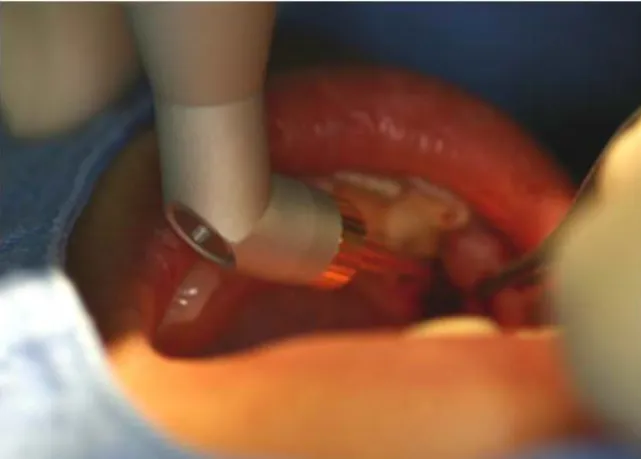
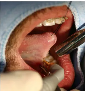
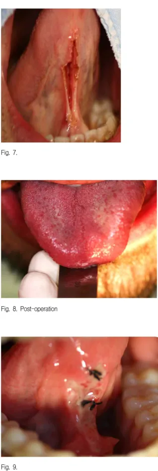
A 36-year-old male was presented with restricted tongue movement. In clinical examination, the lingual frenum was extended to the tip of the tongue, and there was limitation of tongue movement. The maximum range of protrusion of tongue tip was about 14mm from the labial surface of the lower incisor (Fig. 5). The diagnosis was ankyloglossia. So lingual frenectomy was performed with CO2 laser. Under local anesthesia, CO2 laser applied with continuous wave mode and output was 4.0W. The lingual frenum was incised with CO2 laser and the incised space was dilated mechanically (Fig. 6,7). During and after the surgery, maximum range of protrusion of tongue tip was checked. The surgery was performed under a good visibility of the operating site with good bleeding control. Suture was performed to prevent re-attachment of elongated lingual frenum. At 2-days follow-up, the range of movement was increased to about 25mm from the labial surface of the lower incisor and the wound showed good healing state (Fig. 8, 9).
Fig. 5. Pre-operation
Fig. 6. Incision with CO2 laser
Because of extensive bleeding tendency of tongue, conventional surgical methods have some difficulties. In this case, using CO2 laser, the surgical process was much easier with good bleeding control and improved visibility.
Fig. 7.
Fig. 8. Post-operation
Fig. 9.
Ⅲ. DISCUSSION
Because of some characteristics of oral soft tissue conventional surgical methods have some difficulties. Among them, bleeding and difficulty of maintaining visual field are major difficulties to clinicians. The other side, because of many characteristic features, laser can be a good another substitute option for conventional surgical therapy.
Also, lasers are becoming popular due to the advent of office-based lasers, which are small, portable, and easy to manipulate within the oral cavity.
Lasers are becoming the standard of care for many oral and maxillofacial procedures, and they are being introduced as an efficient instrument for a variety of new applications within the speciality.
These case reports show two cases of minor surgery of oral soft tissue using CO2 laser, and emphasize advantages of laser compared to conventional surgical therapies. Compared to the conventional surgical therapies, the laser has some advantages in hemostasis and relative dry field for improved visibility, which might enable it to be an alternative to the conventional surgical therapy.
The laser induces sealing the blood vessels of approximately 500μm or less by increasing platelet activation at the point of the wound and contraction of the vascular wall collagen.7) The reduction in swelling following the use of lasers appears to be related th the sealing of the lymphatic vessels.8) Also, the laser is used in aesthetic procedures such as dental bleaching, and for bio-stimulation by inducing mineralization and osteoblast differentiation, accelerating new bone formation within the marrow cavity, effect on cell proliferation.9,10) Recently, low level laser therapy(LLLT), also known as "soft laser therapy", is used widely for bio-stimulation. Its mechanisms are complex, but LLLT has a potent action that results in stimulation of the normal functions of the cells by increasing supply of the cell's fuel, ATP.11) Because the absorption of laser energy into the tissues facilitates accomplishing the desired result, the CO2 lasers are particularly well suited for soft
tissue surgery: its wavelength is highly absorbed by the water content of the tissues.12) Because soft tissues contain approximately 90% water, especially the swollen, hyperemic tissue would have more water content, they absorb the CO2 wavelength easily. And, the introduction of hollow waveguide technology during the 1980s made CO2 lasers even easier to use.
Although it has many advantages, the laser technique requires some precautions. The CO2 laser is absorbed by the water component of dental hard tissues, which could lead to thermal damage. A significant delivery of laser energy may result in temperature changes enough to compromise the dental pulp, and etching or pitting of the enamel.
Therefore, contact with those tissues must be avoided and modifying the output of the laser may be desirable. Any inadvertent mistake on hard tissue may inhibit the attachment of fibroblasts and delay wound healing.13) So, I think that clinicians experienced in CO2 laser surgery have to be emphasized the need for an adequate shield, such as a blat bladed instrument or silver foil, between the gingiva and teeth.14) If we aware of those cautions, CO2 laser will be very useful method for surgical therapy of oral soft tissue.
REFERENCES
1. Goldman L. Laser cancer research. Berlin, 1996, Springer.
2. Rossman JA. Lasers in periodontics. A position paper by the American Academy of Periodontology. J Periodontol 2002;73:1231-1239.
3. Kaplan I, Gassner S, Shindei Y. Carbon dioxide in laser in head and neck surgery. Am J Surg 1974;128:563-567.
4. Strauss R. Lasers in oral and maxillofacial surgery.
Dent Clin North Am 2000;44:851-873.
5. Wigdor H, Walsh J, Visuri S, Fried D, Waldvogel J.
Lasers in dentistry. Laser Surg Med 1995;16:103-133.
6. Manni JG. Dental applications of advanced lasers.
Burlington(MA), 2000, JGM Associates.
7. Mordon S, Begu S, Buys B, et al. study of platelet behavior in vivo after endithelial stimulation with laser irradiation using fluoresence in trivital videomicsocopy and PEG-ylated liposome staining.
Microvac 2002;64:316-325.
8. Pick PH, Pecaro BC, Silberman CJ. The laser gingivectomy. The use of the CO2 laser for the removal of phenytoin hyperplasia. J Periodontol 1985;56:492-494.
9. Bjordal JM, Couppe C, Ljunggren A. Low level laser therapy for tendinopathies: evidence of a dose- response pattern. Phys Ther Reviews 2000;6:91-100.
10. Sun G, Tuner J. Low-level laser therapy in dentistry.
Dent Clin N Am 2004;48:1061-1076.
11. Coluzzi DJ. An overview of laser wavelengths used in dentistry. Dent Clin N Am 2000;44:753-776.
12. Passarella S. Increase of proton electrochemical potential and ATP synthesis in rat liver mitochondria irradiated in vitro by helium-neon laser. FEBS Lett 1984;175:95-99.
13. Spencer P, Cobb CM, Wieliczka DM, Glaros HG, Morris PJ. Change in temperature of subjacent bone during soft tissue laser ablation. J Periodontol 1998;69:1278-1282.
14. Haytac MC, Ozcelik O. Evaluation of patient perceptions after frenectomy operations: A comparison of carbon dioxide laser and scalpel techniques. J Periodontol 2006;77:1815-1819.
국문초록
연조직 소수술에서 CO2 레이저의 적용 증례
연세대학교 치과대학 구강내과학교실 박주현․전영미․권정승․안형준
구강 내 연조직 소수술 시, 수술도, 열 요법, 전기요법, 한냉요법 등은 현재까지 사용되고 있는 고전적인 수술법이다. 하지만 이들 통상적인 수술적 방법의 문제로 멸균 상태의 유지, 공간의 제한, 출혈, 창상 치유의 문제, 흉터, 전신질환에 의한 수술의 제한 등이 있다. 최근 레이저가 또 하나의 수술법으로 널리 사용되고 있고, 통상적인 외과적 수술법에 비하여 레이저의 장점 으로는 멸균상태 유지, 출혈감소, 통증 감소, 창상의 치유 촉진, 반흔 생성 억제 등이 있다. 특히 탄산가스 레이저는 조직 내 수분에 최대한으로 흡수되어 100μm 정도의 낮은 침투도를 가지며, 주변부 모세 혈관을 응고시켜 구강 내 소수술에 적용 시 우수한 지혈 효과 및 수술 시야의 확보를 얻을 수 있다.
주제어 : 탄산가스레이저, 연조직 소수술