KJTCVS
The Korean Journal of Thoracic and Cardiovascular SurgeryClinical Research The Role of Primary Tumor Resection in Patients with Pleural Metastasis Encountered at the Time of Surgery
Samina Park, M.D., Yongwoo Chung, M.D., Hyun Joo Lee, M.D., In Kyu Park, M.D., Chang Hyun Kang, M.D., Young Tae Kim, M.D.
Department of Thoracic and Cardiovascular Surgery, Seoul National University Hospital, Cancer Research Institute, Seoul National University College of Medicine, Seoul, Korea
ARTICLE INFO
Received October 10, 2019 Revised November 11, 2019 Accepted November 22, 2019 Corresponding author Young Tae Kim Tel 82-2-2072-3161 Fax 82-2-764-3664 E-mail [email protected] ORCID
https://orcid.org/0000-0001-9006-4881
Background: Evidence is lacking on whether the resection of lung parenchymal cancer improves the survival of patients with unexpected pleural metastasis encountered during surgery. We conducted a single-center retrospective study to determine the role of lung resection in the long-term survival of these patients.
Methods: Among 4683 patients who underwent lung surgery between 1995 and 2014, 132 (2.8%) had pleural metastasis. After excluding 2 patients who had incomplete medical records, 130 patients’ data were collected. Only a diagnostic pleural and/or lung biopsy was performed in 90 patients, while the lung parenchymal mass was resected in 40 patients.
Results: The mean follow-up duration was 29.8 months. The 5-year survival rate of the resection group (34.7%±9.4%) was superior to that of the biopsy group (15.9%±4.3%, p=0.016). Multivariate Cox regression analysis demonstrated that primary tumor resection (p=0.041), systemic treatment (p<0.001), lower clinical N stage (p=0.018), and adenocarci- noma histology (p=0.009) were significant predictors of a favorable outcome. Interestingly, primary tumor resection only played a significant prognostic role in patients who received systemic treatment.
Conclusion: When pleural metastasis is unexpectedly encountered during surgical ex- ploration, resection in conjunction with systemic treatment may improve long-term sur- vival, especially in adenocarcinoma patients without lymph node metastasis.
Keywords: Lung neoplasms, Non-small-cell lung carcinoma, Pleural metastasis
Copyright©The Korean Society for Thoracic and Cardiovascular Surgery. 2020. All right reserved.
This is an Open Access article distributed under the terms of the Creative Commons Attribution Non-Commercial License (http://creativecommons.org/licenses/
Introduction
Approximately 40% of cases of non–small cell lung can- cer (NSCLC) are metastatic at the time of diagnosis [1].
Pleural dissemination, which refers to malignant pleural effusion and/or pleural nodules, is identified in 1%–7.5% of NSCLCs and is a component of the M1a descriptor [1,2].
The median survival duration of patients with M1a disease is 8–11.5 months [2,3]. Owing to the poor prognosis of pleural dissemination, with a 2% 5-year survival rate, NS- CLC with pleural dissemination was reclassified from T4 to M1a in the seventh edition of the tumor-node-metasta- sis (TNM) staging system [2].
NSCLC with a solitary metastasis may be treated by re- secting both the primary and metastatic lesions, with a fa-
vorable survival rate [4,5]. Consequently, it was proposed for the eighth edition of the TNM staging system that me- tastasis at a single site or single organ should be distin- guished from other distant metastatic lung cancer [3]. A contralateral lung nodule (the other M1a descriptor) may sometimes also be a surgical candidate. In contrast, NS- CLC with pleural metastasis is not a candidate for surgical resection according to standard treatment guidelines, as pleural dissemination is not amenable to complete resec- tion [6]; therefore, the M1a category was not revised for the eighth edition of the TNM staging system [3]. However, several studies have reported reasonable survival outcomes after surgical resection of the primary tumor in patients with pleural dissemination [7-11]. Therefore, it remains un- clear whether resection of the primary tumor can improve
https://doi.org/10.5090/kjtcs.2020.53.3.114 pISSN: 2233-601X eISSN: 2093-6516
Korean J Thorac Cardiovasc Surg. 2020;53(3):114-120
Samina Park, et al. Primary Tumor Resection in Patients with Pleural Metastasis
KJTCVS
survival in patients with pleural metastasis encountered at the time of surgery [12].
We aimed to evaluate the role of primary tumor resec- tion in patients with pleural dissemination of NSCLC and to identify other predictors of long-term survival.
Methods
Patients
Among 4,683 patients who underwent lung surgery be- tween 1995 and 2014, 132 (2.8%) were identified as having clinical M0 stage disease and found to have unexpected pleural metastasis at the time of surgery. After excluding 2 patients whose medical records were not complete, 130 pa- tients’ data were collected. Survival data were confirmed using medical records, telephone surveys, and the national insurance database. Data were analyzed to test the role of lung resection in long-term survival and to identify other predictors of patients’ prognosis. The primary tumor was not resected in 90 patients, in whom only a diagnostic pleural biopsy was performed, whereas the primary lung mass was resected in 40 patients. Systemic treatment (cyto- toxic chemotherapy and/or tyrosine kinase inhibitors) was administered to 110 patients (84.6%). Several clinical vari- ables were analyzed to identify factors that could have in- fluenced the long-term survival of these 2 groups. This study was reviewed and approved by the Institutional Re- view Board at Seoul National University Hospital (IRB ap- proval no., H-1602-050-739). The recommendations of the Declaration of Helsinki for biomedical research involving human subjects were followed. Informed consent was waived.
Statistical analysis
Categorical variables are presented as numbers and per- centages. Continuous variables are presented as means and standard deviations. Statistical analysis was performed us- ing IBM SPSS ver. 22.0 (IBM Corp., Armonk, NY, USA). In the univariate analysis, categorical variables were com- pared using the chi-square test and the Fisher exact test.
Continuous variables were compared using the Student t-test. Survival curves were obtained using the Kaplan-Mei- er method and compared between the 2 groups using the log-rank test. Prognostic factors for survival were analyzed with a Cox proportional hazard ratio model fit with a backward selection method. The proportional hazards as- sumption, tested by a log minus log plot, was satisfied.
Variables with p≤0.1 in the univariate analysis were includ-
ed in the multivariate analysis. Interactions between any 2 variables in the multivariate models were tested. A p-value of ≤0.05 was considered to indicate statistical significance in both the univariate and multivariate analyses.
Results
Clinicopathologic characteristics
Patients’ characteristics are described in Table 1. The study population had a male predominance (total, 130;
male-to-female ratio, 75:55) and most patients had adeno- carcinoma (n=109, 83.8%). The mean age was 61.5 years.
The mean follow-up duration was 29.8±23.7 months. There were no cases of surgery-related mortality. Of the 65 pa- tients tested for epidermal growth factor receptor (EGFR) mutation status, 50.5% were positive. Clinical variables in- cluding age, sex, tumor size, tumor location, histologic cell type, systemic treatment, clinical T stage, clinical N stage, EGFR mutation status, and preoperative pulmonary func- tion were similar between the 2 groups.
Survival
The overall 5-year survival rate was 20.1% (Fig. 1). The median survival time was estimated at 26.3 months. Pa- tients who received systemic treatment survived longer than those who did not (5-year survival rate, 22.3% versus 6.2%; p<0.001). The 5-year survival rate of the resection group was superior to that of the biopsy group (5-year sur- vival rate, 34.7% versus 15.9%; p=0.016) (Fig. 2). There was no significant difference in survival according to the extent of resection (p=0.719). When patients were stratified ac- cording to whether they received systemic treatment, the survival curves of the 2 groups showed different patterns.
A protective role of primary tumor resection was only ob- served in patients who received systemic treatment (Fig.
3A). In the systemic treatment group, patients who under- went primary tumor resection showed better survival out- comes than those who underwent biopsy (5-year survival rate, 40.0% versus 17.2%; p=0.009). However, among pa- tients who did not receive chemotherapy, the median sur- vival time was extremely poor in both groups (resection group versus biopsy group: 11.8 months versus 8.3 months), indicating that resection failed to improve survival (p=0.775) (Fig. 3B).
https://doi.org/10.5090/kjtcs.2020.53.3.114
KJTCVS
Prognostic factors for long-term survival
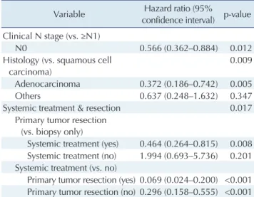
In the univariate analysis, clinical N stage and adenocar- cinoma histology were additional significant factors asso- ciated with favorable survival outcomes, whereas age, sex, and clinical T stage were not significant (Table 2). In the multivariate Cox regression analysis, primary tumor resec- tion, systemic treatment, clinical N0 stage, and adenocarci- noma histology remained significant factors associated with favorable outcomes (Table 3).
Interaction analysis
A significant interaction was found between primary tu-
mor resection and systemic treatment (p=0.017). This demonstrated a differential effect of systemic treatment on the impact of primary tumor resection. After adjusting for interaction between primary tumor resection and systemic treatment, clinical N0 stage (hazard ratio [HR], 1.811; 95%
confidence interval [CI], 1.158–2.832; p=0.009) and adeno- carcinoma histology (HR, 0.344; 95% CI, 0.172–0.689;
p=0.003) remained significant predictors of favorable sur- vival. We then estimated the HRs for mortality according to whether primary tumor resection was performed. The HR of systemic treatment decreased from 0.296 (95% CI, 0.158–0.555; p<0.001) to 0.069 (95% CI, 0.024–0.200;
p<0.001), suggesting that resection of the primary tumor may significantly strengthen the effect of systemic treat- Table 1. Clinicopathological characteristics of the patients
Characteristic Total
(N=130)
Primary tumor resection group (n=40)
Biopsy group
(n=90) p-value
Age (yr) 61.5±11.7 62.7±11.1 61.0±12.0 0.425
Female sex 55 (42.3) 16 (40.0) 39 (43.3) 0.723
Never-smoker 74 (56.9) 21 (52.5) 53 (58.9) 0.566
Forced expiratory volume in 1 second (predicted %) 98.1±21.4 102.6±21.7 95.7±21.0 0.117
Tumor size (cm) 3.2±1.4 3.3±1.7 3.1±1.2 0.428
Tumor location 0.670
Central 35 (26.9) 12 (30.0) 23 (25.6)
Periphery 95 (73.1) 28 (70.0) 67 (74.4)
Histology 130 40 90 0.287
Squamous cell carcinoma 12 (9.2) 6 (15.0) 6 (6.7)
Adenocarcinoma 109 (83.8) 32 (80.0) 77 (85.6)
Others 9 (6.9) 2 (5.0) 7 (7.8)
Systemic treatment (yes) 110 (84.6) 35 (87.5) 75 (83.3) 0.543
Cytotoxic chemotherapy (yes) 96 (73.8) 28 (70.0) 68 (75.6) 0.522
Tyrosine kinase inhibitor (yes) 72 (55.4) 21 (52.5) 51 (56.7) 0.705
T stage 0.503
1 38 (29.2) 14 (35.5) 24 (26.7)
2 61 (46.9) 18 (45.0) 43 (47.8)
3 22 (16.9) 7 (17.5) 15 (16.7)
4 9 (6.9) 1 (2.5) 8 (6.2)
N stage 0.168
0 86 (66.2) 26 (65.0) 60 (66.7)
1 21 (16.2) 10 (25.0) 11 (12.2)
2 21 (16.2) 4 (10.0) 17 (18.9)
3 2 (1.5) 0 2 (2.2)
Surgery 130 40 90
Pleural or lung biopsy - 90 (100.0)
Primary tumor resection 40 (100.0) -
Wedge resection 16 (40.0)
Lobectomy 21 (52.5)
Pneumonectomy 3 (7.5)
Length of hospital stay 8.0±7.9 9.9±11.7 7.1±5.2 0.153
Epidermal growth factor receptor mutation (yes) (n=65) 33/65 (50.5) 16/27 (59.3) 17/38 (44.7) 0.248 Values are presented as mean±standard deviation or number (%).
Samina Park, et al. Primary Tumor Resection in Patients with Pleural Metastasis
KJTCVS
ment (Table 3).
Discussion
Although the mainstay of treatment in patients with ad- vanced-stage cancer is systemic chemotherapy [13], our study showed that surgery can yield an additional survival benefit in carefully selected patients. Metastasis to a single organ, such as the adrenal gland or brain, can be treated with resection of the primary tumor and local manage- ment of the metastatic lesion, either with surgery or with a gamma knife. Therefore, the recent eighth edition of the TNM staging system for lung cancer proposed a subclassi- fication of the M description based on the number of met- astatic lesions and sites [3]. Oligometastasis in the brain,
liver, bone, adrenal gland, skin, and distant lymph nodes, which can be managed with aggressive local treatment, has a survival rate similar to that of M1a disease and better than that of cases with multiple metastases to a single or- gan [3,14]. These outcomes contributed to the subclassifi- cation of the M stage, which was divided into M1a and M1b, corresponding to a distinction between stage IV and stage IVa, in the eighth edition [3].
Pleural dissemination, which accounts for 13%–14% of all cases of metastatic lung cancer, remained in the M1a category [2,3], and the role of primary tumor resection in cases of pleural dissemination has not been established.
Unlike brain or adrenal metastases, complete resection is not possible in pleural dissemination, with the exception of
1.0
0.8
0.6
0.4
0.2
10
Survivalrate
Time (yr) 0
No. at risk 130
2 4 6 8
64 27 6 4 1
0.0
Median survival=26.3 mo 5-yr survival=20.1%
Fig. 1. Kaplan-Meier plot of the overall survival rate of non-small cell lung cancer patients with pleural metastasis.
1.0 0.8 0.6 0.4 0.2
Survivalrate
Time (yr)
0 2 4
0.0
No.
at risk
Tumor resection Biopsy
1 3 5
40
90
32
68
23
41
12
26
8
19 4
9
Fig. 2. Kaplan-Meier plots of survival rates according to primary tumor resection (solid line: primary tumor resection group; dotted line: biopsy group).
1.0 0.8 0.6 0.4 0.2
Survivalrate
Time (yr)
0 2 4
0.0
No.
at risk
Tumor resection Biopsy
1 3 5
35
75
30
63
23
40
12
25
8
18 4
8
A B 1.0
0.8 0.6 0.4 0.2
Survivalrate
Time (yr)
0 2
0.0
No.
at risk
Tumor resection Biopsy
1 3
2
5
0
1 0
1 5
15
Fig. 3. Kaplan-Meier plots of survival rates according to systemic treatment (solid line: primary tumor resection group; dotted line: biopsy group). (A) Patients who received systemic treatment. (B) Patients who did not receive systemic treatment.
https://doi.org/10.5090/kjtcs.2020.53.3.114
KJTCVS
extra-pleural pneumonectomy. Yokoi et al. [15] reported a 5-year survival rate as high as 54.5% after extra-pleural pneumonectomy in carefully selected patients. More recent studies have reported that surgical resection of the primary tumor in NSCLC with pleural dissemination was beneficial in selected patients, particularly in those with N0 status, T1-2 disease, and adenocarcinoma histology [7-10,16,17].
Resection of the primary lesion may provide effective local control because chemoradiation therapy often fails, with a locoregional failure rate of 31%–100% [18,19]. In addition, as minimally invasive surgery has been adopted and expe- rience with lung cancer surgery has been accumulated, surgery-related morbidity and mortality have decreased [20,21], and hence, surgical resection of a parenchymal lung lesion can be safely performed without adding signifi- cant operative complications. In our study, it was evident that primary tumor resection was performed in patients with good physical status, clinical N0 stage, and minimal pleural seeding. However, Li et al. [17] reported that there
were no differences in survival according to performance status, clinical stage, the presence of malignant pleural ef- fusion, or the extent of pleural nodules in cases of adeno- carcinoma. Notwithstanding, it is undeniable that when resection of the primary tumor can be safely performed, surgeons might decide to proceed to curative resection.
In several other reports, in which 57%–72% of patients received systemic treatment, chemotherapy was not found to improve survival [7,8,11]. Those conclusions are contra- dictory to ours. Most patients (85%) received systemic treatment in our series and demonstrated significantly bet- ter survival than those who did not, which may have re- sulted from selection bias. However, the fact that surgical resection of the lung lesion was only beneficial if the pa- tient underwent systemic treatment was interesting. A pos- sible explanation for our observations is that surgery de- creased the tumor burden, while chemotherapy treated microscopic disease. In fact, we found that the best long- term survival was achieved when both the tumor size and nodal stage were low. Although our data do not provide di- rect evidence regarding this possibility, our observations suggest that primary tumor resection may play a more im- pactful role for tumors with actionable mutations that can be treated with molecular target agents. We analyzed the presence of an EGFR mutation as a factor that might affect patients’ outcome. Unfortunately, however, we did not rou- tinely examine EGFR mutation status and we failed to prove that EGFR mutation status had a statistically signifi- cant relationship with survival, as was shown in a previous study [17]. Currently, we routinely evaluate EGFR mutation status and ALK gene rearrangement in all patients, and we Table 2. Univariate analysis of prognostic factors for long-term
survival in non–small cell lung cancer
Variable Hazard ratio (95%
confidence interval) p-value Age (vs. ≥65 yr)
<65 yr 1.225 (0.815–1.840) 0.328
Sex (vs. male)
Female 0.990 (0.660–1.484) 0.961
Smoking (vs. ever-smoker)
Never-smoker 0.700 (0.468–1.045) 0.081 Epidermal growth factor receptor
mutation (vs. wild-type)
Yes 0.621 (0.327–1.180) 0.146
Tumor size (vs. ≥3 cm)
<3 cm 0.951 (0.626–1.445) 0.815
Tumor location (vs. central)
Periphery 0.795 (0.511–1.236) 0.308
Clinical T stage (vs. ≥T2)
T1 0.883 (0.568–1.374) 0.581
Clinical N stage (vs. ≥N1)
N0 0.644 (0.419–0.990) 0.045
Systemic treatment (vs. no)
Yes 0.254 (0.149–0.431) <0.001
Resection (vs. biopsy only)
Primary tumor resection 0.557 (0.343–0.903) 0.018 Extent of resection (vs. wedge
resection) (n=40)
≥ Lobectomy 0.842 (0.330–2.146) 0.719 Histology (vs. squamous cell
carcinoma)
0.039 Adenocarcinoma 0.512 (0.263–0.997) 0.049
Others 0.981 (0.396–2.429) 0.966
Table 3. Multivariate Cox regression analysis of prognostic factors for long-term survival in non–small cell lung cancer
Variable Hazard ratio (95%
confidence interval) p-value Clinical N stage (vs. ≥N1)
N0 0.566 (0.362–0.884) 0.012
Histology (vs. squamous cell carcinoma)
0.009 Adenocarcinoma 0.372 (0.186–0.742) 0.005
Others 0.637 (0.248–1.632) 0.347
Systemic treatment & resection 0.017
Primary tumor resection (vs. biopsy only)
Systemic treatment (yes) 0.464 (0.264–0.815) 0.008 Systemic treatment (no) 1.994 (0.693–5.736) 0.201 Systemic treatment (vs. no)
Primary tumor resection (yes) 0.069 (0.024–0.200) <0.001 Primary tumor resection (no) 0.296 (0.158–0.555) <0.001
Samina Park, et al. Primary Tumor Resection in Patients with Pleural Metastasis
KJTCVS
hope to clarify the role of surgical resection of the primary tumor in patients who harbor a targetable mutation in the near future.
It has been suggested that tumor stage, sex, weight loss, and performance status are predictive factors of the success of systemic treatment. Performance status is an extremely powerful prognostic factor for survival and has been con- sidered as the most important factor in selecting patients eligible for chemotherapy [22,23]. However, we did not in- clude performance status in this study, because surgery was planned for all patients, meaning that their perfor- mance status was good.
We were not able to extract information regarding the number of pleural nodules. Dry pleural dissemination has been reported to have a better prognosis than wet pleural dissemination [8,9,16,24]. As all the patients were in the cM0 stage, the majority of our patients had dry pleural me- tastases. However, a multicenter survey reported that there was no significant difference in prognosis according to M1a descriptors (pleural effusion, pleural nodules, contra- lateral lung nodules) [3].
There are several limitations of this study. Surgical resec- tion is not recommended for cases of extremely advanced lung cancer with overt pleural effusion or pleural nodules;
hence, there was obvious selection bias in our study. Im- portantly, our conclusions should only be applied to pa- tients in whom pleural seeding was unexpectedly diag- nosed at the time of surgery. EGFR mutation data was available for only half of the patients. In addition, we did not analyze patients’ response to chemotherapy or disease progression. As a consequence, we were not able to per- form an interaction analysis to eliminate the effect of tu- mor biology. However, our results suggest that resection of the primary tumor can lead to improved survival if pa- tients can undergo systemic treatment for NSCLC with pleural dissemination that was unexpectedly encountered during surgery, especially in cases of adenocarcinoma without lymph node metastasis. Furthermore, in the era of precision medicine, surgical resection of the primary tu- mor may serve as an effective local treatment in conjunc- tion with appropriate targeted therapy.
In conclusion, when pleural metastasis is encountered during surgical exploration, primary tumor resection can improve long-term survival in conjunction with systemic treatment, especially in selected patients with adenocarci- noma histology without lymph node metastasis.
Conflict of interest
No potential conflict of interest relevant to this article was reported.
Acknowledgments
The authors would like to express their appreciation for the statistical advice provided by the Medical Research Collaborating Center at Seoul National University Hospital and the Seoul National University College of Medicine.
Funding
This study was supported by the National Research Foun- dation: ID (http://dx.doi.org/10.13039/100011512); award no. (NRF-2014R1A2A2A05003665).
ORCID
Samina Park: https://orcid.org/0000-0001-9625-2672 Yongwoo Chung: https://orcid.org/0000-0002-0355-328X Hyun Joo Lee: https://orcid.org/0000-0002-3092-2167 In Kyu Park: https://orcid.org/0000-0003-3550-5554 Chang Hyun Kang: https://orcid.org/0000-0002-1612-1937 Young Tae Kim: https://orcid.org/0000-0001-9006-4881
References
1. Ou SH, Zell JA. Validation study of the proposed IASLC staging re- visions of the T4 and M non-small cell lung cancer descriptors using data from 23,583 patients in the California Cancer Registry. J Thorac Oncol 2008;3:216-27.
2. Postmus PE, Brambilla E, Chansky K, et al. The IASLC Lung Can- cer Staging Project: proposals for revision of the M descriptors in the forthcoming (seventh) edition of the TNM classification of lung can- cer. J Thorac Oncol 2007;2:686-93.
3. Eberhardt WE, Mitchell A, Crowley J, et al. The IASLC Lung Can- cer Staging Project: proposals for the revision of the M descriptors in the forthcoming eighth edition of the TNM classification of lung cancer. J Thorac Oncol 2015;10:1515-22.
4. Billing PS, Miller DL, Allen MS, Deschamps C, Trastek VF, Pairole- ro PC. Surgical treatment of primary lung cancer with synchronous brain metastases. J Thorac Cardiovasc Surg 2001;122:548-53.
5. Mercier O, Fadel E, de Perrot M, et al. Surgical treatment of solitary adrenal metastasis from non-small cell lung cancer. J Thorac Cardio- vasc Surg 2005;130:136-40.
6. Ettinger DS, Wood DE, Akerley W, et al. NCCN guidelines insights:
non-small cell lung cancer, version 4.2016. J Natl Compr Canc Netw
https://doi.org/10.5090/kjtcs.2020.53.3.114
KJTCVS
2016;14:255-64.
7. Ichinose Y, Tsuchiya R, Koike T, et al. Prognosis of resected non- small cell lung cancer patients with carcinomatous pleuritis of mini- mal disease. Lung Cancer 2001;32:55-60.
8. Iida T, Shiba M, Yoshino I, et al. Surgical intervention for non-small- cell lung cancer patients with pleural carcinomatosis: results from the Japanese Lung Cancer Registry in 2004. J Thorac Oncol 2015;10:
1076-82.
9. Fukuse T, Hirata T, Tanaka F, Wada H. The prognostic significance of malignant pleural effusion at the time of thoracotomy in patients with non-small cell lung cancer. Lung Cancer 2001;34:75-81.
10. Go T, Misaki N, Matsuura N, Chang SS, Tarumi S, Yokomise H.
Role of surgery in multi-modality treatment for carcinomatous pleu- ritis in patients with non-small cell lung cancer. Surg Today 2015;45:
197-202.
11. Mordant P, Arame A, Foucault C, Dujon A, Le Pimpec Barthes F, Ri- quet M. Surgery for metastatic pleural extension of non-small-cell lung cancer. Eur J Cardiothorac Surg 2011;40:1444-9.
12. Sawabata N, Matsumura A, Motohiro A, et al. Malignant minor pleu- ral effusion detected on thoracotomy for patients with non-small cell lung cancer: is tumor resection beneficial for prognosis? Ann Thorac Surg 2002;73:412-5.
13. Non-Small Cell Lung Cancer Collaborative Group. Chemotherapy and supportive care versus supportive care alone for advanced non- small cell lung cancer. Cochrane Database Syst Rev 2010;(5):
CD007309.
14. Ashworth AB, Senan S, Palma DA, et al. An individual patient data metaanalysis of outcomes and prognostic factors after treatment of oligometastatic non-small-cell lung cancer. Clin Lung Cancer 2014;
15:346-55.
15. Yokoi K, Matsuguma H, Anraku M. Extrapleural pneumonectomy for lung cancer with carcinomatous pleuritis. J Thorac Cardiovasc Surg 2002;123:184-5.
16. Okamoto T, Iwata T, Mizobuchi T, et al. Pulmonary resection for
lung cancer with malignant pleural disease first detected at thoracot- omy. Eur J Cardiothorac Surg 2012;41:25-30.
17. Li C, Kuo SW, Hsu HH, Lin MW, Chen JS. Lung adenocarcinoma with intraoperatively diagnosed pleural seeding: is main tumor resec- tion beneficial for prognosis? J Thorac Cardiovasc Surg 2018;155:
1238-49.
18. Ball D, Mitchell A, Giroux D, Rami-Porta R; IASLC Staging Com- mittee and Participating Institutions. Effect of tumor size on progno- sis in patients treated with radical radiotherapy or chemoradiotherapy for non-small cell lung cancer: an analysis of the staging project da- tabase of the International Association for the Study of Lung Cancer.
J Thorac Oncol 2013;8:315-21.
19. Nguyen NP, Bishop M, Borok TJ, et al. Pattern of failure following chemoradiation for locally advanced non-small cell lung cancer: po- tential role for stereotactic body radiotherapy. Anticancer Res 2010;
30:953-61.
20. Pages PB, Cottenet J, Mariet AS, Bernard A, Quantin C. In-hospital mortality following lung cancer resection: nationwide administrative database. Eur Respir J 2016;47:1809-17.
21. Paul S, Altorki NK, Sheng S, et al. Thoracoscopic lobectomy is asso- ciated with lower morbidity than open lobectomy: a propensi- ty-matched analysis from the STS database. J Thorac Cardiovasc Surg 2010;139:366-78.
22. Schiller JH, Harrington D, Belani CP, et al. Comparison of four che- motherapy regimens for advanced non-small-cell lung cancer. N Engl J Med 2002;346:92-8.
23. Masters GA, Temin S, Azzoli CG, et al. Systemic therapy for stage iv non-small-cell lung cancer: American Society of Clinical Oncolo- gy Clinical Practice Guideline update. J Clin Oncol 2015;33:3488- 515.
24. Kim YK, Lee HY, Lee KS, et al. Dry pleural dissemination in non- small cell lung cancer: prognostic and diagnostic implications. Ra- diology 2011;260:568-74.