ORIGINAL ARTICLE
전이성 대장암 환자의 원발 종양 위치에 따른 고식적 절제술의 장기 생존율: 단일 기관 후향적 연구
김재현, 진솔, 전민지, 정현엽, 변상환, 정경원, 김성은, 문원, 박무인, 박선자
고신대학교 의과대학 내과학교실
Survival Benefit of Palliative Primary Tumor Resection Based on Tumor Location in Patients with Metastatic Colorectal Cancer: A Single-center Retrospective Study
Jae Hyun Kim, Sol Jin, Min Ji Jeon, Hyun Yeb Jung, Sanghwan Byun, Kyoungwon Jung, Sung Eun Kim, Won Moon, Moo In Park and Seun Ja Park
Department of Internal Medicine, Kosin University College of Medicine, Busan, Korea
Background/Aims: The molecular underpinnings of colorectal cancer (CRC) vary according to the tumor location. The advantages of a palliative primary tumor resection in patients with metastatic CRC are controversial. This study examined the survival out- comes of a palliative primary tumor resection based on the tumor location in patients with metastatic CRC.
Methods: The medical records of 600 patients diagnosed with metastatic CRC between January 2000 and June 2018 were re- viewed retrospectively. Patients undergoing surgery for both the primary tumor and metastatic lesions were excluded. The clinical factors affecting the long-term outcomes were evaluated according to the primary tumor location, and the long-term survival was compared between patients with and without a palliative primary tumor resection. The data were analyzed using the Kaplan-Meier estimator and multivariate Cox regression models.
Results: The median follow-up duration was 18 months (interquartile range, 10-28). Patients with right-sided CRC had a poor overall- and progression-free survival compared to those with left-sided CRC. In multivariate Cox regression analysis, the palliative primary tumor resection was an independent prognostic factor predicting better overall survival in patients with metastatic CRC, regardless of the primary tumor location.
Conclusions: The primary tumor location influences the prognosis, and that a primary tumor resection can improve the overall survival in patients with metastatic CRC, regardless of the primary tumor location. (Korean J Gastroenterol 2020;76:17-27) Key Words: Colorectal neoplasms; Neoplasm metastasis; Tumor location; Surgery; Prognosis
Received April 29, 2020. Revised May 25, 2020. Accepted May 31, 2020.
CC This is an open access article distributed under the terms of the Creative Commons Attribution Non-Commercial License (http://creativecommons.org/licenses/
by-nc/4.0) which permits unrestricted non-commercial use, distribution, and reproduction in any medium, provided the original work is properly cited.
Copyright © 2020. Korean Society of Gastroenterology.
교신저자: 박선자, 49267, 부산광역시 서구 감천로 262, 고신대학교 의과대학 내과학교실
Correspondence to: Seun Ja Park, Department of Internal Medicine, Kosin University College of Medicine, 262 Gamcheon-ro, Seo-gu, Busan 49267, Korea.
Tel: +82-51-990-6103, Fax: +82-51-990-5055, E-mail: [email protected], ORCID: https://orcid.org/0000-0003-3217-5115 Financial support: None. Conflict of interest: None.
INTRODUCTION
Colorectal cancer (CRC) was the third leading cause of worldwide cancer-related death in 2018, with an age-stand- ardized mortality rate of 8.9 per 100,000.1 Approximately one-quarter of patients with newly diagnosed CRC have meta-
static lesions and half of the patients eventually proceed to metastatic CRC.2 Despite the development of new CRC treat- ments, the overall survival (OS) of patients with metastatic CRC is still less than 30 months.3
CRC is a heterogeneous and complex disease resulting from the accumulation of genetic and epigenetic alterations.4,5
The molecular heterogeneity of CRC varies according to the tumor location, and recent biological and clinical data indicate that the molecular pathway differs significantly between right-sided and left-sided CRC.6 In terms of an embryological origin, right-sided and left-sided CRC derives from the embry- onic midgut and embryonic hindgut, respectively.7 Right-sided CRC is characterized more frequently by high microsatellite instability, CpG island methylation, B-type Raf Kinase (BRAF) mutations, and poor differentiation compared to left-sided CRC.8,9 These differences result in different clinical behaviors, with right-sided CRC showing a poorer prognosis.10,11
Patients with a resectable primary tumor and metastatic lesions can be treated with surgery, and the removal of both the primary tumor and metastatic lesions is associated with favorable outcomes. On the other hand, the survival outcomes of a palliative primary tumor resection in patients with un- resectable metastatic CRC remain unclear. A population-based analysis of 37,793 metastatic CRC patients showed that pa- tients who underwent a palliative primary tumor resection had a better overall and disease-free survival.12 On the other hand, an observational cohort study, including 15,154 meta- static CRC patients, showed that a palliative primary tumor resection was not associated with improved survival com- pared to systemic chemotherapy.13 The current National Comprehensive Cancer Network (NCCN) guideline recom- mendations are as follows: consider a colon resection only if there is an imminent risk of obstruction, significant bleeding, perforation, or other significant tumor-related symptoms.14
In this study, it was hypothesized that a palliative primary tumor resection is an important factor affecting the prognosis of patients with metastatic CRC, and it differs according to the primary tumor location. This study investigated the long-term outcomes based on the primary tumor location in patients with metastatic CRC and compared the survival out- comes based on a palliative primary tumor resection.
SUBJECTS AND METHODS
1. Patients and data collection
The medical records of patients diagnosed with metastatic CRC at Kosin University Gospel Hospital (Busan, Korea) be- tween January 2000 and June 2018 were reviewed retro- spectively; patients with histologically confirmed colonic or rectal adenocarcinoma were included. Patients whose medi-
cal records did not include the clinicopathological and fol- low-up data, and patients undergoing surgery for both the primary tumor and metastatic lesions were excluded. This study was approved by the Institutional Review Board of Kosin University Gospel Hospital (KUGH 2020-03-036).
The following detailed clinical data were collected: patient age, sex, BMI, blood type, history of smoking or alcohol, family cancer history, performance status, co-morbidities, primary tu- mor location and metastatic sites, histopathology, biomarkers including microsatellite instability (MSI), KRAS and NRAS, tu- mor stage, laboratory findings, and treatment outcomes, in- cluding chemotherapy, radiotherapy, and surgery.
2. Classification of primary tumor location
The clinicopathological factors were evaluated according to the primary tumor location. Primary tumors originating in the appendix, cecum, ascending colon, hepatic flexure, and trans- verse colon were classified as right-sided CRC. Primary tumors originating in the splenic flexure, descending colon, sigmoid colon, and rectum were classified as left-sided CRC. Patients with tumors originating on both sides were classified as in- determinate-sided and were excluded.
3. Definition of tumor resection
A palliative primary tumor resection was defined as the removal of the primary tumor without removing the metastatic lesions. The palliative primary tumor resection included a left anterior resection, anterior resection, Miles operation, Hartmann’s operation, left hemicolectomy, segmentectomy, right hemicolectomy, and ileocecectomy with a lymph node dissection. No tumor resection was defined when the primary tumor was not removed.
4. Statistical analysis
Statistical analysis was performed using IBM SPSS Statistics version 24.0 (IBM Co., Armonk, NY, USA). A stu- dent’s t-test and chi-square test were performed for the con- tinuous and categorical variables, where appropriate. The OS was measured from the date of the CRC diagnosis to the date of death or final follow-up. The progression-free survival (PFS) was measured from the date of the CRC diagnosis to the date of recurrence or final follow-up. CRC recurrence was diagnosed based on the radiological and endoscopic histo- pathological data. Kaplan-Meier curves were used to con-
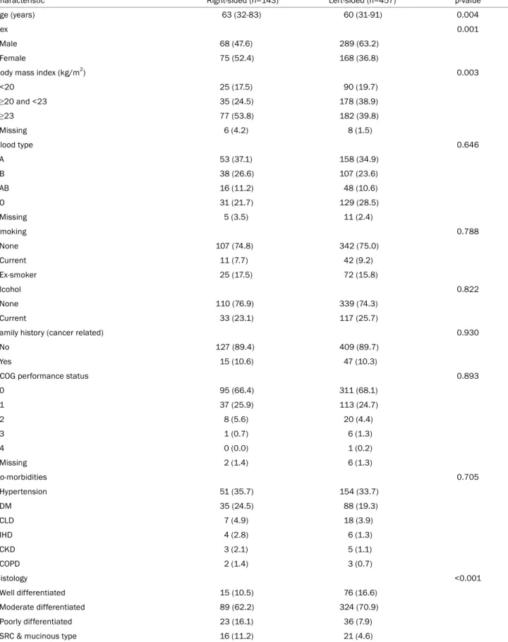
Table 1. Baseline Characteristics according to the Tumor Location
Characteristic Right-sided (n=143) Left-sided (n=457) p-value
Age (years) 63 (32-83) 60 (31-91) 0.004
Sex 0.001
Male 68 (47.6) 289 (63.2)
Female 75 (52.4) 168 (36.8)
Body mass index (kg/m2) 0.003
<20 25 (17.5) 90 (19.7)
≥20 and <23 35 (24.5) 178 (38.9)
≥23 77 (53.8) 182 (39.8)
Missing 6 (4.2) 8 (1.5)
Blood type 0.646
A 53 (37.1) 158 (34.9)
B 38 (26.6) 107 (23.6)
AB 16 (11.2) 48 (10.6)
O 31 (21.7) 129 (28.5)
Missing 5 (3.5) 11 (2.4)
Smoking 0.788
None 107 (74.8) 342 (75.0)
Current 11 (7.7) 42 (9.2)
Ex-smoker 25 (17.5) 72 (15.8)
Alcohol 0.822
None 110 (76.9) 339 (74.3)
Current 33 (23.1) 117 (25.7)
Family history (cancer related) 0.930
No 127 (89.4) 409 (89.7)
Yes 15 (10.6) 47 (10.3)
ECOG performance status 0.893
0 95 (66.4) 311 (68.1)
1 37 (25.9) 113 (24.7)
2 8 (5.6) 20 (4.4)
3 1 (0.7) 6 (1.3)
4 0 (0.0) 1 (0.2)
Missing 2 (1.4) 6 (1.3)
Co-morbidities 0.705
Hypertension 51 (35.7) 154 (33.7)
DM 35 (24.5) 88 (19.3)
CLD 7 (4.9) 18 (3.9)
IHD 4 (2.8) 6 (1.3)
CKD 3 (2.1) 5 (1.1)
COPD 2 (1.4) 3 (0.7)
Histology <0.001
Well differentiated 15 (10.5) 76 (16.6)
Moderate differentiated 89 (62.2) 324 (70.9)
Poorly differentiated 23 (16.1) 36 (7.9)
SRC & mucinous type 16 (11.2) 21 (4.6)
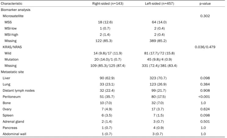
Table 1. Continued
Characteristic Right-sided (n=143) Left-sided (n=457) p-value
Biomarker analysis
Microsatellite 0.302
MSS 18 (12.6) 64 (14.0)
MSI-low 1 (0.7) 2 (0.4)
MSI-high 2 (1.4) 2 (0.4)
Missing 122 (85.3) 389 (85.2)
KRAS/NRAS 0.036/0.479
Wild 14 (9.8)/17 (11.9) 81 (17.7)/72 (15.8)
Mutation 20 (14.0)/1 (0.7) 45 (9.8)/4 (0.9)
Missing 109 (85.3)/125 (87.4) 331 (72.4)/381 (83.4)
Metastatic site
Liver 90 (62.9) 323 (70.7) 0.098
Lung 33 (23.1) 123 (26.9) 0.384
Distant lymph nodes 32 (22.4) 99 (21.7) 0.908
Peritoneum 51 (35.7) 80 (17.5) <0.001
Bone 10 (7.0) 32 (7.0) 1.0
Ovary 7 (4.9) 17 (3.7) 0.624
Spleen 6 (3.5) 7 (1.5) 0.098
Adrenal gland 2 (1.4) 3 (0.7) 0.501
Pancreas 1 (0.7) 4 (0.9) 1.0
Abdominal wall 1 (0.7) 3 (0.7) 1.0
Values are presented as mean (range) or number (%).
ECOG, Eastern Cooperative Oncology Group; DM, diabetes mellitus; CLD, chronic liver disease; IHD, ischemic heart disease; CKD, chronic kidney disease; COPD, chronic obstructive pulmonary disease; SRC, signet ring cell; MSS, microsatellite stable; MSI, microsatellite instability.
struct survival curves based on cumulative incidences and compared using a log-rank test. The Cox proportional hazards regression model was used to assess the factors affecting the OS and PFS. p values <0.05 were considered significant.
RESULTS
1. Baseline characteristics and laboratory findings ac- cording to the tumor location.
Six hundred and six patients were diagnosed with meta- static CRC between January 2000 and June 2018. One hun- dred and forty-three patients (23.8%) had right-sided CRC, and 457 patients (76.2%) had left-sided CRC. Six subjects had tumors located on both the right and left sides, whose origin could not be determined and were excluded. The mean age was 61 years, and 357 subjects (59.5%) were male. The median follow-up duration was 18 months (interquartile range, 10-28). Several differences in the baseline character- istics, including age, sex, BMI, histology, and metastatic sites
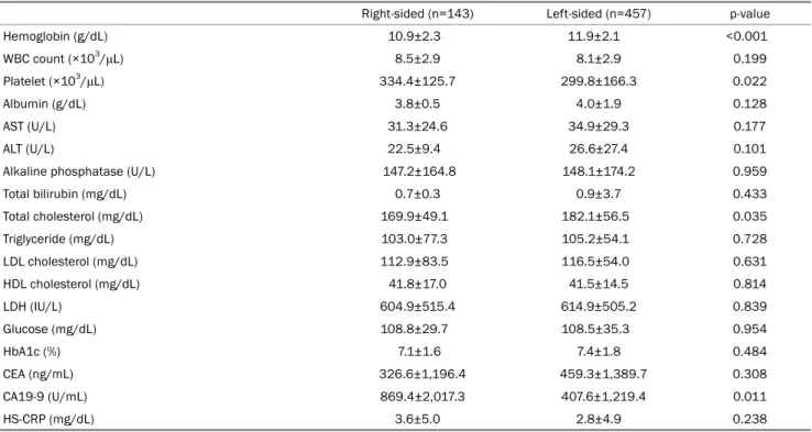
were observed between patients with right-sided and left-sid- ed CRC. Table 1 lists the baseline characteristics. The blood tests showed similar findings between patients with right- and left-sided CRC, except that patients with right-sided CRC had low hemoglobin, high platelet count, and high CA 19-9 levels.
These were assessed at the time of diagnosis and are sum- marized in Table 2.
2. Treatment outcomes according to tumor location Of the 600 patients included, 570 (95.0%) were treated with chemotherapy, and 99 patients (16.5%) were treated with radiotherapy (Table 3). One hundred fifty-three patients (25.5%) were treated with more than three lines of chemo- therapy, which was more common in patients with left-sided CRC compared to right-sided CRC (27.8% vs. 18.2%). The first-line regimen (irinotecan, oxaliplatin, capecitabine, or fluo- rouracil) was similar in patients with right and left-sided CRC, biological treatment (cetuximab or bevacizumab) was not performed. A palliative primary tumor resection was per-
Table 2. Laboratory Findings (at Diagnosis) according to the Tumor Location
Right-sided (n=143) Left-sided (n=457) p-value
Hemoglobin (g/dL) 10.9±2.3 11.9±2.1 <0.001
WBC count (×103/μL) 8.5±2.9 8.1±2.9 0.199
Platelet (×103/μL) 334.4±125.7 299.8±166.3 0.022
Albumin (g/dL) 3.8±0.5 4.0±1.9 0.128
AST (U/L) 31.3±24.6 34.9±29.3 0.177
ALT (U/L) 22.5±9.4 26.6±27.4 0.101
Alkaline phosphatase (U/L) 147.2±164.8 148.1±174.2 0.959
Total bilirubin (mg/dL) 0.7±0.3 0.9±3.7 0.433
Total cholesterol (mg/dL) 169.9±49.1 182.1±56.5 0.035
Triglyceride (mg/dL) 103.0±77.3 105.2±54.1 0.728
LDL cholesterol (mg/dL) 112.9±83.5 116.5±54.0 0.631
HDL cholesterol (mg/dL) 41.8±17.0 41.5±14.5 0.814
LDH (IU/L) 604.9±515.4 614.9±505.2 0.839
Glucose (mg/dL) 108.8±29.7 108.5±35.3 0.954
HbA1c (%) 7.1±1.6 7.4±1.8 0.484
CEA (ng/mL) 326.6±1,196.4 459.3±1,389.7 0.308
CA19-9 (U/mL) 869.4±2,017.3 407.6±1,219.4 0.011
HS-CRP (mg/dL) 3.6±5.0 2.8±4.9 0.238
Values are presented as mean±standard deviation.
WBC, white blood cell; AST, aspartate aminotransferase; ALT, alanine aminotransferase; LDL, low-density lipoprotein; HDL, high-density lipoprotein;
LDH, lactate dehydrogenase; HbA1c, hemoglobin A1c; CEA, carcinoembryonic antigen; CA19-9, carbohydrate antigen 19-9; HS-CRP, high sensitivity C-reactive protein.
Table 3. Treatment Outcomes according to the Tumor Location
Right-sided (n=143) Left-sided (n=457) p-value Chemotherapy
Total number 14 (1-60) 16 (1-142) 0.055
≥Third linea 26 (18.2) 127 (27.8) 0.028
Regimen (first line) 0.604
Irinotecan-based 46 (32.2) 135 (29.5)
Oxaliplatin-based 77 (53.8) 254 (55.6)
Capecitabine-based 5 (3.4) 15 (3.3)
Fluorouracil-based 7 (4.6) 28 (6.1)
No chemotherapy 8 (5.4) 22 (4.8)
Biologics 0.576
Cetuximab 11 (7.7) 44 (9.6)
Bevacizumab 35 (24.5) 108 (23.6)
Radiotherapy 19 (13.3) 80 (17.5) 0.539
Primary tumor 2 (1.4) 29 (6.3)
Metastatic site 17 (11.9) 51 (11.2)
Surgery 0.027
Palliative primary tumor resection 87 (60.8) 228 (49.9)
No tumor resection 56 (39.2) 229 (50.1)
Values are presented as mean (range) or number (%).
aNumber of patients with more than three lines of chemotherapy.
A B
Fig. 1. Kaplan-Meier analysis showing the overall survival (A) and progression-free survival (B) for patients with metastatic colorectal cancer based on primary tumor location. CRC, colorectal cancer.
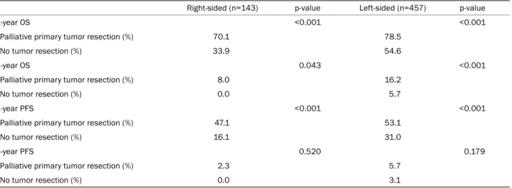
Table 4. Comparison of the Survival Outcomes according to Tumor Location between Patients with and without Palliative Primary Tumor Resection Right-sided (n=143) p-value Left-sided (n=457) p-value
1-year OS <0.001 <0.001
Palliative primary tumor resection (%) 70.1 78.5
No tumor resection (%) 33.9 54.6
3-year OS 0.043 <0.001
Palliative primary tumor resection (%) 8.0 16.2
No tumor resection (%) 0.0 5.7
1-year PFS <0.001 <0.001
Palliative primary tumor resection (%) 47.1 53.1
No tumor resection (%) 16.1 31.0
3-year PFS 0.520 0.179
Palliative primary tumor resection (%) 2.3 5.7
No tumor resection (%) 0.0 3.1
OS, overall survival; PFS, progression-free survival.
formed more in patients with right-sided CRC.
3. OS and disease-free survival according to tumor lo- cation
Fig. 1 shows the Kaplan-Meier curves for OS and PFS ac- cording to the tumor location. Patients with right-sided CRC had a poorer OS and PFS than those with left-sided CRC.
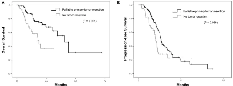
The long-term outcomes for patients with or without a pallia- tive primary tumor resection were evaluated. Table 4 lists the 1-year and 3-year OS/PFS rates for patients with or without a palliative primary tumor resection. Patients who underwent a palliative primary tumor resection had a significantly better
1-year OS and PFS than those without a tumor resection, re- gardless of the primary tumor location. These results were similar compared to the 3-year OS between a palliative pri- mary tumor resection and no tumor resection, regardless of the primary tumor location. Figs. 2, 3 show the Kaplan-Meier curves for the OS and PFS for patients with right-sided and left-sided CRC according to the palliative primary tumor resection. Patients who underwent a palliative primary tumor resection had a better OS and PFS than those without a tumor resection, regardless of the primary tumor location.
A B
Fig. 2. Kaplan-Meier analysis showing the overall survival (A) and progression-free survival (B) for patients with right-sided colorectal cancer based on palliative primary tumor resection.
A B
Fig. 3. Kaplan-Meier analysis showing the overall survival (A) and progression-free survival (B) for patients with left-sided colorectal cancer based on palliative primary tumor resection.
4. Factors affecting long-term outcomes
The factors affecting the long-term outcomes were as- sessed according to the tumor location by multivariate analy- sis using the Cox proportional hazards regression. As shown in Table 5, a palliative tumor resection was a significant prog- nostic factor affecting the better OS (hazard ratio [HR], 0.41;
95% CI, 0.22-0.76; p=0.004), but not PFS (HR, 0.63; 95%
CI, 0.39-1.01; p=0.053) in patients with right-sided CRC.
Poorly differentiated histology and CA 19-9 also influence the survival outcomes. A palliative tumor resection was found to be a significant prognostic factor for a better OS (HR, 0.53;
95% CI, 0.37-0.75; p<0.001) and PFS (HR, 0.76; 95% CI,
0.58-0.99; p=0.041) in patients with left-sided CRC (Table 6).
Poorly differentiated histology, CEA, CA19-9, and LDH also af- fect the survival outcomes.
DISCUSSION
These findings show the prognostic roles of primary tumor location and primary tumor resection in patients with meta- static CRC. The data suggest that a primary tumor resection can improve the OS in patients with metastatic CRC, regard- less of the primary tumor location.
In the present study, patients with right-sided CRC had sev-
Table 5. Prognostic Factors in Patients with Metastatic Colorectal Cancer and a Right-sided Primary Tumor
Predictor OS PFS
HR 95% CI p-value HR 95% CI p-value
Age (years)
<70 1 1
≥70 0.51 0.24-1.08 0.077 1.08 0.65-1.78 0.771
Sex
Female 1 1
Male 1.71 0.92-3.19 0.089 1.26 0.79-2.01 0.328
Histology
Well differentiated 1 1
Moderate differentiated 1.11 0.40-3.09 0.840 2.30 0.95-5.55 0.064
Poorly differentiated 4.90 1.70-16.49 0.006 3.93 1.46-10.61 0.007
SRC & mucinous type 2.36 0.64-8.73 0.195 1.90 0.67-5.37 0.228
CEA (ng/mL)
<20.0 1 1
≥20.0 1.30 0.66-2.54 0.447 0.83 0.50-1.36 0.453
CA 19-9 (U/mL)
<30.0 1 1
≥30.0 1.63 0.79-3.35 0.185 2.14 1.26-3.66 0.005
LDH (IU/L)
<400 1 1
≥400 1.55 0.84-2.85 0.162 1.06 0.68-1.66 0.792
Surgery
No tumor resection 1 1
Palliative primary tumor resection 0.41 0.22-0.76 0.004 0.63 0.39-1.01 0.053
Biologic treatment
Without biologics 1 1
With biologics 0.60 0.32-1.11 0.103 1.40 0.89-2.18 0.145
OS, overall survival; PFS, progression-free survival; HR, hazard ratio; CI, confidence interval; CEA, carcinoembryonic antigen; CA 19-9, carbohydrate antigen 19-9; LDH, lactate dehydrogenase.
eral characteristics that differed from those with left-sided CRC, which included older age, more female patients, BMI 23 kg/m2 or higher, poorly differentiated/signet ring cell/mu- cinous histology, and peritoneal metastasis. These findings are consistent with other recent data,6,8,9 which likely result from different molecular carcinogenesis pathways between right- and left-sided CRC. The primary tumor location is asso- ciated with the prognosis in CRC patients, and generally, right-sided CRC has a poorer outcome.15-17 The data shows that patients with right-sided CRC have poorer survival out- comes than those with left-sided CRC. In propensity score analysis, right-sided CRC was diagnosed at a more advanced state within stage IV disease and showed significantly poorer prognosis than left-sided CRC.18 In this study, the character- istics of patients with right-sided CRC, including older age,
more female patients, and more poorly differentiated/signet ring cell/mucinous histology, are considered as contributors to poorer survival benefits. A primary tumor location influen- ces the selection of the chemotherapy regimen in patients diagnosed with metastatic CRC. A retrospective analysis of the CRYSTAL and FIRE-3 trials analyzed RAS wild-type populations. They showed that first-line FOLFIRI (irinotecan, fluorouracil, and leucovorin) plus cetuximab benefitted pa- tients with left-sided CRC more than those with right-sided CRC.7 A meta-analysis of first-line clinical trials, including PRIME, CRYSTAL, FIRE-3, and the CALGB/SWOG 80405 study, indicated that patients with RAS wild-type left-sided CRC had a significant survival benefit from the addition of an anti-epi- dermal growth factor receptor (EGFR) antibody to conventional chemotherapy.19 Based on these results, the current NCCN
Table 6. Prognostic Factors in Patients with Metastatic Colorectal Cancer and a Left-sided Primary Tumor
Predictor OS PFS
HR 95% CI p-value HR 95% CI p-value
Age (years)
<70 1 1
≥70 0.89 0.57-1.38 0.605 0.78 0.57-1.08 0.139
Sex
Female 1 1
Male 1.12 0.78-1.62 0.546 1.20 0.91-1.58 0.191
Histology
Well differentiated 1 1
Moderate differentiated 1.51 0.85-2.67 0.158 0.98 0.60-1.60 0.924
Poorly differentiated 4.28 2.05-8.92 <0.001 0.78 0.35-1.75 0.541
SRC & mucinous type 2.66 1.01-7.02 0.048 0.82 0.30-2.25 0.703
CEA (ng/mL)
<20.0 1 1
≥20.0 1.23 0.80-1.90 0.340 1.45 1.09-1.92 0.010
CA 19-9 (U/mL)
<30.0 1 1
≥30.0 1.53 1.07-2.18 0.020 1.22 0.91-1.65 0.182
LDH (IU/L)
<400 1 1
≥400 1.17 0.80-1.71 0.416 1.44 1.08-1.91 0.012
Surgery
No tumor resection 1 1
Palliative primary tumor resection 0.53 0.37-0.75 <0.001 0.76 0.58-0.99 0.041
Biologic treatment
Without biologics 1 1
With biologics 0.95 0.67-1.34 0.762 0.96 0.74-1.25 0.780
OS, overall survival; PFS, progression-free survival; HR, hazard ratio; CI, confidence interval; CEA, carcinoembryonic antigen; CA 19-9, carbohydrate antigen 19-9; LDH, lactate dehydrogenase.
guidelines recommend the addition of anti-EGFR antibody to conventional chemotherapy for metastatic CRC patients with KRAS/NRAS/BRAF wild type genes and left-sided tumors.14
The data revealed a survival benefit of palliative primary tumor resection. In this study, a palliative primary tumor re- section was performed more in patients with right-sided CRC than those with left-sided CRC (60.8% vs. 49.9%, Table 3).
Multivariate analysis revealed a palliative primary tumor re- section to be an independent prognostic factor for a better OS, regardless of the primary tumor location. Surgical re- section of the primary tumor is a curative treatment for CRC patients without metastasis. On the other hand, the role of primary tumor resection for CRC patients with metastases is unclear. A reduction of the tumor burden may lead to a better response to systemic therapy in patients with metastatic
CRC.20 A reduction of the tumor burden has a survival benefit for primary renal or ovarian tumors with metastatic lesions.21,22 A recent study suggested that a primary tumor resection can prevent micro-metastases to the liver paren- chyma by increasing the angiogenic markers, including vas- cular endothelial growth factor (VEGF) A, VEGF receptor 1, VEGF receptor 2, and placental growth factor.23 On the other hand, some studies reported worsening liver or lung meta- stases after a primary tumor resection, which was attributed to the depletion of anti-angiogenic proteins, such as angiosta- tin and endostatin produced by the primary tumor.24,25 Further studies will be needed to clarify the role of the primary tumor resection for patients with metastatic CRC.
This study had several limitations. First, this study had a retrospective, single-center design, which means the selection
bias could not be avoided, even though an attempt was made to minimize bias by repeatedly reviewing the medical records.
Second, the efficacy of an anti-EGFR antibody plus conven- tional chemotherapy depending on the location of the primary tumor could not be confirmed, because the number of tests for KRAS/NRAS and the proportion of patients treated with biologics were not high enough to assess the efficacy. Third, tumors originating from the transverse colon were classified as right-sided CRC for the convenience of analysis, but classi- fying transverse colon cancer as right-sided vs. left-sided is controversial. To overcome this limitation, further well-de- signed studies for the characterization of transverse colon cancer will be needed.
In conclusion, this study showed that the primary tumor location has a prognostic effect on patients with metastatic CRC. Moreover, a palliative primary tumor resection is a sig- nificant prognostic factor affecting better OS, regardless of the primary tumor location. Based on these results, a pallia- tive primary tumor resection is a suitable option for patients with metastatic CRC to improve the long-term outcomes, re- gardless of the primary tumor location.
REFERENCES
1. Cancer Today: data visualization tools for exploring the global cancer burden in 2018. [Internet]. Lyon: International Association of Cancer Registries; c2018 [cited 2020 Mar 30]. Available from:
https://gco.iarc.fr/today/home.
2. Muratore A, Zorzi D, Bouzari H, et al. Asymptomatic colorectal cancer with un-resectable liver metastases: immediate color- ectal resection or up-front systemic chemotherapy? Ann Surg Oncol 2007;14:766-770.
3. Loupakis F, Cremolini C, Masi G, et al. Initial therapy with FOLFOXIRI and bevacizumab for metastatic colorectal cancer. N Engl J Med 2014;371:1609-1618.
4. Bardhan K, Liu K. Epigenetics and colorectal cancer pathogenesis.
Cancers (Basel) 2013;5:676-713.
5. Hong SN. Genetic and epigenetic alterations of colorectal cancer. Intest Res 2018;16:327-337.
6. Loupakis F, Yang D, Yau L, et al. Primary tumor location as a prog- nostic factor in metastatic colorectal cancer. J Natl Cancer Inst 2015;107:dju427.
7. Tejpar S, Stintzing S, Ciardiello F, et al. Prognostic and predictive relevance of primary tumor location in patients with RAS wild-type metastatic colorectal cancer: retrospective analyses of the CRYSTAL and FIRE-3 trials. JAMA Oncol 2017;3:194-201.
8. Nishihara R, Glass K, Mima K, et al. Biomarker correlation net- work in colorectal carcinoma by tumor anatomic location. BMC Bioinformatics 2017;18:304.
9. Yamauchi M, Morikawa T, Kuchiba A, et al. Assessment of color-
ectal cancer molecular features along bowel subsites challenges the conception of distinct dichotomy of proximal versus distal colorectum. Gut 2012;61:847-854.
10. Modest DP, Schulz C, von Weikersthal LF, et al. Outcome of pa- tients with metastatic colorectal cancer depends on the primary tumor site (midgut vs. hindgut): analysis of the FIRE1-trial (FuFIRI or mIROX as first-line treatment). Anticancer Drugs 2014;25:
212-218.
11. NCCN Clinical Practive Guidelines in Oncology. Colon Cancer ver 2.2020. [Internet]. Pennsylvania: National Comprehensive Cancer Network; 2020 [updated 2020 Mar 3; cited 2020 Apr 7].
Available from: https://nccn.org/professionals/physician_gls/
default.aspx#colon.
12. Tarantino I, Warschkow R, Worni M, et al. Prognostic relevance of palliative primary tumor removal in 37,793 metastatic color- ectal cancer patients: a population-based, propensity score-ad- justed trend analysis. Ann Surg 2015;262:112-120.
13. Alawadi Z, Phatak UR, Hu CY, et al. Comparative effectiveness of primary tumor resection in patients with stage IV colon cancer.
Cancer 2017;123:1124-1133.
14. NCCN Clinical Practive Guidelines in Oncology. Colon Cancer ver 2.2020. [Internet]. Philadelphia: National Comprehensive Cancer Network; 2020 [updated 2020 March 3; cited 2020 Apr 7]. Available from: https://nccn.org/professionals/physician_gls/
default.aspx#colon.
15. Yahagi M, Okabayashi K, Hasegawa H, Tsuruta M, Kitagawa Y.
The worse prognosis of right-sided compared with left-sided colon cancers: a systematic review and meta-analysis. J Gastrointest Surg 2016;20:648-655.
16. Peng J, Li C, Wang F, et al. Right- and left-sided stage III colon can- cers present different prognostic outcomes of oxaliplatin-based adjuvant chemotherapy after curative resection. Cancer Manag Res 2018;10:2095-2103.
17. Kishiki T, Kuchta K, Matsuoka H, et al. The impact of tumor loca- tion on the biological and oncological differences of colon can- cer: multi-institutional propensity score-matched study. Am J Surg 2019;217:46-52.
18. Ishihara S, Nishikawa T, Tanaka T, et al. Prognostic impact of tu- mor location in stage IV colon cancer: a propensity score analysis in a multicenter study. Int J Surg 2014;12:925-930.
19. Holch JW, Ricard I, Stintzing S, Modest DP, Heinemann V. The rele- vance of primary tumour location in patients with metastatic col- orectal cancer: a meta-analysis of first-line clinical trials. Eur J Cancer 2017;70:87-98.
20. de Mestier L, Manceau G, Neuzillet C, et al. Primary tumor re- section in colorectal cancer with unresectable synchronous metastases: a review. World J Gastrointest Oncol 2014;6:156-169.
21. van der Burg ME, Vergote I; Gynecological Cancer Group of the EORTC. The role of interval debulking surgery in ovarian cancer.
Curr Oncol Rep 2003;5:473-481.
22. Flanigan RC, Salmon SE, Blumenstein BA, et al. Nephrectomy fol- lowed by interferon alfa-2b compared with interferon alfa-2b alone for metastatic renal-cell cancer. N Engl J Med 2001;345:
1655-1659.
23. van der Wal GE, Gouw AS, Kamps JA, et al. Angiogenesis in syn- chronous and metachronous colorectal liver metastases: the liv-
er as a permissive soil. Ann Surg 2012;255:86-94.
24. Holmgren L, O'Reilly MS, Folkman J. Dormancy of micro- metastases: balanced proliferation and apoptosis in the pres- ence of angiogenesis suppression. Nat Med 1995;1:149-153.
25. Peeters CF, de Waal RM, Wobbes T, Ruers TJ. Metastatic dor- mancy imposed by the primary tumor: does it exist in humans?
Ann Surg Oncol 2008;15:3308-3315.