저작자표시-비영리-변경금지 2.0 대한민국 이용자는 아래의 조건을 따르는 경우에 한하여 자유롭게 l 이 저작물을 복제, 배포, 전송, 전시, 공연 및 방송할 수 있습니다. 다음과 같은 조건을 따라야 합니다: l 귀하는, 이 저작물의 재이용이나 배포의 경우, 이 저작물에 적용된 이용허락조건 을 명확하게 나타내어야 합니다. l 저작권자로부터 별도의 허가를 받으면 이러한 조건들은 적용되지 않습니다. 저작권법에 따른 이용자의 권리는 위의 내용에 의하여 영향을 받지 않습니다. 이것은 이용허락규약(Legal Code)을 이해하기 쉽게 요약한 것입니다. Disclaimer 저작자표시. 귀하는 원저작자를 표시하여야 합니다. 비영리. 귀하는 이 저작물을 영리 목적으로 이용할 수 없습니다. 변경금지. 귀하는 이 저작물을 개작, 변형 또는 가공할 수 없습니다.
Doctoral Thesis in Medicine
EpCAM as a Predictive Marker of
Tumor Recurrence and Survival in
Patients Who Underwent Surgical
Resection for Hepatocellular
Carcinoma
Graduate School of Ajou University
Department of Gastroenterology
EpCAM as a Predictive Marker of
Tumor Recurrence and Survival in
Patients Who Underwent Surgical
Resection for Hepatocellular
Carcinoma
Jae Youn Cheong, Advisor
I submit this thesis as the
Doctoral thesis in Medicine.
February 2019
Graduate School of Ajou University
Department of Gastroenterology
Choong Kyun, Noh
i - ABSTRACT -
EpCAM as a Predictive Marker of Tumor Recurrence and Survival in Patients Who Underwent Surgical Resection for Hepatocellular Carcinoma Background/Aims: Epithelial cell adhesion molecule (EpCAM) is expressed in hepatic progenitor cells and hepatocellular carcinoma (HCC), and is considered a marker of liver cancer stem cells. We evaluated the immunohistochemical expression of EpCAM and other stemness-related markers as prognosticators of tumor recurrence and survival in patients who underwent surgical resection for HCC
Materials and Methods: A total of 262 patients were enrolled who had undergone surgical resection for HCC, with immunohistochemical staining results for EpCAM. The alpha-fetoprotein (AFP), cytokeratin 19 (CK19), CK7 and glypican-3 expressions were also analyzed in resected tumor specimens via immunohistochemistry.
Results: A multivariate Cox regression analysis showed that tumor size [hazard ratio (HR)=2.26, p=0.005], intrahepatic metastasis (HR=2.31, p=0.011), and EpCAM positivity (HR=1.74, p=0.038) were associated with tumor recurrence. In a Kaplan–Meier survival analysis, patients with EpCAM-positive tumors had a significantly higher tumor recurrence rate and a reduced overall survival compared to those with EpCAM-negative tumors.
Conclusion: Immunohistochemical expression of EpCAM was identified as a poor prognosticator of recurrence and survival after surgical resection in patients with HCC.
iii
TABLE OF CONTENTS
LIST OF TEXT
ABSTRACT ··· i
TABLE OF CONTENTS LIST OF TEXT ··· iii
LIST OF FIGURES ··· iv
LIST OF TABLES ··· v
Ⅰ. INTRODUCTION ··· 1
Ⅱ. MATERIALS AND METHODS ··· 3
A. Patients ··· 3
B. Samples and pathological data ··· 4
C. IHC staining ··· 4
D. Statistical analysis ··· 5
Ⅲ. RESULTS ··· 7
A. Baseline clinicopathological characteristics ··· 7
B. Factors predictive of HCC recurrence after surgical resection ··· 8
C. Clinicopathological characteristics and outcomes according to IHC expression of EpCAM ··· 9
D. EpCAM and serum AFP level as a predictor of HCC recurrence and survival ··· 12
E. Diagnostic accuracy of EpCAM and serum AFP level as a predictor of HCC recurrence ··· 13
Ⅳ. DISCUSSION ··· 15
Ⅴ. CONCLUSION ··· 18
REFERENCES ··· 19
LIST OF FIGURES
Figure 1. Flow diagram of study enrollment, allocation, and analysis ··· 3 Figure 2. Immunohistochemical staining of resected hepatocellular carcinoma
specimens for epithelial cell adhesion molecule and other stemness-related markers ··· 5
Figure 3. Kaplan–Meier analyses of recurrence and overall survival among patients with EpCAM-positive HCC vs. those with EpCAM-negative HCC ··· 12 Figure 4. Kaplan–Meier analyses of recurrence and overall survival among
patients with EpCAM-positive HCC and high expression of alpha-fetoprotein vs. other patients with HCC ··· 13 Figure 5. Receiver-operating characteristic curve of EpCAM expression for the
v
LIST OF TABLES
Table 1. Clinicopathological characteristics of patients who had undergone resection for hepatocellular carcinoma ··· 7 Table 2. Univariate and multivariate analyses of clinicopathological
parameters associated with cumulative recurrence of
hepatocellular carcinoma ··· 9 Table 3. Clinicopathological characteristics and hepatocellular carcinoma
I. INTRODUCTION
Hepatocellular carcinoma (HCC) is one of the most frequently occurring cancers and the third leading cause of cancer-related death in Asia (1). A high prevalence of hepatitis B (HBV) and hepatitis C virus infections strongly predisposes the population to chronic liver disease and subsequent development of HCC in Asian countries (2-4). Although treatments for HCC have evolved, only those limited to early HCC have satisfactory treatment outcomes, and the mortality rate remains high despite the application of surgical resection, liver transplantation, and various other treatment modalities. Curative resection is widely considered as the first choice of therapy for patients with resectable HCC, however, the relatively high postoperative recurrence of HCC remains an unsolved problem, as up to 70% of patients will experience a recurrence within 5 years after curative resection (5-7). Prognostic predictions regarding recurrence and mortality should be made soon after surgical resection, as this information can help to direct the postoperative management strategies in patients undergoing surgical resection.
Two basic models have been proposed to explain the initiation and development of tumors in vivo. In the first model, the accumulation of mutations in somatic cells leads to uncontrolled proliferation and, finally, to carcinogenic transformation (8-10). The second model assumes the existence of a hierarchical tumor cell arrangement, wherein only a subpopulation of cells harbors the capacity for tumorigenesis (11, 12). In the latter model, the cells of interest possess two main characteristics, self-renewal and multipotency, and express stemness-related markers such as epithelial cell adhesion molecule (EpCAM), cytokeratin (CK) 7 and 19, glypican-3 and cluster of differentiation (CD)133 (13, 14).
2
EpCAM is expressed on a subset of normal epithelial cells and can be overexpressed on malignant cells from a variety of different tumor entities. This overexpression is even more pronounced on so-called tumor-initiating cells associated with many carcinomas (15). EpCAM is also expressed on hepatic progenitor cells and fetal hepatoblasts, and is considered a biomarker of hepatic cancer stem cell. Notably, EpCAM-positive HCC cells derived from cell lines and tumor specimens were found to be highly invasive and tumorigenic, suggesting that EpCAM might be a useful biomarker of HCC-initiating cells (16-18).
Given the importance of prognostic prediction for patients with resected HCC, clinicians should identify those who are at a high risk of recurrence following treatment in order to consider the need for additional therapies or more careful follow-up. Although previous studies have described various prognostic indicators in patients with HCCs, reliable prognosticators are limited in number. In this study, the immunohistochemical (IHC) expression of stemness-related markers as prognosticators of tumor recurrence and survival was evaluated in patients who underwent surgical resection for HCC, with a particular focus on the potential of EpCAM as a predictive biomarker.
II. MATERIALS AND METHODS
A. Patients
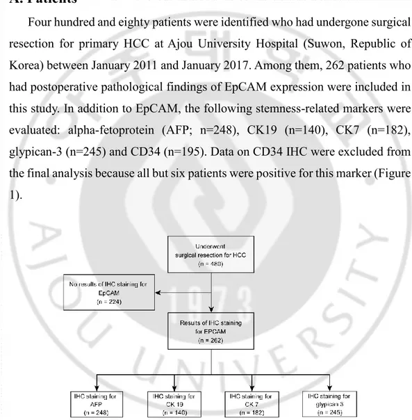
Four hundred and eighty patients were identified who had undergone surgical resection for primary HCC at Ajou University Hospital (Suwon, Republic of Korea) between January 2011 and January 2017. Among them, 262 patients who had postoperative pathological findings of EpCAM expression were included in this study. In addition to EpCAM, the following stemness-related markers were evaluated: alpha-fetoprotein (AFP; n=248), CK19 (n=140), CK7 (n=182), glypican-3 (n=245) and CD34 (n=195). Data on CD34 IHC were excluded from the final analysis because all but six patients were positive for this marker (Figure 1).
4
Patients were followed up at 3-month intervals in the first year after surgery, at 4-month intervals in the second year, and then, every six months thereafter. A contrast-enhanced abdomen computed tomography or magnetic resonance imaging scan was performed every visit at the out-patient department during the follow up period. All processes regarding access to their medical records were conducted in accordance with the guidelines of institutional Review Board (approval no. AJIRB-MED-MDB-17-005).
B. Samples and pathological data
Tumor size, presence of vascular invasion, tumor stage and serum AFP level, in addition to patient age and sex, were analyzed. Resected surgical specimens were subjected to IHC to identify the protein expression of EpCAM, AFP, CK19, CK7, and glypican-3. Tumors were graded using the criteria proposed by Edmondson and Steiner: I, Well-differentiated; II, moderately differentiated; III, poorly differentiated; and IV, undifferentiated (19). Tumor staging was based on the modified Union for International Cancer Control (UICC) staging system (20, 21). The analysis included patients who were followed-up for at least 6 months and for whom recurrence and death had been confirmed. The mean follow-up period was 29.9 ± 17.15 (median=26, range=6–75) months.
C. IHC staining
HCC specimens collected from patients who had undergone resection were fixed in 10% formalin and embedded in paraffin. Four-micrometer-thick sections of the paraffin-embedded tissue blocks were used for IHC studies. All slides were scored by two pathologists blinded to the patients' clinical information (Kim YB and Roh J). The proportion of cell staining was used to evaluate the IHC results, ranging from 0 to 100%. The extent of positivity was assigned a score of 0 when
no positive cells were observed; other results were semi-quantitatively scored as follows: negative, <5%; weak (1+), 5–30%; moderate (2+), 30–60%; and strong staining (3+), >60%. The expression of EpCAM and other stemness-related markers was considered positive if >10% of cells received a final score of moderate or strong staining (Figure 2).
Figure 2. Immunohistochemical staining of resected hepatocellular carcinoma (HCC) specimens for epithelial cell adhesion molecule (EpCAM) and other stemness-related markers. A: Hematoxylin and eosin-stained HCC (magnification: ×100). B: Specimen positive for EpCAM expression (magnification: ×200). C: Specimen negative for EpCAM expression (magnification: ×200). D: Specimen positive for CK19 expression (magnification: ×200). E: Specimen positive for CK7 expression (magnification: ×200). F: Specimen positive for glypican-3 expression (magnification: ×200).
6
The clinicopathological characteristics of the patients with HCC were evaluated using the chi-square test and Fisher’s exact test. A multivariate Cox regression hazards model with forward stepwise entry was used to identify independent predictors of survival. The classification accuracy was measured using the area under the receiver-operating characteristic curve (AUROC). Using this curve, the cut-off value was determined as the point where the value of the sum yielded the greatest sensitivity and specificity, and was used to estimate the sensitivity, specificity, positive predictive value, negative predictive value, and overall accuracy. Kaplan–Meier survival curves were used to evaluate the cumulative recurrence and overall survival outcomes, and statistical differences in survival curves between the low-risk and high-risk groups were determined using the log-rank test. Recurrence and overall survival time were defined as the interval between the dates of curative resection and first recurrence or death, respectively. All statistical analyses were performed using the open-source statistical programming environment R (R software program ver. 3.1.2; R Project for Statistical Computing, Vienna, Austria). A p-value of less than 0.05 was considered statistically significant.
III. RESULTS
A. Baseline clinicopathological characteristics
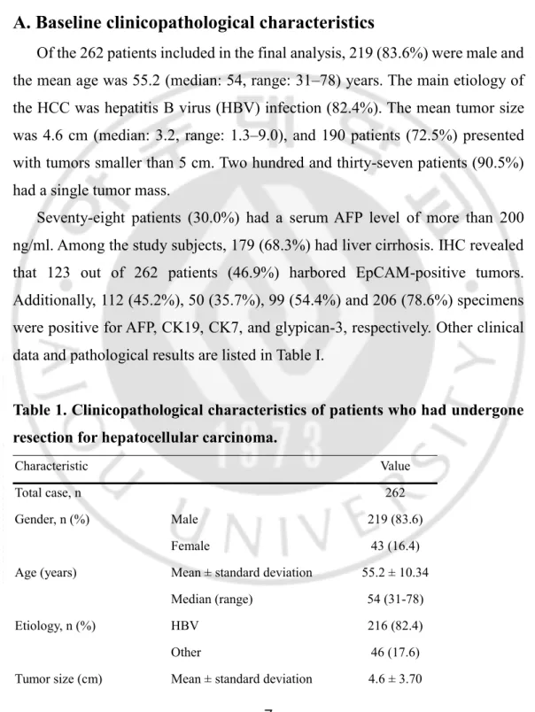
Of the 262 patients included in the final analysis, 219 (83.6%) were male and the mean age was 55.2 (median: 54, range: 31–78) years. The main etiology of the HCC was hepatitis B virus (HBV) infection (82.4%). The mean tumor size was 4.6 cm (median: 3.2, range: 1.3–9.0), and 190 patients (72.5%) presented with tumors smaller than 5 cm. Two hundred and thirty-seven patients (90.5%) had a single tumor mass.
Seventy-eight patients (30.0%) had a serum AFP level of more than 200 ng/ml. Among the study subjects, 179 (68.3%) had liver cirrhosis. IHC revealed that 123 out of 262 patients (46.9%) harbored EpCAM-positive tumors. Additionally, 112 (45.2%), 50 (35.7%), 99 (54.4%) and 206 (78.6%) specimens were positive for AFP, CK19, CK7, and glypican-3, respectively. Other clinical data and pathological results are listed in Table I.
Table 1. Clinicopathological characteristics of patients who had undergone resection for hepatocellular carcinoma.
Characteristic Value
Total case, n 262
Gender, n (%) Male 219 (83.6)
Female 43 (16.4)
Age (years) Mean ± standard deviation 55.2 ± 10.34
Median (range) 54 (31-78)
Etiology, n (%) HBV 216 (82.4)
Other 46 (17.6)
8
Median (range) 3.2 (1.3-9.0)
Size (cm), n (%) <5 190 (72.5)
≥5 72 (27.5)
Number of tumors, n (%) Single 237 (90.5)
Multiple 25 (9.5)
Modified tumor stage T1, T2 171 (65.3)
T3, T4 91 (34.7)
AFP (ng/ml)*, n (%) <200 182 (70.0)
≥200 78 (38.0)
Liver cirrhosis, n (%) No 83 (31.7)
Present 179 (68.3)
Pathological data, n (%) Microvessel invasion (n=251) 58 (39.4) Portal vein invasion (n=247) 82 (33.2) Intrahepatic metastasis (n=245) 58 (23.7)
Edmonson grade**, n (%) I, II 103 (40.9)
III, IV 149 (59.1)
Stemness-related marker EpCAM (n=262) 123 (46.9)
AFP (n=248) 112 (45.2)
CK19 (n=140) 50 (35.7)
CK7 (n=182) 99 (54.4)
Glypican-3 (n=245) 206 (78.6) *n=260, **n=252.
n, number; HBV, hepatitis B virus; AFP, alpha-fetoprotein; EpCAM, epithelial cell adhesion molecule; CK, cytokeratin
B. Factors predictive of HCC recurrence after surgical resection
For the univariate analyses, a Cox proportional hazards model was used to investigate factors predictive of HCC recurrence after surgical resection. Of theinvestigated variables, tumor size ≥5 cm), tumor stage >2, microvessel invasion, intrahepatic metastasis, serum AFP ≥200 ng/ml, and EpCAM positivity were identified as significant predictors of HCC recurrence (p<0.05). In the multivariate analysis, however, tumor size [hazard ratio (HR)=3.69, 95% CI=1.310–10.409, p=0.013], intrahepatic metastasis (HR=5.06, 95% CI=1.387– 18.490, p=0.014), and EpCAM positivity (HR=2.94, 95% CI=1.054–8.173, p=0.039) remained significant predictors of HCC recurrence (Table II). EpCAM positivity was the only significant stemness-related immunohistochemical marker identified as a predictor of HCC recurrence in the multivariate analysis. Table 2. Univariate and multivariate analyses of clinicopathological parameters associated with cumulative recurrence of hepatocellular carcinoma.
Univariate analysis Multivariate analysis
Variable HR 95% CI p-Value HR 95% CI p-Value
Age>55 years 1.10 0.728-1.653 0.658 Male gender 1.64 0.871-3.072 0.126 Tumor size ≥5 cm 3.45 2.273-5.222 <0.001 3.69 1.310-10.409 0.013 Tumor stage >2 3.43 2.270-5.194 <0.001 Microvessel invasion 3.72 2.416-5.724 <0.001 Intrahepatic metastasis 4.66 2.998-7.245 <0.001 5.06 1.387-18.490 0.014 AFP≥200 ng/ml 2.05 1.355-3.101 <0.001 EpCAM (IHC) 2.15 1.413-3.268 <0.001 2.94 1.054-8.173 0.039 AFP (IHC) 1.31 0.864-1.974 0.206 CK19 (IHC) 1.44 0.791-2.621 0.232 CK7 (IHC) 1.30 0.813-2.087 0.272 Glypican-3 (IHC) 0.76 0.444-1.313 0.330
10
HR, Hazard ratio; CI, confidence interval; AFP, alpha-fetoprotein; CK, cytokeratin; EpCAM, epithelial cell adhesion molecule; IHC, immunohistochemical stain.
C. Clinicopathological characteristics and outcomes according to
IHC expression of EpCAM
To determine the clinical significance of EpCAM after surgical resection for HCC, the patients were divided into two groups according to EpCAM expression (Table III). The two groups did not differ in terms of sex, age, etiology of HCC, tumor size, tumor multiplicity, tumor stage, presence of cirrhosis, or serum total bilirubin levels. However, significant intergroup differences were observed in serum AFP level (p<0.001) and various pathological factors (microvessel invasion, p=0.003; portal vein invasion, p=0.006; intrahepatic metastasis, p=0.041; Edmonson grade, p=0.002) (Table III).
Table 3. Clinicopathological characteristics and hepatocellular carcinoma recurrence according to epithelial cell adhesion molecule (EpCAM) expression. Variable EpCAM-negative (n=139), n (%) EpCAM-positive (n=123), n (%) p-Value Gender Male 116 (83.5) 103 (83.7) 0.950 Female 23 (16.5) 20 16.3) Age <55 Years 69 (49.6) 75 (61.0) 0.066 ≥55 Years 70 (50.4) 48 (39.0) Etiology, n HBV 111 (79.9) 105 (85.4) 0.242 Other 28 (20.1) 18 (14.6) Tumor size <5 cm 106 (76.3) 84 (68.3) 0.149 ≥5 cm 33 (23.7) 39 (31.7)
Number of tumor mass Single 123 (88.5) 114 (92.7) 0.446
Multiple 16 (11.5) 9 (7.3)
Modified tumor stage T1, T2 98 (70.5) 73 (59.3) 0.058
T3, T4 41 (29.5) 50 (40.7)
AFP (n=260) <200 ng/ml 112 (81.2) 70 (57.4) <0.001 ≥200 ng/ml 26 (18.8) 52 (42.6)
Liver cirrhosis Absent 47 (33.8) 36 (29.3) 0.430
Present 92 (66.2) 87 (70.7)
Total bilirubin <1.2 mg/dl 110 (79.1) 97 (78.9) 0.644 ≥1.2 mg/dl 29 (20.9) 26 (21.1)
Microvessel invasion (n=251) Absent 92 (69.2) 60 (50.8 0.003
Present 41 (30.8) )58 (49.2)
Portal vein invasion (n=247) Absent 97 (74.6) 68 (58.1 0.006
Present 33 (25.4) )49 (41.9)
Intrahepatic metastasis (n=245) Absent 106 (81.5) 81 (70.4) 0.041
Present 24 (18.5) 34 (29.6)
Edmonson grade (n=252) I, II 67 (49.6) 36 (30.8) 0.002
III, IV 68 (50.4) 81 (69.2)
EpCAM, epithelial cell adhesion molecule; n, number; HBV, hepatitis B virus; AFP, alpha-fetoprotein
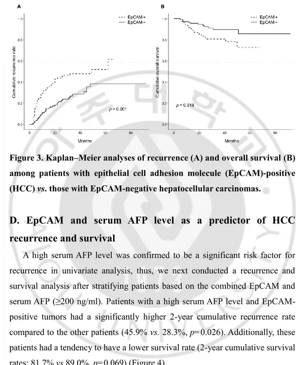
The cumulative recurrence rate was higher among those with EpCAM-positive tumors than those with EpCAM-negative tumors (2-year cumulative recurrence: 44.0% vs. 21.5%, p<0.001). Furthermore, patients with EpCAM-positive tumors had a worse overall survival rate compared to their counterparts with EpCAM-negative tumors (2-year overall survival: 82.1% vs. 92.4%, p=0.018; Figure 3).
12
Figure 3. Kaplan–Meier analyses of recurrence (A) and overall survival (B) among patients with epithelial cell adhesion molecule (EpCAM)-positive (HCC) vs. those with EpCAM-negative hepatocellular carcinomas.
D. EpCAM and serum AFP level as a predictor of HCC
recurrence and survival
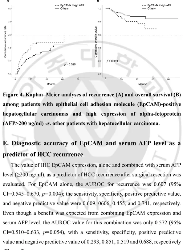
A high serum AFP level was confirmed to be a significant risk factor for recurrence in univariate analysis, thus, we next conducted a recurrence and survival analysis after stratifying patients based on the combined EpCAM and serum AFP (≥200 ng/ml). Patients with a high serum AFP level and EpCAM-positive tumors had a significantly higher 2-year cumulative recurrence rate compared to the other patients (45.9% vs. 28.3%, p=0.026). Additionally, these patients had a tendency to have a lower survival rate (2-year cumulative survival rates: 81.7% vs 89.0%, p=0.069) (Figure 4).
Figure 4. Kaplan–Meier analyses of recurrence (A) and overall survival (B) among patients with epithelial cell adhesion molecule (EpCAM)-positive hepatocellular carcinomas and high expression of alpha-fetoprotein (AFP>200 ng/ml) vs. other patients with hepatocellular carcinoma.
E. Diagnostic accuracy of EpCAM and serum AFP level as a
predictor of HCC recurrence
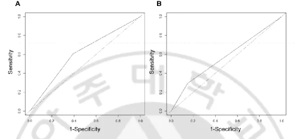
The value of IHC EpCAM expression, alone and combined with serum AFP level (≥200 ng/ml), as a predictor of HCC recurrence after surgical resection was evaluated. For EpCAM alone, the AUROC for recurrence was 0.607 (95% CI=0.545–0.670, p=0.004); the sensitivity, specificity, positive predictive value, and negative predictive value were 0.609, 0606, 0.455, and 0.741, respectively. Even though a benefit was expected from combining EpCAM expression and serum AFP level, the AUROC value for this combination was only 0.572 (95% CI=0.510–0.633, p=0.054), with a sensitivity, specificity, positive predictive value and negative predictive value of 0.293, 0.851, 0.519 and 0.688, respectively (Figure 5).
14
Figure 5. Receiver-operating characteristic curve of epithelial cell adhesion molecule (EpCAM) expression for the detection of hepatocellular carcinoma recurrence. A: EpCAM only. B: EpCAM combined with serum alpha-fetoprotein (AFP) level.
IV. DISCUSSION
In our study, we found that tumor expression of EpCAM, alone and in combination with the serum AFP level, may serve as a prognostic marker in patients with HCC who have undergone surgical resection. Specifically, the IHC expression of EpCAM was found to be associated with a higher risk of recurrence and shorter overall survival in patients with HCC. Additionally, the combination of positive EpCAM expression and a high serum AFP level was associated with rapid recurrence after surgical resection.
EpCAM is expressed on several types of epithelial cells and gastrointestinal carcinomas, where it acts as a homotypic calcium-independent cell adhesion molecule (22, 23). EpCAM strongly influences both the cell cycle and proliferation and contributes to up-regulation of the proto-oncogenes c-MYC and cyclins A and E (23). Furthermore, the inhibition of EpCAM expression was shown to reduce proliferation and metabolism in human cancer cells (24). In our study, the results obtained from a relatively simple IHC analysis of resected surgical specimens were consistent with these earlier findings. The EpCAM-positive group had a higher recurrence rate and lower survival rate compared to their EpCAM-negative HCC counterparts. These results suggest that a useful analysis of the resected surgical specimen can be conducted and that a patient's prognosis might be sufficiently predicted even from a preoperative biopsy.
A recent study compared peritumoral EpCAM expression and CD13 as a prognostic marker in patients who underwent curative resection of HBV-related HCC, using samples of liver parenchyma located within 20 mm of the tumor (25). The authors concluded that peritumoral EpCAM and CD13 expression were associated with a poor prognosis, but EpCAM may be a better prognostic marker than CD13 in HBV-related patients with HCC (25).
16
Another recent study showed an association between the preoperative
presence of EpCAM-expressingcirculating tumor cells (CTC) and T-regulatory
cell level with the recurrence of HCC after radical hepatic resection (26). In that study, the group with a high level of CTCs had a significantly higher 1-year recurrence rate compared with the group with lower CTC level (50.0% vs. 10.3%, p=0.004) in patients with HCC (26). Another investigation reported a correlation of survival outcomes with EpCAM-positive CTCs from patients with HCC. Patients with EpCAM-positive CTCs had a significantly reduced overall survival compared to patients without these cells (p=0.017), and the presence of CTCs was also found to correlate with a high serum AFP level (p=0.050) (27). The same group also reported a correlation of EpCAM-positive CTCs with a high recurrence rate (HR=2.3, p=0.027) and shorter recurrence-free survival among patients who underwent curative resection for HCC (5.0 ± 1.5 vs. 12.0 ± 2.6 months, p=0.039) (28). To our knowledge, there are no previous studies addressing the correlation between the IHC expression of EpCAM and the level of EpCAM-positive CTCs, thus further studies are needed to determine the relationship between these two measures.
CK19 and CK7 are induced in biliary marker-positive hepatic progenitor cells undergoing carcinogenesis (29, 30). These markers are not always expressed in HCCs. A previous study divided patients with HCC into three groups according to the CK19 and glypican-3 IHC results: CK19+/glypican-3+, CK19−/glypican-3+,
CK19−/glypican-3−. The researchers recorded a significantly lower cumulative
recurrence-free survival rate among the CK19+/glypican-3+ group relative to the
other groups (31). Furthermore, Uenishi et al. reported that both CK19 and CK7 were associated with tumor-free survival, although only the association with CK19 remained significant in a multivariate analysis (risk ratio=2.2, p=0.0082) (32). Although we also analyzed the associations of stemness-related markers
(AFP, CK19, CK7, and glypican-3) with recurrence and overall survival, no differences in recurrence and survival rates were found. In contrast to previous studies, the Cox proportional analysis failed to identify these markers as significant risk factors for recurrence in patients with HCC who underwent surgical resection.
Serum AFP is a known prognostic marker in patients with HCC. We investigated whether the serum AFP level can be a complimentary prognostic marker when combined with IHC expression of EpCAM of resected HCC specimens. However, better prediction of recurrence or survival was not obtained when an elevated serum AFP level (>200 ng/ml) was combined with EpCAM expression compared to use of EpCAM expression alone.
In the current study, analysis yielded an AUROC of EpCAM for recurrence of 0.607. Among patients with urothelial carcinoma, IHC-detected EpCAM expression was reported to be associated with worse overall survival (p=0.033) and an AUROC of 0.604 (33). Given that both studies divided patients into EpCAM-positive and -negative groups, the AUROC values would be expected to improve if patients were classified by IHC staining intensity with specific grade.
Our study had some limitations. Firstly, our IHC findings could not be compared with those of CTCs from patient blood samples, given the retrospective design. As noted, previous studies have identified EpCAM-positive CTCs as a poor prognostic factor. Future studies should determine whether CTC findings correlate with IHC results. Secondly, this study included many patients with short postoperative follow-up periods after surgery (mean follow-up period=29.9 months). A longer follow-up period may increase the value of EpCAM as a prognostic marker. Finally, we evaluated only patients for whom EpCAM IHC data were available, rather than all patients who underwent surgical resection. We
18
would expect more informative results from data that included all patients, regarding the usefulness of other stemness-related markers.
V. CONCLUSION
In conclusion, we identified EpCAM expression as a marker of poor prognosis of recurrence and survival in those undergoing surgical resection for HCC even after clinicopathological factors for prognosis were taken into account. We expect that IHC expression of EpCAM might be used to predict outcomes in patients with HCC who underwent percutaneous biopsy as well as hepatic resection.
20
REFERENCES
1 Yang JD and Roberts LR: Hepatocellular carcinoma: a global view. Nature
Reviews Gastroenterology and Hepatology 7: 448, 2010.
2 El–Serag HB and Rudolph KL: Hepatocellular carcinoma: epidemiology
and molecular carcinogenesis. Gastroenterology 132: 2557-2576, 2007.
3 Mittal S and El-Serag HB: Epidemiology of HCC: consider the population.
Journal of clinical gastroenterology 47: S2, 2013.
4 Chen CJ, Wang LY and Yu MW: Epidemiology of hepatitis B virus
infection in the Asia–Pacific region. Journal of gastroenterology and hepatology 15: S3-6, 2000
5 Ikeda K, Saitoh S, Tsubota A, Arase Y, Chayama K, Kumada H, Watanabe
G and Tsurumaru M: Risk factors for tumor recurrence and prognosis after curative resection of hepatocellular carcinoma. Cancer 71: 19-25, 1993.
6 Ang SF, Ng ES-H, Li H, Ong Y-H, Choo SP, Ngeow J, Toh HC, Lim KH,
Yap HY and Tan CK: The Singapore Liver Cancer Recurrence (SLICER) Score for relapse prediction in patients with surgically resected hepatocellular carcinoma. PLoS One 10: e0118658, 2015.
7 Llovet JM, Fuster J and Bruix J: Intention‐to‐treat analysis of surgical treatment for early hepatocellular carcinoma: resection versus transplantation. Hepatology 30: 1434-1440, 1999.
8 Tsai W and Chung R: Viral hepatocarcinogenesis. Oncogene 29: 2309,
2010.
9 Martincorena I and Campbell PJ: Somatic mutation in cancer and normal
cells. Science 349: 1483-1489, 2015.
10 Yamashita T, Honda M, Nakamoto Y, Baba M, Nio K, Hara Y, Zeng SS, Hayashi T, Kondo M and Takatori H: Discrete nature of EpCAM+ and
CD90+ cancer stem cells in human hepatocellular carcinoma. Hepatology 57: 1484-1497, 2013.
11 Beachy PA, Karhadkar SS and Berman DM: Tissue repair and stem cell
renewal in carcinogenesis. Nature 432: 324, 2004.
12 Guo W, Yang X-R, Sun Y-F, Shen M-N, Ma X-L, Wu J, Zhang C-Y, Zhou Y, Xu Y and Hu B: Clinical Significance of EpCAM mRNA-Positive Circulating Tumor Cells in Hepatocellular Carcinoma by an Optimized Negative Enrichment and qRT-PCR–Based Platform. Clinical cancer research 20: 4794-4805, 2014.
13 Kim H and Park YN: Hepatocellular Carcinomas Expressing ‘Stemness'-Related Markers: Clinicopathological Characteristics. Digestive diseases 32: 778-785, 2014.
14 Yamashita T, Ji J, Budhu A, Forgues M, Yang W, Wang HY, Jia H, Ye Q, Qin LX and Wauthier E: EpCAM-positive hepatocellular carcinoma cells are tumor-initiating cells with stem/progenitor cell features. Gastroenterology 136: 1012-1024. e1014, 2009.
15 Sun YF, Xu Y, Yang XR, Guo W, Zhang X, Qiu SJ, Shi RY, Hu B, Zhou J and Fan J: Circulating stem cell–like epithelial cell adhesion molecule– positive tumor cells indicate poor prognosis of hepatocellular carcinoma after curative resection. Hepatology 57: 1458-1468, 2013.
16 Martowicz A, Seeber A and Untergasser G: The role of EpCAM in physiology and pathology of the epithelium. Histol Histopathol 31: 349-355, 2016.
17 Oishi N and Wang XW: Novel therapeutic strategies for targeting liver cancer stem cells. International journal of biological sciences 7: 517, 2011. 18 Park H, Lee H, Seo AN, Cho JY, Choi YR, Yoon Y-S, Han H-S, Park YN and Kim H: SALL4 expression in hepatocellular carcinomas is associated
22
with EpCAM-positivity and a poor prognosis. Journal of pathology and translational medicine 49: 373, 2015.
19 Edmondson HA and Steiner PE: Primary carcinoma of the liver. A study of 100 cases among 48,900 necropsies. Cancer 7: 462-503, 1954.
20 Ueno S, Tanabe G, Nuruki K, Hamanoue M, Komorizono Y, Oketani M, Hokotate H, Inoue H, Baba Y and Imamura Y: Prognostic performance of the new classification of primary liver cancer of Japan for patients with hepatocellular carcinoma: a validation analysis. Hepatology research 24: 395-403, 2002.
21 Kudo M, Kitano M, Sakurai T and Nishida N: General rules for the clinical and pathological study of primary liver cancer, nationwide follow-up survey and clinical practice guidelines: the outstanding achievements of the Liver Cancer Study Group of Japan. Digestive Diseases 33: 765-770, 2015. 22 Dollé L, Theise ND, Schmelzer E, Boulter L, Gires O and van Grunsven LA: EpCAM and the biology of hepatic stem/progenitor cells. American Journal of Physiology-Gastrointestinal and Liver Physiology 308: G233-G250, 2014.
23 Münz M, Kieu C, Mack B, Schmitt B, Zeidler R and Gires O: The carcinoma-associated antigen EpCAM upregulates c-myc and induces cell proliferation. Oncogene 23: 5748, 2004.
24 Imrich S, Hachmeister M and Gires O: EpCAM and its potential role in tumor-initiating cells. Cell adhesion & migration 6: 30-38, 2012.
25 Dai X-M, Huang T, Yang S-L, Zheng X-M, Chen GG and Zhang T: Peritumoral EpCAM is an independent prognostic marker after curative resection of HBV-related hepatocellular carcinoma. Disease markers 2017: e8495326, 2017.
J: Association of preoperative EpCAM Circulating Tumor Cells and peripheral Treg cell levels with early recurrence of hepatocellular carcinoma following radical hepatic resection. BMC cancer 16: 506, 2016. 27 Schulze K, Gasch C, Staufer K, Nashan B, Lohse AW, Pantel K, Riethdorf S and Wege H: Presence of EpCAM‐positive circulating tumor cells as biomarker for systemic disease strongly correlates to survival in patients with hepatocellular carcinoma. International journal of cancer 133: 2165-2171, 2013.
28 von Felden J, Schulze K, Krech T, Ewald F, Nashan B, Pantel K, Lohse AW, Riethdorf S and Wege H: Circulating tumor cells as liquid biomarker for high HCC recurrence risk after curative liver resection. Oncotarget 8: 89978, 2017.
29 Wu P-C, Fang J, Lau V, Lai C-L, Lo C-K and Lau J: Classification of hepatocellular carcinoma according to hepatocellular and biliary differentiation markers. Clinical and biological implications. The American journal of pathology 149: 1167, 1996.
30 Uenishi T, Kubo S, Hirohashi K, Yamamoto T, Ogawa M, Tanaka H, Shuto T and Kinoshita H: Expression of bile duct-type cytokeratin in hepatocellular carcinoma in patients with hepatitis C virus and prior hepatitis B virus infection. Cancer letters 178: 107-112, 2002.
31 Feng J, Zhu R, Chang C, Yu L, Cao F, Zhu G, Chen F, Xia H, Lv F and Zhang S: CK19 and glypican 3 expression profiling in the prognostic indication for patients with HCC after surgical resection. PLoS One 11: e0151501, 2016.
32 Uenishi T, Kubo S, Yamamoto T, Shuto T, Ogawa M, Tanaka H, Tanaka S, Kaneda K and Hirohashi K: Cytokeratin 19 expression in hepatocellular carcinoma predicts early postoperative recurrence. Cancer science 94:
851-24 857, 2003.
33 Brunner A, Prelog M, Verdorfer I, Tzankov A, Mikuz G and Ensinger C: EpCAM is predominantly expressed in high grade and advanced stage urothelial carcinoma of the bladder. Journal of clinical pathology 61: 307-310, 2008.
- 국문요약 -
간암의 수술적 절제를 시행한 환자에서 종양 재발 및 생존에
대한 예측 인자로서 EpCAM 에 대한 연구
아주대학교 대학원 의학과 노 충 균 (지도교수: 정 재 연)배경/목적: Epithelial cell adhesion molecule (EpCAM)은 간 전구세포와 간암에서 발현하며, 간암 줄기 세포의 표지자로 알려져 있다. 우리는 간암을 수술적 절제한 환자에서 종양 재발과 생존의 예후 예측 인자로서 EpCAM 과 다른 줄기세포관련 표지자의 면역조직화학적 발현을 평가하였다. 방법: 간암을 수술적 절제를 했으면서 EpCAM 의 면역조직화학염색 결과가 있는 262 명의 환자들이 연구에 등록이 되었다. 알파태아단백, 시토케라틴 19, 시토케라틴 17, 글리피칸-3 의 발현 또한 절제된 검체에서 면역조직화학 염색을 통해 분석하였다. 결과: 다변량 콕스 희귀분석을 시행한 결과 종양의 크기 (위험비=2.26, p=0.005), 간내 전이 (위험비=2.31, p=0.011)), 그리고 EpCAM 양성 (위험비=1.74, p=0.038)이 종양 재발과 관련이 있음을 확인하였다. 카플란-마이어 생존 분석을 시행한 결과 EpCAM 양성이 확인된
26 환자에서 EpCAM 음성 환자보다 높은 재발율과 낮은 생존율이 확인되었다. 결론: EpCAM 의 면역조직화학적 발현은 간암 환자의 수술적 절제 후 재발과 생존에 나쁜 예후 예측 인자임을 확인하였다.