생쥐 정소에서 비스페놀 에이 (Bisphenol A)가 Leydig Cell의 유전자 발현과 세포자멸사에 주는 영향
건국대학교 의생명과학과 어 진 원·임 현 정*
Effects of Bisphenol A on Gene Expression and Apoptosis of Leydig Cells in the Mouse Testis
Jinwon Eo, Hyunjung Lim
*Department of Biomedical Science & Technology, Konkuk University
Objective: Environmental chemicals alter reproduction, growth, and survival by changing the normal function of the endocrine
system. Bisphenol A (BPA), one of the endocrine disruptors, is known to be an estrogen receptor agonist. Therefore, we hypothesized that BPA may affect male reproduction including spermatogenesis in the mouse testis.Methods: We used 7-week-old ICR mice. The first experiment group received BPA in sesame oil (vehicle, 1 mg/kg, 10 mg/kg,
and 100 mg/kg) by i.p. injection and mice were sacrificed 24 hr later. The second experiment group received BPA (vehicle, 10 μg/kg, 1 mg/kg, and 100 mg/kg) daily for 14 days by subcutaneous injection. Expression of cell type-specific marker genes in the testis was evaluated by RT-PCR. Histological analysis, immunofluorescence staining, and TUNEL staining were also performed.Results: RT-PCR analyses showed that expression of luteinizing hormone receptor (LHR), a marker gene for the Leydig cell, was
notably decreased in the testes of high dose-exposed mice. No obvious difference in the histology of testes was noted among treatment groups. Immunostaining of LHR in the first experiment group did not show noticeable difference in LHR protein expression in Leydig cells. Immunohistochemistry also revealed heightened expression of the immunoreactive Bax in the treatment group, and this was accompanied by positive TUNEL staining in the interstitial area within testis where Leydig cells reside.Conclusions: Our result suggests that BPA affects Leydig cell functions by altering gene expression and by increasing apoptosis
in the mouse testis.[Korean. J. Reprod. Med. 2008; 35(3): 181-191.]
Key Words: Bisphenol A, Mouse, Testis, Leydig cell, Apoptosis
최근 들어 불임 부부의 증가가 계속적으로 보고 되고 있다. 불임의 원인은 정자의 양적 · 질적 결함 으로 인한 남성불임이 40%, 난소 · 자궁 등 여성에 게 문제가 있는 여성불임이 40%, 부부 모두의 복 합적 원인이 10%, 원인을 알 수 없는 불임이 10%
로 밝혀지고 있다. 남성의 경우, 정소 내에서 정자 가 만들어지는 정자형성과정, 남성 생식기 내부에 서의 정자수송과정, 여성 생식기로의 사정과정, 난 자와 수정하는 과정 중 한 단계에라도 문제가 생기 면 불임이 초래되는데 이 중 60~70%를 차지하는 정자형성장애에는 정자가 아예 만들어지지 않는 무정자증, 정자수가 적게 만들어지는 감정자증, 정 자의 활동성이 약한 약정자증 등이 있다. 남성은 1회 사정 시 보통 1억~2억개 정도의 정자가 배출
주관책임자: 임현정, 우) 143-701 서울특별시 광진구 화양동 1, 건국대학교 의생명과학과
Tel: (02) 450-4087, Fax: (02) 446-9001 e-mail: [email protected]
*이 논문은 2007년도 정부재원(교육인적자원부 학술조성사업비)으로 학술진흥재단의 지원을 받아 연구되었음 (KRF2007-313-C00607).
된다고 알려져 있으나, 최근에는 정자의 수가 거의 반 정도로 줄어들어 정자수 감소가 불임의 원인이 되는 경우가 증가하고 있다. 정자를 만드는 정소와 성염색체에 선천적 장애가 있거나 다른 병 때문에 염증이 생길 경우, 정계정맥류로 고환의 온도가 올 라갈 경우 등에 정자가 잘 만들어지지 않으며 흡 연, 음주, 방사선 노출 등도 불임의 원인이 된다.
최근에는 합성 화학물질에 노출된 야생동물들의 발생과 생식장애가 보고되면서 내분비계 교란물질 (Endocrine Disruptors, EDs)에 의한 남성불임연구가 활발히 진행되고 있다.
1내분비계 장애물질은 생체 외부에서 들어와 내 분비 기관 안에서 호르몬의 생리 작용을 교란시키 는 화합물을 말하며, diethysilbesterol (DES)과 같은 약제성 인공 호르몬과 다이옥신 (dioxin), polychlori- nated biphenyls (PCBs), polycyclic aromatic hydrocarbon (PAH), furan, phenol, 그리고 disambiguation (DDT)와 같은 일부 살충제 등이 잘 알려져 있다. 살충제와 여러 환경성 내분비계 교란물질들 (Environmental Endocrine Disruptors)은 많은 척추동물에서 estrogen 유사 효과를 갖는다.
2~8DDT와 PCBs 등과 같은 물질에 대한 노출은 어류, 양서류 등에서는 vitello- genin을 증가시키고,
9,10부화하는 거북이의 성비를 불균형하게 만든다.
11또한 DDT는 포유류에서 정 소성숙과 남성 2차 성징의 발달을 저해하며,
12다 이옥신은 gonadotropin을 증가시키고 testosterone을 감소시킨다.
13비스페놀 에이 (Bisphenol A, BPA)도 잘 알려진 내분비계 교란물질 중 하나로 1891년 A. P. Dianin에 의해 최초로 합성되었고, 1930년대에 합성 에스트로젠 (synthetic estrogens)에 관한 연구를 하면서 함께 연구되기 시작하였다. 그러나 다른 합 성 화합물인 diethylstilbestrol이 estrogen보다 더 강 한 효과를 가진다는 보고로 인해 BPA는 합성 에 스트로젠으로 사용되지 않고 플라스틱 제조에서 antioxidant과 PVC에서 polymerization inhibitor로 사 용되어 플라스틱 제품은 물론, 선글라스, 컴팩트 디스크, 캔용기 내부 코팅제, 젖병, 식탁용 식기류, 치과치료용 봉합제 등 일상에서 많이 사용되고 있
다.
14에스트로젠 수용체에 결합하여 생체 내 에스 트로젠과 유사한 생리학적 효과를 일으키는 에스 트로젠 수용체 작용제로 알려져 있으며,
15프로제 스테론 수용체의 발현을 유도한다.
16실험동물에 대 한 BPA의 영향에 대해서는 많은 이견들이 있다.
일부는 BPA가 수컷 생식계에 아무런 문제가 되지 않는다고 보고하고 있다.
17~21그러나, BPA가 낮은 농도에서 개구리의 성비에 영향을 주며,
22불임에 의한 달팽이 멸종의 원인이 된다는 보고가 있다.
23또한, 생쥐에서 태아기 때 저농도 BPA에 노출된 수컷은 전립선의 무게가 증가하며, 매일 만들어지 는 정자수도 감소된다는 보고도 있다.
24또 다른 연 구팀들은 BPA가 fisher 344 rat에서 프로락틴의 분 비를 자극하고
25생쥐 Leydig tumor cells에서 human chorionic gonadotropin 자극에 의한 steroidogenesis를 저해한다고 보고하고 있다.
26이 같은 보고들을 통해 BPA가 수컷 정소에서 정자형성과정을 비롯한 생식과정에 영향을 주는 것으로 생각될 수 있으며, 본 연구에서는 BPA의 단일 주사로 인한 급성 영향과 2주간 주사로 인한 만성 영향을 보는 두 가지 실험 scheme을 통해 생 식세포와 정소 내 체세포들의 표지유전자 발현 및 세포자멸자 표지의 발현을 살펴보고 BPA가 정소 내에서 어떤 세포에 영향을 주며 정자형성과정에 영향을 주는지의 여부를 알아보았다.
연구대상
및 방법1. 실험동물 및 BPA treatment
실험동물은 ICR strain의 6주령 수컷 생쥐를 (주) 오리엔트바이오 (경기도, 한국)에서 구입하여 건국 대학교 수의과대학 동물사육실에서 명 12시간, 암 12시간으로 광주기가 조절되고 물과 먹이가 충분 히 공급되는 표준 사육 환경에서 사육하였다. 생쥐 들을 아래의 두 군으로 나누어 실험을 진행하였다.
Group I에서는 일주일간의 안정화 기간이 지난 7주
령 생쥐들에게 각 농도별 (1 mg/kg, 10 mg/kg, 100
mg/kg)로 sesame oil에 희석된 BPA를 복강주사하고
24시간 후에 정소를 적출하였고, Group II에서는 각 농도별 (10 μg/kg, 1 mg/kg, 100 mg/kg)로 희석된 BPA 를 목 뒤쪽 피하에 2주간 하루에 한 번씩 매일 같 은 시간대에 주사하였다. 두 실험에서 대조군으로 sesame oil을 같은 양 주사하였다. 대조군과 각 농 도별 실험군은 각 5마리씩 사용되었다.
2. RNA 분리
모든 대조군과 실험군 생쥐의 정소 2개 중 하나 는 TRIzol Reagent (Molecular Research center, Inc)을 사용하여 균질화시킨 후 상온에서 5분간 두었다가 클로로포름을 처리하여 vortex 후 상온에서 20분간 두고 4℃에서 12,000 prm으로 15분간 원심분리한 뒤, RNA를 포함한 무색의 상층액을 새로운 튜브로 옮겼다. 여기에 동일한 양의 isopropanol을 첨가하 여 -20℃에서 1시간 둔 후 4℃에서 12,000 rpm으 로 10분간 원심분리하여 RNA를 침전시켰다. 상층 액을 버린 후 70% ethanol을 넣고 4℃에서 10분간 12,000 rpm으로 원심분리하여 RNA 침전물을 공기 중에서 건조시킨 후 완전 건조되기 전에 20 μl Ultra Pure Water (바이오세상)에 녹여 RNA를 분리 하였다.
3. RT-PCR
추출한 RNA에 ribonuclease (RNase)-free deoxyri- bonuclease (DNase I, Takara) 2 μl를 처리하여 37℃에 서 1시간 둔 후 95℃에서 5분간 두어 DNase I을 불활성화하였다. Spectrophotometer (ND-1000)으로 농도 및 순도를 측정하고 2 μg의 total RNA를 주 형으로 SuperSript III (Invitrogen, Califonia, USA)와 Oligo (dT)를 이용하여 cDNA를 합성하였다. 역전사 반응 후 Prime Taq Polymerase Premix (Genet Bio, 대 전)에 cDNA와 각각의 유전자에 대한 primer를 혼 합하여 PCR을 수행하였다. 정자형성과정에 대한 영향을 살펴보기 위해서 정원세포의 특이 유전자인 Stra8, Dazla의 발현과 primary spermatocyte의 특이 유전자인 Rbm의 발현을 보았으며, 생식세포 외에 정소 내 체세포인 Leydig cell의 표지유전자인 mRlf,
LHR와 Sertoli cell의 표지유전자인 ERM의 발현 도 함께 확인하였다. 정량을 위한 대조군 PCR로는 GAPDH를 사용하였다. 각 조건의 PCR 산물들은 1.5% agarose gel (Bio-rad)로 전기영동하여 분리하였 으며, 0.5 μg/ml의 ethidium bromide (Et-Br)로 염색하 였다. 각 실험군 당 3개의 샘플이 사용되었으며 사 용한 primer의 염기서열은 Table 1에 명시되어 있다.
4. 조직학적 분석 시료 준비
면역염색을 위한 시료는 샘플링 후 4% parafor- madehyde에 넣고 4℃에서 하루 동안 고정시킨 뒤 30% sucrose in PBS에 넣어 가라앉은 후에 O.C.T 용액에 embedding하여 액체공기로 동결하였다. 냉 동절편기로 조직을 12 μm로 절편하여 slide에 붙여 사용 시까지 -20℃에서 보관하였다. 조직학적 분석 을 위한 시료는 샘플링 후 4% paraformadehyde에 넣고 4℃에서 하루 동안 고정한 뒤 후 Leica tissue processor로 탈수 및 paraffin embedding 과정을 거쳐 준비하였다.
5. 조직학적 분석
Paraffin embedding된 정소조직은 microtome으로 절편하여 slide에 붙인 후 56℃ incubator에 10분 동 안 넣어 둔 후 xylene에 30초씩 두 번 처리하여 파 라핀을 제거하고 100%, 95%, 85%, 70%, 50%, 30%
ethanol을 30초씩 순차적으로 처리하여 rehydration 하였다. Meyer's Hematoxylin으로 핵을 염색하고 eosin으로 세포질을 염색하였다. 탈수과정을 거치고 Xylene으로 알코올을 제거한 뒤 Permount solution (Fisher Scientific, St. Louis, MO, USA)으로 mounting 하였다.
6. 면역형광염색
동결절편한 정소조직은 4% paraformaldehyde에서 20분간 고정한 후 PBS로 5분씩 세 번 수세하였다.
0.1% Tween-20에 20분간 넣어 permeabilization한 후
PBS로 5분씩 세 번 수세하였다. Blocking을 위해
2% BSA in PBS에 1시간 넣어둔 후 4 μg/ml의 rabbit
polyclonal anti-Bax (Santa Cruz Technologies, Santa Cruz, CA, USA)와 0.5 μg/ml rabbit polyclonal anti-LHR (Santa Cruz Technologies)에 1시간 동안 반응시켰다.
2% BSA in PBS에 5분씩 세 번 수세한 후 1:250으 로 희석된 goat anti-rabbit Alexa 488 secondary anti- body (Invitrogen, Carlsbad, CA, USA)에 1시간 반응 시켰다. 반응 후에 2% BSA in PBS에 5분간 두 번 수세하고 PBS에 5분간 한 번 수세한 후 TO-PRO- 3-Iodide (1:500)으로 15분간 counterstaining을 하고 PBS로 5분간 세 번 수세하였다. Antifade (Invitrogen) 로 mounting을 하고 coverslip을 덮고 confocal Laser Microscope (Olympus, Tokyo, Japan)으로 관찰하였다.
7. TUNEL ASSAY
세포자멸사를 관찰하기 위해 DeadEnd Fluoro- metric TUNEL system (Promega, Madison, WI, USA) 키트를 사용하였다. Paraffin embedding된 정소조직 을 절편하여 슬라이드에 붙인 후 56℃ incubator 에 10분 동안 넣어 둔 후 xylene에 2분씩 두 번 incubation 하고 100%, 95%, 85%, 70%, 50%, 30%
ethanol에 30초씩 순차적으로 넣어 deparaffinization 하였다. PBS에 5분간 수세 후 4% paraformaldehyde 에서 15분간 고정한 후 PBS로 5분씩 두 번 수세하 였다. 20 μg/ml의 Proteinase K를 20분 처리하고 PBS 로 5분 동안 수세한 후 4% paraformadehyde에 10 Table 1. Sequences of oligonucleotide primers and reaction conditions for RT-PCR
Name Sequences Annealing Temp. (℃) Size(bp)
Spermatogonia
5'-GCC AGA ATG TAT TCC GAG AA-3' Stra8
5'-CTC ACT CTT GTC CAG GAA AC-3' 54 649
5'-CCT CCA ACC ATG ATG AAT CC-3' Dazla
5'-TCT GTA TGC TTC GGT CCA CA-3' 62 301
Primary spermatocyte
5'-TGG AAG AGG AGG AAG AAG CA-3' mRbm
5'-TTT TAG AAG GGG GAC CAT CC-3' 62 158
Sertoli cell
5'-AAG AGG TTG CTC GCC GTT G-3'
ERM 5'-CCT TCT GCA TGA TGC CCT TTT-3' 62 250
Leydig cell
5'-TTT GGA AGA ATT GCC TGA TGA T-3'
LHR 5'-CAT GAC AAA CTT GTC TAG ACT A-3' 59 313
5'-CGC GCC GCT GCT ACT GAT GC-3' mRlf
5'-GGG CCT GTG GTC CTT GCT TAC TGC-3' 62 495
Housekeeping
5'-TGC CCC CAT GTT TGT GAT G-3' GAPDH
5'-TGT GGT CAT GAG CCC TTC C-3' 62 150
분간 고정하였다. PBS로 5분간 수세한 후에 equili- bration buffer에 10분간 반응시키고 rTdT incubation buffer를 더한 후 Plastic coverslip을 덮고 37℃에서 반응시켰다. Plastic coverslip을 제거하고 탈이온수 에 희석된 2X SSC에 15분간 반응 후에 PBS에 5 분씩 세 번 수세하였다. TO-PRO-3-iodide (1:500)로 counterstain을 하고 탈이온수에 5분씩 세 번 수세하 고 Antifade solution으로 mounting을 한 후 coverslip 을 덮고 Confocal Laser Microscope (Leica, Japan)으로 관찰하였다.
결
과1. BPA가 정소 내 세포 특이적 표지유전자들의 발현에 미치는 영향
BPA가 정소에 미치는 급성 영향과 만성 영향을 비교하기 위해 Group I은 1회의 복강주사를 맞았 고 Group II는 총 14회의 피하주사를 맞은 뒤 정소 를 추출하였다. 생쥐 정소의 다양한 세포 종류에 서 특이적으로 발현되는 마커유전자들을 Figure 1 에서 보여진 바와 같이 선별하여 그들의 발현양상 을 대조군과 실험군에서 비교하였다. 정원세포의 특이 유전자인 Stra8, Dazla
27~29의 발현과 primary spermatocyte의 특이 유전자인 Rbm의 발현을 보았 으며,
30생식세포 외에 정소 내 체세포인 Sertoli cell 의 표지유전자인 ERM,
31Leydig cell의 표지유전자 인 LHR와 mRlf의
32발현도 함께 확인하였다. 농도 별 각 3개의 샘플을 검증하였고 Figure 1의 Group I 은 이 중 2세트의 결과를 보여준다. Figure 1에서 본 바, Group I에서는 대부분의 유전자들의 변화가 관찰되지 않았는데 이 중 고농도의 BPA를 처리한 생쥐들에서 Leydig cell의 마커유전자인 LHR의 경 우 현저하게 감소되어 있었다. Group II의 경우, 1 mg/kg와 100 mg/kg군에서 정원세포의 마커인 ERM 의 감소가 관찰되었다. Leydig cell의 마커유전자인 LHR와 mRlf는 100 mg/kg group에서 감소가 관찰 되었다. 한 가지 특이한 결과는 LHR의 발현이 저 농도, 즉 10 μg/kg로 2주간 투여받은 생쥐들에서
일시적 증가현상을 보이는 것이다. BPA는 여러 동 물 실험을 통해 적은 농도로 오랜 기간 노출되었 을 때의 "저농도 효과"가 잘 연구되어 있으며, 본 실험시스템에서 관찰된 LHR의 한시적 증가는 BPA 가 적은 농도에서 LH의 분비를 저해한다는 결과와 연관 있을 가능성이 있다.
33Stra8
Dazla
mRbm
ERM
LHR
mRlf
GAPDH
Control 1 mg/kg 10 mg/kg 100 mg/kg
Group I
Stra8
Dazla
mRbm
ERM
LHR
mRlf
GAPDH
Control 10 μg/kg 1 mg/kg 100 mg/kg
Group II
Figure 1. Effects of BPA on expression of cell type-
specific marker genes in the mouse testis. Expression of
cell type-specific markers was confirmed in the mouse
testis treated with BPA by using RT-PCR. Group I
received a single intraperitoneal injection, while Group
II received 14 daily subcutaneous injections. Stra8 and
Dazla, spermatogonia markers; Rbm, a primary sper-
matocyte marker; ERM, a Sertoli cell marker; LHR and
mRlf, Leydig cell markers. LHR, luteinizing hormone
receptor. Primer sequences are shown in Table 1.
이번 RT-PCR 결과는 BPA가 Leydig cell과 정원 세포의 유전자 발현을 변화시킬 수 있음을 보여 준다.
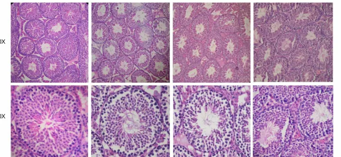
2. BPA가 생쥐 정소의 조직 및 세포 형태에 미 치는 영향
Group I의 생쥐들에서 정소를 채취하여 조직학적 형태 변화를 관찰하였다. 파라핀 절편을 hemato- xylin과 eosin으로 염색하여 관찰한 결과, Figure 2 에서 보여진 것처럼 BPA를 1회 저농도 또는 고농 도를 주사 받은 생쥐의 정소에서는 별다른 조직학 적 형태 변화가 관찰되지 않았다. 이 결과는 1회의 BPA 처리로 LHR 등의 유전자 발현을 변화시킬 수 있으나 조직학적 변화를 가져오지는 않음을 시사 해준다.
3. BPA가 Leydig cell의 마커인 LHR 발현에 미 치는 영향
RT-PCR 결과, Leydig cell의 표지유전자인 LHR의
감소가 관찰되었으므로 이 변화가 단백질 레벨에 도 영향을 주는지 조사하기 위해 면역형광염색을 실시하였다. Figure 3에서 보여진 바, immunoreactive LHR는 Leydig cell들의 세포질 부분에서 특이적으 로 관찰되는데, vehicle을 처리한 대조군과 BPA 100 mg/kg group 간 눈에 띠는 큰 차이는 없는 것으로 관찰되었다.
4. BPA가 정소 내 세포자멸사에 미치는 영향
BPA가 정소 내 Leydig cell의 유전자 발현 변화와 더불어 세포자멸사를 초래할 수 있는지 조사하기 위해 세포자멸사를 직접 확인할 수 있는 TUNEL 염색을 실시하였다. 또한 pro-apoptotic factor인 Bax 의 발현도 면역형광염색법으로 조사하였다 (Figure 4). 이 실험은 1회의 BPA를 복강주사 받은 group I 의 조직에서 수행되었다. 세포자멸사 과정 중 잘려 지는 DNA를 직접 labeling하는 TUNEL assay 결과, 100 mg/kg의 고농도 BPA 처리군의 정소에서 다수 의 Leydig cell들에서 시그널이 관찰되었다. 또한
10X
60X
Control 1 mg/kg 10 mg/kg 100 mg/kg
Group I
Figure 2. Acute effects of BPA on the histological features of the mouse testis. Testes of Group I mice were paraffin-
embedded, sectioned at 6 μm, and stained with hematoxylin and eosin. Photomicrographs are shown at 10× or 60×,
as indicated.
위치상으로 정원세포 또는 Sertoli cell에 해당하는 Seminiferous tubule의 basal side에서도 몇몇 세포가 TUNEL staining을 나타내었다 (Figure 4A). Immuno- reactive Bax의 존재는 세포자멸사가 일어나고 있음 을 시사하는데, Figure 4B에서처럼 100 mg/kg BPA group의 정소에서 Bax protein이 Leydig cell들이 존 재하는 interstitial space의 많은 세포들에서 관찰되 었다. 이 같은 결과는 내분비계 교란물질인 BPA가 생쥐 정소의 내분비 세포인 Leydig cell을 표적으로 하며 세포자멸사를 유발하여 정소 내 생식학적 기 능에 영향을 줄 가능성을 시사한다.
고
찰포유동물에서 정자형성과정은 생식세포의 분열 과 분화가 매우 활발하게 일어나는 과정으로 생식 세포의 특정 유전자 발현과 체세포인 Leydig cell
Control
10 mg/kg
100 mg/kg
10X 20X
Group 1 LHR
Figure 3. Immunofluorescence staining of LHR. Expres- sion of immunoreactive LHR protein is examined by using rabbit polyclonal anti-LHR antibody and visualized by anti-rabbit Alexa 488 secondary antibody. Nuclei are counterstained with TO-PRO-3-Iodide. Immunoreactive LHR is shown in green and nucleus in red. Photo- micrographs are shown at 10× or 20×, as indicated.
Figure 4. Acute effects of BPA on apoptosis of the mouse testis. (A) TUNEL staining was performed on paraffin sections of the testes of mice treated with vehicle or 100 mg/kg. Photomicrographs are shown at 20×. For the 100 mg/kg-treated group, two separate regions showing positive TUNEL staining are shown.
Fluorescein TUNEL staining is in green and nuclear counterstain is in red. (B) Immunofluorescence staining of pro-apoptotic protein Bax in the testes of mice treated with vehicle or 100 mg/kg. Mice received one i.p. injec- tion and were sacrified 24 hr later. Control mice received sesame oil injection. Anti-Bax rabbit polyclonal antibody at 0.5 μg/ml was used and visualized by anti-rabbit Alexa 488 secondary antibody. Expression of immunoreactive Bax protein is shown in green and nucleus is counter- stained in red with TO-PRO-3-iodide. Arrows indicate positive Bax staining in the cytoplasm of Leydig cells within the interstitial regions of the testis. Photomicro- graphs are shown at 20×.
Control
10 mg/kg
100 mg/kg
Group 1 TUNEL
Group 1 Bax
A B
및 Sertoli cell과의 상호작용, 그리고 이들로부터 분 비되는 호르몬들의 영향을 받아 진행된다. 이러한 호르몬들의 기능을 방해하는 내분비계 교란물질의 하나인 BPA가 유전자 발현 및 세포자멸사에 어떠 한 영향을 주는가 조사하였다.
본 연구에서는 내분비계 교란물질인 BPA가 다 양한 농도에서 정소 내 어떠한 세포 종류의 유전 자 발현에 영향을 주는지 관찰하기 위해 정자형성 과정의 각 단계별 특정 세포들에서 발현되는 표지 유전자들과 정소 내 체세포들의 표지유전자들의 발현을 조사하였는데, 특히 Leydig cell의 표지유전 자인 LHR의 mRNA 양이 감소되는 경향을 보였다.
정소의 조직학적 관찰 결과 1회의 BPA 처리가 세 포들의 형태나 LHR 단백질 발현에는 영향을 주지 않는 것으로 보였고, pro-apoptotic Bax의 면역형광 염색은 대조군보다 고농도의 BPA를 주사한 정소의 Leydig cell에서 높게 발현되는 것이 관찰되었다. 마 우스를 포함한 여러 종에서 정소의 다양한 세포 들이 ERα 또는 ERβ를 발현한다는 보고들이 있다.
특히 정소 내 유일한 내분비 세포인 Leydig cell은 ERα, ERβ, 그리고 testosterone을 estrogen으로 바꾸 는 aromatase를 모두 발현한다.
34즉 이 결과는 ER 작용제로 알려진 BPA가 Leydig cell의 ER에 직접 작용하여 세포자멸사 및 유전자 발현에 영향을 줄 가능성을 시사한다. 생쥐의 Leydig tumor cell line에 서는 BPA가 Nur77과 같은 고아핵수용체의 발현을 조절하고 스테로이드 합성의 변화를 가져온다는 결과가 보고된 바 있다.
35따라서 BPA는 내분비 기 능을 갖는 Leydig cell의 기능을 변화시킴으로서 호 르몬 밸런스를 교란시키는 것으로 사료된다.
정소 내 세포에서 비정상적으로 세포자멸사가 일어날 경우에는 정상적인 정자형성과정에 이상이 생길 뿐 아니라 정소의 발달에도 문제가 생길 수 있다. 세포자멸사의 유발은 세포 손상과 관련된 중 요한 현상 중 하나로 보고되고 있으며, 내분비계 교란물질에 의한 세포 손상 시에도 세포자멸사 관 련 분자들이 관련될 것으로 보고되었다.
36,37세포 의 자멸사는 크게 extrinsic pathway와 intrinsic path-
way를 통해 일어나는데, 특히 이 중 intrinsic path- way에서는 미토콘드리아에 Bax가 증가하면서 Bax homodimer를 형성하고 이것이 channel이 되어 cyto- chrome C가 미토콘드리아 밖으로 방출된다. 방출된 cytochrome C는 caspase 9를 활성화시키고 활성화된 caspase 9이 caspase 3를 활성화시켜 세포자멸사를 일으킨다.
38본 연구에서는 BPA가 Leydig cell에서 Bax 단백질의 발현을 증가시켜 세포자멸사를 유발 함으로서 정소 내 생식학적 기능에 영향을 줄 가 능성을 보여주었다. 생쥐 난소의 granulosa cell을 이용한 연구에서 BPA 처리가 세포자멸사를 초래 하며 이는 Bax의 증가를 수반한다는 보고는 BPA 가 세포자멸사를 유도한다는 본 결과를 뒷받침해 준다.
39그러나 본 실험 결과 1회의 BPA 주사 후에 는 Leydig cell의 LHR 단백질 발현 감소나 세포자 멸사적인 조직학적 변화가 수반되지 않았고 Bax의 발현 증가와 TUNEL 염색으로 세포자멸사를 확 인할 수 있었다. BPA에 수 회 노출되는 경우에는 LHR 단백질 발현이나 조직학적 변화를 수반할 수 있는지는 장기 효과를 보는 실험군에서 차후 확인 하고자 한다.
또한 rat의 Sertoli cell을 체외 배양한 모델에서 는 BPA 처리가 이들의 세포자멸사를 유발한다는 결과가 있다.
40본 연구에서도 TUNEL 염색 결과, Seminiferous tubule의 basal side, 즉 Sertoli cell과 정 원세포가 존재하는 부분에서 BPA가 세포자멸사를 유발하는 것으로 보이나 (Figure 4) 세포특이적 마 커들과의 동시 염색 등으로 이 세포들의 종류를 확인해야 할 것이다.
결론적으로 본 연구에서는 BPA가 생쥐 정소에
서 Leydig cell을 주요 타깃으로 하며 이 세포들의
유전자 발현과 세포자멸사를 조절할 수 있음을 보
여준다. 앞으로 BPA가 어떤 기작으로 Leydig cell
의 유전자 발현과 세포자멸사에 영향을 주는지에
대해서는 분자적으로 좀 더 깊은 연구가 되어야
할 것이다.
참
고 문 헌1. Anway MD, Cupp AS, Uzumcu M, Skinner MK. Epigenetic transgenerational actions of endocrine disruptors and male fertility. Science 2005; 308: 1466-9.
2. Krishnan AV, Stathis P, Permuth SF, Tokes L, Feldman D.
Bisphenol-A: and estrogenic substance is released from poly- carbonate flasks during autoclaving. Endocrinology 1993; 132:
2279-86.
3. Steinmetz R, Brown NG, Allen DL, Bigsby RM, Ben-Jonathan N. The environmental estrogen bisphenol A stimulates prolactin release in vitro and in vivo. Endocrinology 1997; 138: 1780-6.
4. Gould JC, Leonard LS, Maness SC, Wagner BL, Conner K, Zachalewski T, Safe S, McDonnell DP, Gaido KW. Bisphenol A interacts with the estrogen receptor alpha in a distinct manner from estradiol. Mol Cell Endocrinol 1998; 142: 203 -14.
5. Ashby J, Tinwell H. Uterotrophic activity of bisphenol A in the immature rat. Environ Health Perspect 1998; 106: 719-20.
6. Bergeron RM, Thompson TB, Leonard LS, Pluta L, Gaido KW. Estrogenicity of bisphenol A in a human endometrial carcinoma cell line. Mol Cell Endocrinol 1999; 150: 179-87.
7. Schafer TE, Lapp CA, Hanes CM, Lewis JB, Wataha JC, Schuster GS. Estrogenicity of bisphenol A and bisphenol A dimethacrylate in vitro. J Biomed Mater Res 1999; 45: 192-7.
8. Papaconstantinou AD, Umbreit TH, Fisher BR, Goering PL, Lappas NT, Brown KM. Bisphenol-A induced increase in uterine weight and alterations in uterine morphology in ovariectomized B6C3F1 mice: role of the estrogen receptor.
Toxicol Sci 2000; 56: 332-9.
9. Palmer BD, Palmer SK. Vitellogenin induction by xenobiotic estrogens in the red-eared turtle and African clawed frog.
Environ. Health Perspect 1995; 103(Suppl.4): 19-25.
10.Anderson MJ, Olsen H, Matsumura F, Hinton DE. In vitro modulation of 17β-estradiol-induced vitellogenin synthesis and estrogen receptor in rainbow trout (Oncorhynchus mykiss) liver cells. Aquatic Toxicol 1996; 137(2): 210-8.
11. Crews D, Bergeron JM, McLachlan JA. The role of estrogen in turtle sex determination and the effect of PCBs. Environ Health Perspect 1995; 103(Suppl.7): 73-7.
12. Egeland GM, Sweeney MH, Fingerhut MA, Halperin WE, Wille
KK, Schnorr TM. Serum dioxin 2,3,7,8 Tetrachlorodibenzo- p-dioxin 2,3,7,8-Tcdd and total serum testosterone and gonado- tropins in occupationally exposed men. Am J Epidem 1992;
136: 1014.
13.Egeland GM, Sweeney MH, Fingerhut MA, Wille KK, Schnorr TM, Halperin WE. Total serum testosterone and gonadotropins in workers exposed to dioxins. Am J Epidem 1994; 139(3):
272-81.
14. Haishima Y, Hayashi Y, Yagami T, Nakamura A. Elution of bisphenol-A from hemodialyzers consisting of polycarbonate and polysulfone resins. J Biomed Mater Res 2001; 58: 209-15.
15. Hiroi H, Tsutsumi O, Momoeda M, Takai Y, Osuga Y, Taketani Y. Differential interactions of bisphenol A and 17 beta- estradiol with estrogen receptor alpha (ER alpha) and ER beta.
Endocr J 1999; 46(6): 773-8.
16. Funabashi T, Sano A, Mitsushima D, Kimura F. Bisphenol A increases progesterone receptor immunoreactivity in the hypothalamus in a dose-dependent manner and affects sexual behaviour in adult ovariectomized rats. J Neuroendocrinol 2003; 15: 134-40.
17. Ashby J, Tinwell H, Haseman J. Lack of effects for low lose levels of bisphenol A and diethylstilbestrol on the prostate gland of CF1 mice exposed in utero. Regul Toxicol Pharmacol 1999; 30: 156-66.
18. Cargen SZ, Waechter JM Jr, Dimond SS, Breslin WJ, Butala JH, Jekat FW, Joiner RL, Shiotsuka RN, Veenstra GE, Harris LR. Normal reproductive organ development in Wistar rats exposed to bisphenol A in the drinking water. Regul Toxicol Pharmacol 1999; 50: 36-44.
19. Nagao T, Saito Y, Usumi K, Kuwagata M, Imai K. Repro- ductive function in rats exposed neonatally to bisphenol A and estradiol benzoate. Reprod Toxicol 1999; 13(4): 303-11.
20. Nagao T, Saito Y, Usumi K, Yoshimura S, Ono H. Low dose bisphenol A does not affect reproductive organs in estrogen- sensitive C57BL/6N mice exposed at the sexually mature, juvenile, or embryonic stage. Reprod Toxicol 2002; 16: 123 -30.
21. Atanassova N, McKinnell C, Turner KJ, Walker M, Fisher JS, Morley M, Millar MR, Groome NP, Sharpe RM. Comparative effects of neonatal exposure of male rats to potent and weak (environmental) estrogens on spermatogenesis at puberty and the relationship to adult testis size and fertility: evidence for stimulatory effects of low estrogen levels. Endocrinology 2000;
141: 3898-907.
22. Kloas W, Lutz I, Einspanier R. Amphibians as a model to study endocrine disruptors: II Estrogenic activity of environ- mental chemicals in vitro and in vivo. The Sci of the Total Env 1999; 225(1-2): 59-68.
23. Welshons WV, Nagel SC, Thayer KA, Judy BM, Vom Saal FS. Low-dose bioactivity of xenoestrogens in animals: fetal exposure to low doses of methoxychlor and other xeno- estrogens increases adult prostate size in mice. Toxicol Ind Health 1999; 15(1-2): 12-25.
24. vom Saal FS, Cooke PS, Buchannan DL, Palanza P, Thayer KA, Nagel SC, Parmigiani S, Welshons WV. A physiolog- ically based approach to the study of bisphenol A and other estrogenic chemicals on the size of reproductive organs, daily sperm production, and behaviour. Toxicol Ind Health 1998;
14: 239-60.
25. Steinmetz R, Brown NG, Allen DL, Bigsby RM, Ben-Jonathan N. The environmental estrogen bisphenol A stimulates prolactin release in vitro and vivo. Endocrinology 1997; 138: 1780-6.
26. Delbes G, Levacher C, Duguemec C, Racine C, Pakarinen P, Habert R. Endogenous Estrogens Inhibit Mouse Fetal Leydig Cell Development via Estrogen Receptor α. Endocrinology 2005; 146(5): 2454-61.
27. Oulad-Abdelqhani M, Bouillet P, Decimo D, Gansmuller A, Heyberger S, Dolle P, Bronner S, Lutz Y, Chambon P. Char- acterization of a premeiotic germ cell-specific cytoplasmic protein encoded by Stra8, a novel retinoic acid-reponsive gene.
J Biol Chem 1996; 135(2): 469-77.
28. Schrans-Stassen BH, Saunders PT, Cooke HJ, de Rooij DG.
Nature of the spermatogenic arrest in Dazl-/- Mice. Biol Reprod 2001; 65(3): 771-6.
29. Venables JP, Ruggiu M, Cooke HJ. The RNA-binding speci- ficity of the mouse Dazl protein. Nucleic Acids Research 2001; 29(12): 2479-83.
30. Lee J, Hong J, Kim E, Kim K, Kim SW, Krishnamurthy H, et al. Developmental stage-specific expression of Rbm suggests its involvement in early phases of spermatogenesis. Mol Hum
Reprod 2004; 10(4): 259-64.
31. Chen C, Ouyang W, Griqura V, Zhou Q, Carnes K, Lim H, et al. ERM is required for transcriptional control of the spermatogonial stem cell niche. Nature 2005; 436: 1030-4.
32.Balvers M, Spiess AN, Domagalski R, Hunt N, Kinic E, Mukhopadhyay AK, Hanks E, Charlton HM, Ivell R. Relaxin- like factor expression as a marker of differentiation in the mouse testis and ovary. Endocrinology 2006; 139: 2960-70.
33.vom Saal FS, Hughes C. An extensive new literature con- cerning low-dose effects of bisphenol A shows the need for a new risk assessment. Environ Health Perspect 2005; 113(8):
926-33.
34. O'Donnell L, Robertson KM, Jones ME, Simpson ER. Estrogen and spermatogenesis. Endocr Rev 2001; 22(3): 289-318.
35. Song KH, Lee K, Choi HS. Endocrine disrupter bisphenol a induces orphan nuclear receptor Nur77 gene expression and steroidogenesis in mouse testicular Leydig cells. Endocrinology 2002; 143(6): 2208-15.
36. Nair R, Shaha C. Diethylstilbestrol induces rat spematogenic cell apoptosis in vivo through increased expression of sper- matogenic cell Fas/FasL system. J Biol Chem 2003; 278(8);
6470-81.
37. Fritz WA, Cotroneo MS, Wang J, Eltoum IE, Lamartiniere CA. Dietary diethylstilbestrol but not genistein adversely affects rat testicular development. J Nutr 2003; 133(7): 2287 -93.
38. Zha H, Reed JC. Heterodimerization-independent Functions of Cell Death Regulatory Proteins Bax and Bcl-2 in Yeast and Mammalian Cells. J Biol Chem 1997; 272(50): 31482-8.
39. Xu J, Osuga Y, Yano T, Morita Y, Tang X, Fujiwara T, Takai Y, Matsumi H, Koga K, Taketani Y, Tsutsumi O. Bisphenol A induces apoptosis and G2-to-M arrest of ovarian granulosa cells. Biochem Biophys Res Commun 2002; 292(2): 456-62.
40. Iida H, Maehara K, Doiguchi M, Mori T, Tamada F. Bisphenol A-induced apoptosis of cultured rat Sertoli cells. Reprod Toxicol 2003; 17(4): 457-64.
= 국문초록 =