大 韓 不 妊 學 會 誌 : 第 28 卷 第 3 號 2 001 Kor. J. Fertil. Steril., Vol. 28, No. 3, 2001, 9
생쥐 1-세포기 수정란의 동결방법에 있어서 초자화동결과 완만동결의 비교
상지대학교 생명과학과1, 연세대학교 원주기독병원 산부인과2
이지향1・한혁동2・구혜영1
Comparison of Vitrification and Slow Freezing-thawing Method on 1-cell Zygotes
Ji-Hyang Lee1, Hyuck-Dong Han2, Hye-Young Koo11Department of Biological Sciences, Sangji University, 2Department of Obstetrics and Gynecology, Yonsei University, Wonju College of Medicine, Wonju, Korea
Objective : This study was conducted to examine the effect of vitrification on the survival and in vitro development of mice 1-cell zygotes.
Method: Effects of exposure to vitrification solution and vitrification, with different concentrations of the cryoprotectant solution, were examined. The 1-cell zygotes were also subjected to a slow freezing- thawing method to compare with vitrification method. Solution composed of ethylene glycol (6.0 M, 5.0 M, 4.0 M) and sucrose (1.0 M) were used as cryopropectant. The experiments employed the method loading the embryos on electron microscope grids.
Results:
I. The effects of exposure in vitrification solution
1-cell zygotes were non-toxic at all concentrations of the vitrification solution showing the survival rate between 88.1% and 97.5%. Development into 2-cell was more successful in the higher concentrations of the vitrification solution. Therefore, higher concentrations of the vitirification solution do not seem to cause any problems in vitrification procedure.
II. The effects of vitrification method
1-cell zygotes showed the survival rate between 78.8% and 92.4%. The lowest and the highest survival rate was observed in the 6.0 M and 4.0 M vitrification solution, respectively. 2-cell development rates varied from 77.6% to 91.3%. Blastocyst development rate was shown highest in 5.0 M and the lowest in 4.0 M solution. Therefore, the highest 2-cell and blastocyst development rate was observed in 5.0 M solution.
III. Comparison of vitrification and slow freezing-thawing method on 1-cell zygotes
This experiment showed that 1-cell zygotes had the highest survival and development rates in 5.0 M vitrification solution. Vitrified group of 1-cell zygotes, in the 5.0 M vitrification solution, were compared with the group processed in slow freezing-thawing method. The development rate into 2-cell and blastocyst as well as the survival rate were higher in the vitrified group than in the slowly freezed group.
Conclusion: 1. The results demonstrate that the best cryoprotectant is a 5.0 M vitrification solution for 1-cell zygotes. 2. Vitrification method significantly increases the survival rate of the 1-cell zygote
and its development into 2-cell and blastocyst. Equilibration and exposure time during the vitrification was remarkerbly short in this experiment. Total time, from the exposure to vitirification solution to storage in the liquid nitrogen, was taken only 90 seconds. In contrast, the slow freezing-thawing method have taken more than four hours. Taken together, we presume that the overall time used for the procedure contributes to the results as an important parameter. 3. The loading of 1-cell zygotes on the EM grid is technically more simple and takes less time than the straw or cryo vial method.
Key Words: Cryoprotectant, Vitrification, Ethylene glycol, Electron microscope grid
배아의 냉동에 관한 연구는 1972년 Whittingham이 DMSO (dimethyl sulfoxide)를 사용하여 완만동결과 완만융해로 생쥐 배아를 빙결점 이하의 온도에서 장 기간 동결보존에 성공하여 산자를 탄생시킨 이후 급속히 가속화 되었다.7,21 냉동보존 실험에서는 일 반적으로 동해를 막기 위해 항동해제 (cryoprotective agents : CPAs)를 사용하고 있으며, 그 동안 항동해제 로는 대개 투과성 항동해제인 DMSO, PROH, glyce- rol을 사용하였으나, 최근에 이르러 ethylene glycol을 사용하는 경우가 늘고 있으며, 비투과성 보호제로는 glucose 또는 sucrose를 사용하고 있다.1,6,10,17~20,22,23
동결보존 후 배아의 생존율은 동결, 융해과정 중 에 사용되는 항동해제의 종류와 농도, 동결방법과 융해속도 등에 많은 영향을 받는다고 알려져 있으며
5,7,12,15
또한 동결되는 배아의 세포주기, 발생시기,11,12 배아의 종 (species)의 차이7도 동결보존 후의 생존율 과 깊은 관계를 갖는다고 한다.
1985년 Rall과 Fahy16가 초자화동결 (vitrification)을 보고한 이후 초자화동결방법에 대한 항동해제의 종 류, 농도, 평형시간, 융해방법 등에 대한 많은 연구 가 보고되어 왔다.1,2,17,22,23
초자화동결은 고농도의 항 동해제를 사용하여 초급속으로 냉각시켜 높은 냉각 률과 응집력을 가지게 하는 방법으로서 완만동결과 는 달리 동결과정 중에 발생하여 세포 손상의 주원 인이 되는 빙결정 (ice crystallization) 형성을 막으며, 동결에 소요되는 시간을 감소시키고, 기존의 automa- tic freezer가 필요 없이 액화질소에 곧바로 침지함으 로써 매우 경제적이면서 간편하다는 장점이 있다.
본 실험은 생쥐 1-세포기 수정란을 이용하여 경 제적이고 간편한 초자화동결방법을 사용하여 가장 적절한 항동해제의 농도를 검토하고, 종전에 주로 사용해왔던 완만동결방법과 비교하여 수정란의 생 존율 및 포배기로의 발달률을 비교하였다.
연구 대상 및 방법
1. 실험동물
실험동물로는 생쥐 제1대 잡종 BDF1 (C57BL/DBA) 의 생후 6~8주령의 암컷과 생식능력이 확인된 생후 10~12주령의 DBA 수컷을 선택하였으며, 10시간/14 시간 light cycle을 유지시켰다. 과배란 유도는 preg- nant mare's serum gonadotropin (PMSG; G-4877, Sigma, USA) 7.5 IU를 복강 내 주사하고 48시간 후 human chorionic gonadotropin (HCG; C-8554, Sigma, USA) 7.5 IU를 다시 복강 내 주사하였다. HCG 주사 후 수컷 생쥐와 1:1로 합사시켜 자연교미를 유도하였으며 다 음날 질전이 확인된 개체만을 실험에 사용하였다.
2. 1-세포기 수정란의 회수
HCG 주사 후 18~22시간에 난관 팽대부로부터 난 구세포에 싸여있는 1-세포기 수정란을 회수하였다.
회수된 수정란을 67 IU/ml 농도의 hyaluronidase (Si- gma, H-3506)로 처리하여 난구세포를 제거하고 두 번 D-PBS에서 세척하였다. 그 후 50 mg/ml bovine serum albumin (BSA; A-9647, Sigma, USA)이 첨가된 Dulbecco's phosphate buffered saline (D-PBS; 21300-017, GibcoBL, USA)이 담긴 배양접시에서 세척 후 37℃, 5.0% CO2, 100% 습도로 조절된 배양기 (Forma Sci- entific, 1058)에서 10% 태아제대혈청이 첨가된 mo- dified HTF (Basal XI) 배양액에 넣어 약 1시간 동안 배양한 후 정상적인 형태의 것만 실험에 사용하였 고, 대조군, 독성검사를 위한 동결보존액 노출군, 초 자화동결군 및 완만동결군으로 나누어 사용하였다.
3. 대조군의 배양
회수된 수정란을 10% 태아제대혈청이 첨가된 mo-
dified HTF (Basal XI) 배양액에 넣어 배양기에서 배 양하여 체외발달률을 구하였다.
4. 독성검사를 위한 초자화 동결보존액 노출군 초자화 동결보존액은 ethylene glycol (Sigma, E- 9129)을 기본 용액으로 하여 6.0 M, 5.0 M, 4.0 M의 농도를 사용하였으며, 각 농도의 ethylene glycol에 1.0 M sucrose (Sigma, S-1888)를 첨가하였다. Ethylene glycol과 sucrose는 기본액 10% fetal bovine serum (FBS)이 첨가된 D-PBS (GibcoBRL. 11500-022)로 희 석하였으며, 제조된 용액은 0.2 µm filter (Gelman Sci- ence, 4192)로 여과하고 냉장보관 후 다음날 사용하 였다. 또한 초자화 융해액은 sucrose 0.5 M을 사용하 였으며, 10% FBS가 첨가된 D-PBS로 희석하여 0.25 M 과 0.125 M을 만들었다.
1시간 동안 배양기에 두었던 수정란을 각각 10~
15개씩 mouth pipet으로 pipetting하여 10% FBS가 첨 가된 D-PBS에서 씻은 후 즉시 초자화 동결보존액 6.0 M, 5.0 M, 4.0 M에 각각 넣어서 20초 동안 실온 (25℃)에서 평형시켰다. Pipetting으로 수정란을 채집 하여 여과지 위에 놓인 electron microscope grids (EM grids; Gilder 400 M, Cu, v/100, Ted Pella, Inc, Redding, CA) 위로 뿜었다. EM grid 상에서 동결보존액이 여 과되는 것을 확인한 후 0.5 M, 0.25 M, 0.125 M의 초 자화 융해액 및 10% FBS가 포함된 D-PBS의 순서 로 각 1분씩 노출시킨 후 다시 10% FBS가 포함된 D-PBS에서 5분간 정치하고 배양기에서 배양하는 방법으로 독성 실험을 하였다.
5. 동결보존액의 초자화동결군
초자화 동결보존액에 대한 노출군과 같은 방법으 로 EM grid 상에서 동결보존액이 여과되는 것을 확 인한 후 곧바로 -196℃의 액화질소에 넣었다. 동결 보존액에 평형시키는 시간부터 액화질소에 넣기까 지 걸린 시간은 1분 30초를 초과하지 않았다.
24시간에서 약 4주 가량까지 액화질소에 냉동보 존되었던 EM grid를 액화질소에서 다시 꺼내 공기 중에서 약 10초간 유지하였다. 37℃의 warmmer plate 위에서 준비해 놓은 융해액 0.5 M, 0.25 M, 0.125 M, 10% FBS가 첨가된 D-PBS 순서로 각각 1분씩 정치 하고, 다시 한번 10% FBS가 첨가된 D-PBS의 융해
액에서 5분 동안 유지하였다. 37℃, 5.0% CO2, 100%
습도의 배양기에 보관한, 10% 태아제대혈청이 첨가 된 modified HTF (Basal XI) 배양액으로 1회 세척한 다음 정상적인 형태의 것만 실험에 사용하였다.
6. 완만동결군
완만동결액으로는 3 mg/ml의 BSA 가 첨가된 D- PBS에 1.5 M PROH를 첨가하여 사용하였으며, 융해 액은 1.5 M PROH를 3 mg/ml의 BSA가 첨가된 D- PBS로 희석하여 1.0 M, 0.5 M 을 만들어 사용하였다.
동결을 위해 1-세포기 수정란을 0.3 ml의 1.5 M PROH를 넣은 cryo via l (Nunc, 375299)로 이송하여 실온에서 30분 동안 평형시킨 후, 냉각기 내에서 cryo vial을 상온에서부터 -6℃까지 -1℃/min의 속도 로 냉각하고 다시 5분간 평형시킨 다음 빙정형성을 위해 식빙 (seeding)을 시행하였다. 다시 5분 동안 평 형시킨 후 -0.5℃/min의 속도로 -80℃까지 냉각시켜 서 -196℃의 액화질소 탱크에 넣어 냉동보존하였다.
동결보존액에 평형시키기 시작한 시간부터 액화질 소에 넣기까지 약 4시간이 소요되었다. 융해과정에 서는 냉동보존되었던 cryo vial을 꺼내어 -100℃의 냉각기로 이송하여 5분간 평형시키고 8.0℃/min 의 융해속도로 상온 (20℃)까지 융해시킨 뒤 5분간 평 형시켜서 융해를 완료하였다. 항동해제를 제거하기 위해 1.0 M PROH를 첨가한 용액에서 5분, 0.5 M 용액에서 5분, 기본액인 3 mg/ml의 BSA가 첨가된 D-PBS 용액에서 5분간 평형시켰으며 항동해제를 제거한 1-세포기 수정란은 현미경으로 관찰하여 형 태학적 특징이 잘 보존되어 있는 배아를 선택하여 배양하였다.
7. 생존판정
동결-융해된 수정란은 배율 200의 역반사 현미경 (Leitz Labovert FS)에서 형태적인 생존을 판정하였 다. 세포질에 응집된 현상이 나타나지 않고 깨끗하 며 위란강 (perivitelline space)이 넓지 않고, 투명대가 손상되지 않은 것을 생존한 것으로 판정하였다.
8. 사진촬영
역반사 현미경에 부착되어 있는 CCD-colo r TV carmera system (Polaroid video recorder)을 사용하여
200배의 배율에서 촬영하였다.
9. 통계처리
전체 수에 대한 생존율, 2-세포기로의 발달률 및 포배기로의 발달률을 백분율로 표시하였으며, 통계 검증은 SPSS pc version 7.5를 이용하여 one way AN- OVA 를 시행하였으며, p<0.05 수준에서 노출군, 초 자화동결군, 완만동결군 간에 통계학적 유의성이 있 는 것으로 판정하였다.
결 과
1. 대조군과 노출군에서 생쥐 1-세포기 수정란 의 생존율과 배아발달률
대조군의 실험에서 1-세포기 수정란의 2-세포기
로의 발달률은 93.9±4.9%와 포배기로의 발달률은 90.4±3.7%를 나타냈다 (Table 1).
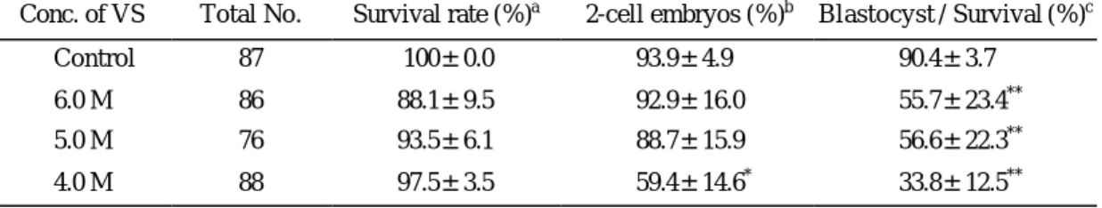
1-세포기 수정란을 초자화 동결보존액에 노출시 켰을 때의 독성 여부에 대한 것은 모든 농도에서 생존율이 88.1~97.5% 로 대조군과 생존율의 차이를 보이지 않았고 6.0 M에서만 대조군에 비하여 낮게 나타났다. 2-세포기로의 발달률은 4.0 M에서 59.4±
14.6% 의 발달률을 보여 대조군 및 6.0 M과 5.0 M에 비해 유의하게 낮은 발달률을 보였다 (p<0.05). 생존 율에 대한 포배기로의 발달률에서는 4.0 M의 33.8±
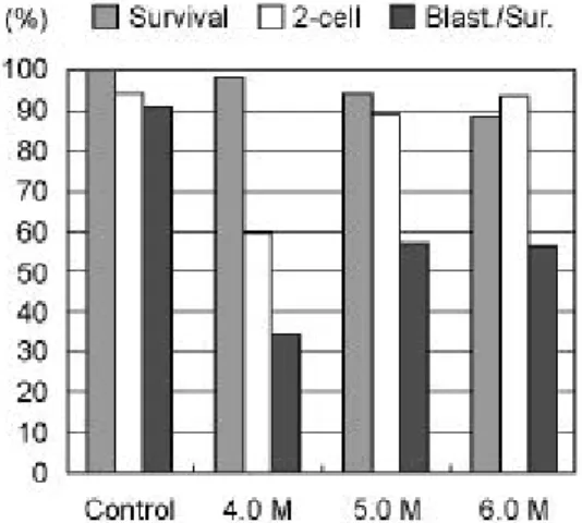
12.5%에 비하여 5.0 M과 6.0 M에서 56.6±22.3%와 55.7±23.4% 로 다소 높게 나타났으나 각 농도 사이 에 큰 유의성은 없었다. 하지만 대조군에 비하여 현 저하게 낮은 발달률을 보였다 (p<0.05) (Table 1, Fig- ure 1).
Table 1. In vitro development of 1-cell zygotes exposed to vitrification solutions (VS)
Conc. of VS Total No. Survival rate (%)a 2-cell embryos (%)b Blastocyst / Survival (%)c
Control 87 100±0.0 93.9±4.9 90.4±3.7
6.0 M 86 88.1±9.5 92.9±16.0 55.7±23.4**
5.0 M 76 93.5±6.1 88.7±15.9 56.6±22.3**
4.0 M 88 97.5±3.5 59.4±14.6* 33.8±12.5**
* Development rate into 2-cell embryos; p<0.05, ** Development rate into blastocyst; p<0.05
a Mean±SD of survival rate (%); No. of survived 1-cell zygotes/No. of total zygotes
b Mean±SD of 2-cell development rate (%); No. of 2-cell embryos/No. of survived 1-cell zygotes
c Mean±SD of development rate into blastocyst (%); No. of blastocyst embryos/No. of survived 2-cell zygotes
Table 2. In vitro development of 1-cell zygotes stored at -196℃ by vitrification
Conc. of VS Total No. Survival rate (%) 2-cell embryos (%) Blastocyst / Survival (%)
6.0 M 108 78.8±22.6 77.6±23.2 55.3±17.6
5.0 M 85 87.0±15.3 91.3±11.7 62.0±14.7
4.0 M 99 92.4±11.2 78.3±20.3 33.8±8.9*
* Develo pment rate into blastocyst; p<0.05
Table 3. In vitro development of 1-cell zygotes. They are exposed to or vitrified in vitrification solution or processed in slow freezing-thawing method
Group Total No. Survival rate (%) 2-cell embryo (%) Blastocyst / Survival (%) Vitrified (5.0 M ) 85 87.0±15.3 91.3±11.7 62.0±14.7
Slow freezing 64 62.7±4.2 49.5±4.2 19.0±5.1
Vitrified and slow freezing group show significantly different results (p<0.001).
2. 초자화동결-융해 후 생쥐 1-세포기 수정란 의 생존율 및 배아발달률
초자화동결-융해 후 생존율은 6.0 M에서 78.8±
22.6%, 5.0 M에서 87.0±15.30%, 4.0 M에서 92.4±
11.2%를 보여 유의한 차이를 보이지 않았으나, 2- 세포기로의 발달률은 6.0 M에서 77.6±23.2%, 5.0 M 에서 91.3±11.7%, 4.0 M에서 78.3±20.3%로 5.0 M에 서 가장 높게 나타났다. 포배기로의 발달률은 4.0 M 에서 33.8±8.9%로 5.0 M에서 62.0±14.7%와 6.0 M 에서 55.3±17.6% 에 비하여 현저하게 낮게 나타났다 (p<0.05) (Table 2, Figure 2). Figure 4는 초자화동결-융
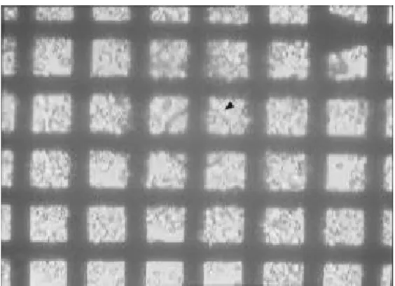
해시킨 1-세포기 수정란이 EM grid 위에 있는 모습 으로서 hyaluronidase를 약하게 처리하여 난구세포가 붙어있다. Figure 5는 24시간 후 2-세포기로 난할된 상태를 보여주며, Figure 6은 4일 후 포배기로 발달된 상태를 보여준다.
3. 1-세포기 수정란의 초자화동결군과 완만동결 -융해군의 비교
초자화동결에서 가장 좋은 결과를 나타낸 농도 5.0 M 과 완만동결-융해군을 비교한 실험에서 완만동결 -융해군은 생존율 62.7±4.2%와 2-세포기로의 발달 률 49.5±4.2%를 나타냈으며 포배기로의 발달률은 19.0±5.1%를 나타냈다. 이는 생존율, 2-세포기로의 발달률 및 포배기로의 발달률 모두 완만동결-융해 군이 유의하게 낮음을 나타내는 수치이다 (p<0.001) (Table 3, Figure 3).
고 찰
1972년 Whittingham에 의해 개발된 완만동결법에 의한 동결보존 실험 이후 지금까지 동결 융해 후의 생존율 향상과 다른 종에의 적용 등에 대한 많은 연 구가 진행되어 왔다. 그 결과로 생쥐, 토끼, 양 등의 배아 냉동보존이 가능하게 되었으며, 냉동보존되었 던 사람의 배아에서 정상출산이 이루어지는 결과까 지 보고되었다.4,20 완만동결법은 배아 및 세포내의 Figure 1. In vitro development of 1-cell zygotes ex-
posed to vitrification solution.
Figure 2. In vitro development of 1-cell zygotes sto- red at -196℃ by vitrification.
Figure 3. In vitro development of 1-cell zygotes. They are exposed to or vitrified in 5.0 M vitrification solution or processed in slow freezing-thawing method.
탈수를 유도한 다음, 천천히 냉각시켜서 일정온도에 도달한 후 세포질 내에 빙결정이 이루어지도록 식빙 을 하고 일정온도에 도달하면 보존을 하는 방법으로, 융해 역시 일정한 온도의 조건에서부터 융해를 시 작한다. 하지만 매우 복잡한 단계를 거치는 냉동과 융해과정 동안 세포 및 배아가 손상될 가능성이 매 우 높다. 세포내의 빙정형성, 용액 자체의 영향과 세포의 삼투성 응축 등 냉동 동안 세포에 손상을 주 는 요인들이 많이 있으며, 그 외에 얼음 결정체에 의해 세포가 물리적으로 손상될 수도 있다.14
초자화동결은 세포가 동결되는 동안 동결용액의 점성을 점차 증가시켜 세포내외의 빙정형성을 방지 하는 동결법이다. 그러나 초자화된 동결용액은 낮은 온도에서 과냉각 (supercooled) 되기 때문에 융해과정 동안 결정화가 일어나 세포에 손상을 줄 수 있다.
따라서 융해되는 동안 결정화가 일어나지 않도록
투과력이 높은 고농도의 항동해제를 사용하여야 한 다.17 일반적으로 완만동결은 저농도의 항동해제 (1.5 M에서 2 M 정도)를 사용하지만 초자화동결에서는 3.5 M 이상에서 약 7.5 M 까지의 항동해제를 사용 한다. 항동해제로는 오랫동안 DMSO, 1.2-PROH, gly- cerol을 주로 사용하였으나, 최근에는 가장 독성이 약한 것으로 알려진 ethylene glycol을 많이 사용하고 있다.3
본 실험에서 사용한 완만동결법은 10년 이상 원 주기독병원에서 불임환자를 대상으로 시행하였던 동결방법으로서 실온에서부터 시작하여 -80℃까지 점차 냉각시켜 냉동보존한 후, -100℃부터 시작하여 실온으로 올리면서 융해시키는 방법이다.
초자화 동결보존액은 Ali와 Shelton1,2이 독성이 약 하다고 발표한 VS14 (5.5 M ethylene glycol에 1.0 M sucrose가 혼합된 용액)를 기초로 하였으며, 항동해제 로 6.0 M에서 4.0 M 까지의 ethylene glycol을 사용하 여 아직까지 생쥐의 초자화동결에서 보고되지 않은, 직접 액화질소에 EM grid 를 노출시키는 Martino13 방법 (EM grid를 사용하여 소의 성숙난자를 냉동시 키는데 성공함)으로 실험하였다. Ali와 Shelton의 실 험에서는 Swis s outbred 생쥐 2-세포기 배아에서 81%
의 생존율을 보였고 동결-융해 후에는 71% 의 생존 율을 보고하였다. 이에 비해 본 실험은 BDF1 쥐를 사용하여 5.0 M의 ethylene glycol과 1.0 M sucrose가 혼합된 군에서 독성 실험한 결과 93.5±6.1%의 생존 율 및 초자화동결-융해 후 87.0±15.3% 의 생존율을 보임으로써 Ali와 Shelton의 실험보다 크게 향상된 결과를 얻었다.
Figure 4. 1-cell zygotes (arrowhead) on EM grid after vitrification and thawing (×200).
Figure 5. 2-cell stage embryos after vitrification and thawing (×200).
Figure 6. Blastocyst after vitrification and thawing (×200).
1990년 Kasai 등10은 실온에서 40% ethylene glycol, 30% ficoll, 20% sucrose (EFS40)를 혼합하여 straw에 장진한 생쥐 상실배에서 동결 융해 후 98% 의 생존 율과 51% 의 산자 생산을 보고하였다. 그 이후 EFS- 40을 사용하여 생쥐,22~24 소,13 토끼 등에서 초자화동 결법에 대한 많은 연구가 이루어졌다. 또한 1996년 Martino 등13은 5.5 M 과 4.0 M 의 ethylene glycol에 1.0 M과 0.5 M의 sucrose가 혼합된 용액을 동결보존액 으로 사용하여 소의 성숙난자를 35℃에서 20초 동 안 노출 후 동결을 시행하였으며, sample container로 straw와 EM grid를 사용하였다. 이때 동결-융해 후 생존율은 straw가 34% , EM grid 가 51~72% 로 나타나 EM grid에서 높은 생존율을 얻었으며 이 방법으로 융해 후 수정되고, 포배기로 발달된 난자는 15%였다.
생쥐의 1-세포기 수정란을 EM grid에 장진하고 동결보존액으로 ethylene glycol과 1.0 M의 sucrose가 혼합된 용액을 사용한 본 초자화동결 연구 결과를 straw에 장진하는 Kasai (1990)의 방법을 변형시킨 1996년 Kim23의 EFS40 (ethylene glycol 40%가 혼합 됨) 방법으로 초자화동결한 연구 자료와 비교해 보 았다. Kim의 EFS40을 사용하여 체외수정시킨 1-세 포기 수정란을 1단계 동결법으로 초자화동결 하였을 때 생존율 및 2-세포기로의 발달률 85.5%, 포배기 로의 발달률 53.2%를 나타냈는데, 본 실험에서는 수정란을 채집하여 초자화동결 하였을 때 생존율 78.8~92.4% , 2-세포기로의 발달률 77.6~91.3%로 나 타났으며 모든 농도에서 EFS40과 비슷한 결과를 보 이거나 더 높은 결과를 얻었다. 그러나 본 연구의 적정농도로 생각된 5.0 M에서는 EFS40 보다 높은 포배기로의 발달을 보였다.
1-세포기 수정란을 5.0 M의 초자화 동결보존액 을 사용해 초자화동결 했을 때와 완만동결-융해군 을 비교하면 생존율, 2-세포기로의 발달률과 포배 기로의 발달률 모두 초자화동결이 유의하게 높았다 (p<0.001). 그러나 ethylene glycol에 난자를 오랫동안 노출시키면 배아의 기포형성 원인이 된다는 보고가 있으며,9 ethylene glycol이 쥐의 착상 전 발생 중 기 관형성 (organogenesis)에 해로운 영향을 준다는 보고 도 있다.8 그러므로 실온에서 가능한 한 짧은 시간 동안 동결보존액 내에 평형시켜 액화질소에 냉동시 킬수록 융해 후 높은 생존율과 발달률을 가져오리라
고 판단된다. 본 실험에 따르면 1분 동안 초자화 동 결보존액에 노출시킨 후 6.0 M부터 4.0 M까지 노출 군과 동결군을 비교한 결과, 동결-융해 후 5.0 M 이 상의 고농도를 사용한 실험군에서 포배기로의 발달 률이 가장 높게 관찰되었다. 2-세포기로의 발달률에 서는 완만동결-융해군 49.5±4.2%에 비하여 초자화 동결군이 모든 농도에서 77.6~91.3% 의 범위로 나타 나 현저하게 높은 결과를 보였다.
이상과 같은 연구 결과로 본 실험에서 채택한 ethylene glycol을 기본 동결보존액으로 사용하여 초 자화동결-융해하는 방법은 완만동결-융해방법 보다 시간이 단축되고 비싼 장비가 필요 없어 경제적이고 간단하면서도 동결-융해 후 높은 생존율과 체외발 달률을 가져왔고, 특히 EM grid 를 사용하는 방법은 straw나 cryo vial을 사용하는 방법보다 조작이 간단 하면서도 효과적인 결과를 가져왔다. 앞으로 이와 같 은 결과를 토대로 생쥐 1-세포기 수정란의 초자화 동결방법을 더욱 다양하게 접근해 볼 수 있을 것이 다. 항동해제의 농도, 노출시간, 노출온도와 노출 단 계를 달리 사용하여 평형시키는 방법 등이 함께 연 구된다면 생존율과 체외발달률을 더욱 높일 수 있 는 성공적인 냉동보존법이 개발될 수 있을 뿐 아니 라 이는 인간을 포함한 포유동물 전반에 걸쳐, 그리 고 1-세포기 수정란 뿐 아니라 난자세포 단계에도 적용될 수 있을 것이다.
참 고 문 헌
1. Ali J, Shelton N. Vitrification on pre implantation stages of mouse embryos. Reprod Fertil 1993a; 98:
459-65.
2. Ali J, Shelton N. Design of vitrification solutions for the cryopreservation of embryos. J Reprod Fertil 1993b; 99: 471-7.
3. Bautista JAN, Dela Pena EC, Katagiri S, Takahashi Y, Kanagawa H. In vitro viability of mouse oocytes vitrifed in an ethylene glycol-based solution, Jpn. J Res 1998; 46(1): 13-8.
4. Cohen J, Kort HI, Devane GW, Massey JB, Elsner CW, Turner TG, Fehilly CB. Cryopreservation of zygotes and early cleaved human embryos. Fertil
Steril 1988; 49: 283-9.
5. Dumoulin JC, Bergers -Janssen JM, Pieters MH, Enginsu ME, Geradts JP, Evers JL. The protective effects of polymers in the cryopreservation of hu- man and mouse zona pellucida and embryos. Fertil Steril 1994; 62: 793-8.
6. Fehilly CB, Cohen J, Simons RF, Fishel SB, Ed - wards RG. Cryopreservation of cleaving embryos and expanded blastocysts in the human: a compa- rative study. Fertil Steril 1985; 44: 638-44.
7. Friedler S, Giudice LC, Lamb EJ. Cryopreservation of embryos and ova. Fertil Steril 1988; 88: 743-64.
8. Grafton TF, Hansen DK. Invitro embryotoxic effects of ethylene glycol in rats. Teratog Carcineg Mutagen 1987; 7: 480-9.
9. Hotamisligils S, Toner M, Powers RD. Changes in membrane integrity, cytoskeletal structure and de- velopmental potential of murine oocytes after vitri- fication in ethylene glycol. Biol Reprod 1996; 55:
161-8.
10. Kasai M, Komi JH, Takamo A, Tsudera HM, Sa- kurai T, Machida T. A simple methods for mouse embryo cryopreservation in a low toxic vitrification solution, without appreciable loss of viability. J Re - prod Fertil 1990; 89: 91-7.
11. Liu J, Van der Abbeel E, Van Steirteghem AC.
Assessment of ultra -rapid and slow freezing pro - cedures for 1-cell and 4-cell mouse embryos. Hum Reprod 1993; 8: 1115-9.
12. Mandelbaum J, Junca AM, Plachot M, Alnot MO, Salat-Baroux, Alvarez S, Tibi C, Cohen J, Debache C, Tesquier L. Cryopreservation of human embryos and oocytes. Human Reprod 1988; 3: 117-9.
13. Martino A, Songsasen N, Leibo SP. Development into blastocysts of bovine oocytes cryopreserved by ultra -rapid cooling. Biol Reprod 1996; 54: 1059-69.
14. Miyake T, Kasai M, Zhu SE, Sakurai T, Machida T. Vitrification of mouse oocytes and embryos at various stages of development in an ethylene-glycol
based solution by a simple method. Theriogenology 1993; 40: 121-34.
15. Nowshari MA, Nayudu PL, Hodges JH. Effect of cryoprotectants and their concentration on post-thaw survival and development of rapid frozen-thawed pronuclear stage mouse embryos. Human Reprod 1995; 10: 3237-42.
16. Rall WF, Fahy GM. Ice-free cryopreservation of mouse embryos at -196℃ by vitrification. Nature 1985; 313: 573-5.
17. Rall WF, Wood MJ, Kirby C, Whittimgham DG.
Development of mouse embryos cryopreserved by vitrification. J Reprod Fert 1987; 80: 499-504.
18. Rayos AA, Takahashi Y, Hishinuma M, Kanagawa H. Quick freezing of unfertilized mouse oocytes using ethylene glycol with sucrose or trehalose. J Reprod Fertil 1994; 100: 123-9.
19. Testart J, Allart JB, Lassalle B, Hazout A, Forman R, Rainbhorn JD, Gazengel A, Frydman R. High pregnancy rate after early human embryo freezing.
Fertil Steril 1986; 46: 268-72.
20. Trounson A, Mohr L. Human pregnancy following cryopreservation, thawing and transfer of an eight- cell embryo. Nature 1983; 305: 707-9.
21. Whittingham DG, Leibo SP, Mazur P. Survival of mouse embryos frozen to -196℃ and -269℃. Sci- ence NY 1972; 187: 411-4.
22. Zhu SE, Kasai M, Otoge H, Sakurai T, Machida T.
Cryopreservation of expanded mouse blastocysts by vitrification in ethylene gly col-based solutions. J Reprod Fert 1993; 98: 139-45.
23. Kim MK, Lee SJ, Uhm EY, Yoon SH, Park SP, Chung KS, Lim JH. Cryopreservation of mouse IVF zygotes by vitrification. Korean J Animal Re - prod 1996; 20(2): 119-26.
24. Kim MK, Yi SH, Yoon SH, Park SP, Chung KS, Lim JH. In Vtro/In Vivo Development if mouse oocytes vitrified by EFS. Korean J Animal Reprod 1998; 25(1): 87-92.