대한소화기학회지 2006;47:89-91 □IMAGE OF THE MONTH □
증례: 24세 여자 환자가 내원 1년 전부터 시작해 최근 10 일 전부터 악화된 상복부 통증과 식후 5분 이내에 발생하는 구토를 주소로 내원하였다. 과거력과 가족력에서 특이 소견 은 없었고 최근 체중이 6 kg 감소하였다. 활력징후는 정상 이었으며, 신체검사에서 상복부에 약간의 압통이 있었다. 말 초혈액검사에서 백혈구 7,400/mm3, 혈색소 13.2 g/dL, 혈소판 322,000/mm3이었고, 혈액응고검사는 정상이었다. 혈청생화 학검사는 총 단백 7.1 g/dL, 알부민 4.2 g/dL, AST 24 IU/dL, ALT 18 IU/L, 총 빌리루빈 0.7 mg/dL, BUN 9 mg/dL, 크레아 티닌 0.8 mg/dL이었고 적혈구침강속도, CRP는 정상이었다.
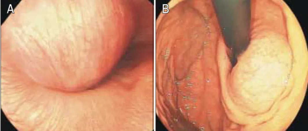
입원 2일째 시행한 상부위장관 내시경에서 위식도 접합부 와 하부 식도에 5×4 cm 크기의 점막하 종양이 발견되었고 종괴에 의해 식도내강은 좁아져 있었으며 생검 겸자로 눌러 보았을 때 단단하고 움직임이 없었다(Fig. 1). 흉부 컴퓨터단 층촬영에서 간 좌엽 후방, 위식도 접합부에 4.8×4.1 cm 크 기의 주변 조직과 경계가 뚜렷하고 조영 증강이 되지 않는 저밀도의 낭종성 종괴가 발견되었으며 이 종괴는 식도 내부 에서 기원하는 것으로 판단하였다(Fig. 2). 수술로 종괴를 제 거하였고 환자는 합병증 없이 수술 후 14일째 퇴원하였다.
진단은?
진단: 식도 중복 낭종(esophageal duplication cyst) 수술 후 조직의 육안 소견에서 종괴는 낭종이었고 낭종 내벽은 두 개의 근육층으로 이루어져 있었으며 낭종의 내강 은 호흡상피로 둘러싸여 있었다(Fig. 3).
식도 낭종 질환은 종격동 종양의 약 10%를 차지하며 주 로 태생기 전장(foregut)의 이상 발육으로 발생하는 선천 질 환이다.1,2 발생 장기와 위치, 내피 구성세포 등에 따라 일반 적으로 식도 중복 낭종, 기관지 낭종(bronchogenic cyst), 장 낭종(enteric cyst) 등으로 분류한다.2,3 식도 중복 낭종은 극히 드문 질환으로 1711년 Blassium이 처음 기술한 이래 1990년 까지 약 50예 정도가 보고되었고 국내에서는 4예4-7가 보고 되었다. 중복 낭종의 발생 위치는 회장이 가장 흔하고 식도 는 두 번째로 흔하다. 다른 선천 기형으로 소장 중복 낭종, 식도 무형성증, 기관식도루, 척추 기형 등이 동반될 수 있
다.2,8-11 임신 4주 이후 전장이 길게 늘어나고 임신 6주째에
늘어난 전장 내에 소강(vacuole)이 발생하면서 식도 내강을 형성하는데 하나의 소강이 따로 분리되어 지속될 때 식도 중복 낭종이 발생한다. 태아가 성장하면서 흉강 내 장기 성 장과 위의 우회전으로 인하여 60%는 하부식도 우측에서 발 생하는데 17%는 중부식도, 23%는 상부식도에서 발생한 다.2,3 식도 중복 낭종은 대개 증상이 없지만 크기와 발생 위 치에 따라 연하 곤란, 심와부 불쾌감, 흉골하 통증, 호흡곤 란, 역류증상, 기침 등이 나타날 수 있다.2,3,8,9,12 대부분은 주 변 조직과 분리되어 있지만 10% 이하에서 정상 소화기관과 연결된다. 합병증으로는 감염이 가장 흔하고, 낭종 파열, 낭 종 내 출혈 등이 있으며 낭종 파열로 인한 심낭 압진(cardiac tamponade)도 보고되었다. 악성으로의 변화는 극히 드문데 현재까지 두 예가 보고되었다.3
식도 중복 낭종
성균관대학교 의과대학 강북삼성병원 내과학교실
박동일․유태우
Esophageal Duplication Cyst
Dong Il Park, M.D., and Tae Woo Yoo, M.D.Department of Internal Medicine, Kangbuk Samsung Hospital, Sungkyunkwan University School of Medicine, Seoul, Korea
ꠏꠏꠏꠏꠏꠏꠏꠏꠏꠏꠏꠏꠏꠏꠏꠏꠏꠏꠏꠏꠏꠏꠏꠏꠏꠏꠏꠏꠏꠏꠏꠏꠏꠏ Correspondence to: Dong Il Park, M.D.
Department of Internal Medicine, Kangbuk Samsung Hospital 108 Pyeong-dong, Jongno-gu, Seoul 110-746, Korea
Tel: +82-2-2001-2059, Fax: +82-2-2001-2049 E-mail: diksmc.park@samsung.com
ꠏꠏꠏꠏꠏꠏꠏꠏꠏꠏꠏꠏꠏꠏꠏꠏꠏꠏꠏꠏꠏꠏꠏꠏꠏꠏꠏꠏꠏꠏꠏꠏꠏꠏ
연락처: 박동일, 110-746, 서울시 종로구 평동 108번지 강북삼성병원 소화기내과
Tel: (02) 2001-2059, Fax: (02) 2001-2049 E-mail: diksmc.park@samsung.com
90 대한소화기학회지: 제47권 제2호, 2006
진단은 방사선 검사가 도움을 준다. 흉부 단순촬영에서 종격동 종양으로 나타나거나 식도조영술에서 부드러운 원 형 종괴에 의한 음영결손으로 발견되기도 한다. 상부위장관 내시경 소견은 정상 점막으로 덮인 평활한 점막하 종양으로 보이는데 대부분 생검 겸자로 잘 눌린다. 생검 겸자가 병변 까지 도달할 수 없고 점막에 궤양이나 염증반응을 유발할 수 있기 때문에 조직 생검은 권유하지 않는다. 초음파내시 경으로 낭종과 고형 종괴의 감별이 어려울 때 MRI가 도움 이 될 수 있다.
비록 증상이 없어도 감염, 출혈, 천공 등의 합병증을 유발 할 수 있기 때문에 수술 절제를 고려해야 한다.13,14 국내에 서 보고된 4예도 모두 수술 절제로 치료하였다.
Fig. 1. Esophagogastroduodenoscopic findings. (A) It shows a huge, round mass bulging into the eso- phageal lumen on the gastro- esophageal junction. The overlying mucosa looks normal. (B) When endoscope is retroflexed, the lesion looks more prominent.
Fig. 2. Chest CT finding. A 4.8×4.1 cm sized, round, well defined and cystic mass is seen on the medial aspect of esophagus, at the gastroesophageal junction level.
Fig. 3. Histologic findings. (A) It is an esophageal duplication cyst, 3×3×2.8 in size. (B) The cystic wall is lined by ciliated respiratory epithelium surrounded by circular and longitudinal smooth muscle layers (H&E stain, ×100).
A B
박동일 외 1인. 식도 중복 낭종 91
참고문헌
1. Morrison IM. Tumors and cysts of the mediastinum. Thorax 1958;13:294-307.
2. Kirwan WO, Walbaum PR, McCormack RJ. Cystic intratho- racic derivatives of the foregut and their complications. Thorax 1973;28:424-428.
3. Arbona JL, Fazzi JG, Mayoral J. Congenital esophageal cyst:
case report and review of literature. Am J Gastroenterol 1984;
79:177-182.
4. Hong JS. Intramural esophageal cyst. Korean J Thorac Car- diovasc Surg 1981;14:95-97.
5. Hur Y, Lee KS, Kang KH, Yu HS, Sur JI, Ma SD. Surgical treatment of esophageal duplication. Korean J Thorac Car- diovasc Surg 1988;21:787-792.
6. Kim PN, Kim IY, Lee BH. CT finding of an esophageal duplication cyst-a case report. J Korean Radiol Soc 1989;25:
294-296.
7. Lee HS, Jeon HJ, Song CW, et al. Esophageal duplication cyst complicated with intramural hematoma-case report. J Korean Med Sci 1994;9:188-196.
8. Whitaker JA, Deffenbaugh LD, Cooke AR. Esophageal dupli- cation cyst. Case report. Am J Gastroenterol 1980;73:329- 332.
9. Desforges G, Strieder JW. Esophageal cysts. N Engl J Med 1960;262:60-64.
10. Nakahara K, Fujii Y, Miyoshi S, Yoneda A, Miyata M, Kawashima Y. Acute symptoms due to a huge duplication cyst ruptured into the esophagus. Ann Thorac Surg 1990;50:
309-311.
11. Gatzinsky P, Fasth S, Hansson G. Intramural oesophageal cyst with massive mediastinal bleeding. A case report. Scand J Thorac Cardiovasc Surg 1978;12:143-145.
12. Vithespongse P, Blank S. Ciliated epithelial esophageal cyst.
Am J Gastroenterol 1971;56:436-440.
13. Nobuhara KK, Gorski YC, La Quaglia MP, Shamberger RC.
Bronchogenic cysts and esophageal duplications: common origins and treatment. J Pediatr Surg 1997;32:1408-1413.
14. Cioffi U, Bonavina L, De Simone M, et al. Presentation and surgical management of bronchogenic and esophageal dupli- cation cysts in adults. Chest 1998;113:1492-1496.