<접수일:2008년 2월 29일, 심사통과일:2008년 3월 13일>
※통신저자:김 성 호
경북 경주시 석장동 1090-1번지 동국대학교 경주병원 류마티스내과
Tel:054) 770-8563, Fax:054) 770-8378, E-mail:[email protected] 지식경제부 지정 동국대학교 해양심층수 및 소재 RIS 사업단의 지원으로 기술개발되었음.
섬유근통 동물모델의 특정 뇌영역에서 심층수 음용후 세로토닌의 변화
동국대학교 의과대학 내과학교실, 대구가톨릭대학교 자연대학 의생명과학과*, 동국대학교 한의과대학 해부학교실**, 동국대학교 의과대학 약리학교실 및 해양심층수/소재 RIS 사업단***
김성호ㆍ최난희*ㆍ박인식**ㆍ남경수***
= Abstract =
Serotonin Changes in Specific Brain Regions of Fibromyalgia Animal Model after Deep-sea Water Drinking
Seong-Ho Kim, Nan Hee Choi*, In-Sick Park**, Kyung Soo Nam***
Department of Internal Medicine, Dongguk University College of Medicine, Gyeongju, Department of Medical Life Science, College of Natural Science, Catholic University
of Daegu*, Daegu, Department of Anatomy, College of Oriental Medicine, Dongguk University**, Department of Pharmacology, College of Medicine and
Regional Innovation System, Dongguk University***, Gyeongju, Korea
Objective: The acidic saline animal model of pain has been suggested to mimic fibromyalgia (FM). In this model, repeated intramuscular (IM) injections of acidic saline produce a widespread hyperalgesia that persists without evidence of significant peripheral tissue damage or inflammation, and is believed to be centrally maintained. We examined the changes of pain-related neurotransmitters in specific brain regions of this model after deep-sea water (DSW) drinking.
Methods: Rats were injected with 100μL of acidic saline (pH 4.0) at days 0 and 5 into the left gastrocnemius muscle. Control rats received identical injections of physiological saline (pH 7.2) on the same schedule. Two acidic saline rats were given DSW from 1 week following the last IM injection to sacrifice. All rats were sacrificed on day 20. All regions of interest were examined for the changes of pain-related neurotransmitters with immunohistochemistry.
Results: Preliminary results showed that compared to controls, acid injected rats demon- strated strong expression of serotonin in red and raphe nucleus. Acid injected rats showed
significant reductions of the serotonin expression in red and raphe nucleus after DSW drinking.
Conclusion: IM acid injections increased the expression of serotonin in red and raphe nucleus of rats. The overwhelming reduction of serotonin expression in the nuclei after DSW drinking suggests DSW might be helpful for pain and anxiety. These preliminary data support the validity of acidic saline treatment as a model of FM, and provide a foundation for future analyses of specific brain regions that contribute to this syndrome.
Key Words: Fibromyalgia, Serotonin, Acidic saline model, Deep-sea water
서 론
섬유근통 증후군은 만성 통증 증후군으로 전신적 인 비관절성 근골격계 통증 및 압통점들이 특징이다 (1). 섬유근통 증후군에서는 두통, 피로, 수면 장애, 과민성 대장 증후군, 감각이상 등이 나타날 수 있다.
만성통증의 신경생물학적 연구에 의하면 잘못된 통 증 조정, 즉 비정상적인 ‘중추성 민감화' 현상이 섬 유근통 증후군의 병인에 중요한 역할을 한다고 알려 졌다 (2,3). ‘중추성 민감화'에 중요한 신경전달물질 인 N-Methyl-D-Aspartate (NMDA)는 그 수용체가 활 성화되면 수용체내의 마그네슘 차단을 제거하여, 결 과적으로 신경세포로 칼슘이 유입되어 통각세포의 활성이 증폭된다 (4). 다른 요인들로는 일부 섬유근 통 증후군 환자들의 뇌척수액에서 통각의 매개물인 substance P의 증가 (5) 및 변형된 serotonin 대사 (6) 등이 보고되고 있다. 세로토닌계가 섬유근통의 병태 생리에 관여된다는 주장들이 많다 (7). 세로토닌(5- hydroxytryptamine, 5-HT)은 신경전달물질로서 24시간 주기 및 신경내분비 리듬뿐만 아니라 기분, 감정 및 인지와 운동 기능의 구성에서 중요한 역할을 한다 (8). 낮은 세로토닌 수준이 항진된 통각과 불면의 원 인일 수 있다는 가정도 있다 (9).
기능적 뇌영상법(functional brain imaging)을 포함한 많은 증거들에 의하면, 만성 전신성 통증 및 섬유근통 증후군의 증상들이 중추신경계의 통증 처리 과정의 기 능장애로 생각된다 (10). 자극받지 않은 섬유근통 증후 군 환자들에서 SPECT 영상을 사용해서 국소적 대뇌 혈류량이 감소한 것을 증명한 보고들이 있다 (11-13).
1. 산성 식염수 모델(Acidic saline model)
현재까지는 섬유근통 증후군이나 만성 광범위 통
증에 대한 동물 모델이 없는 실정이었으나, 최근 산 성 식염수 모델(acidic saline model)이 적절한 모델로 대두되고 있다 (14). 이 산성 식염수 모델에서는 low pH (pH 4.0) saline이 5일 간격으로 쥐의 한쪽 장딴 지(gastrocnemius) 근육에 두 번 주사된다. 기계적 자 극에 대한 발 회피반사(paw withdrawal reflex)의 양 측 과반응이 두 번째 주사 후 24∼48 시간에 약 70
∼80%에서 발생되어 유의한 말초 조직 손상이나 염 증없이 4주까지 지속된다. 이는 섬유근통과 매우 유 사하다. 섬유근통에서는 신체적 손상 또는 초기의 염 증성 자극에 노출되면, 심지어 그 초기 손상이나 염 증이 사라진 후에도 만성 광범위 통증과 과통증이 유 발된다. 산성 식염수 모델의 섬유근통에 대한 관련성 은 최근의 기능적 영상법 자료로 더 확인할 수 있다 (15). 이 자료에서 대조군과 뇌활성도의 차이가 두드 러진 여러 부위들(감각, 운동, 정서 및 항통각에 관여 한다고 알려진 부위들)을 본 연구에서도 조사했다.
2. 심층수(Deep-Sea Water)
요즘 사람들의 관심이 높아지고 있는 심층수(deep- sea water)는 태양광선이 다다르지 못하므로 식물 플 랑크톤의 광합성이 적고 유기 분해가 많아 마그네 슘, 칼슘 및 칼륨과 같은 무기 영양소가 풍부하다 (16). 최근에 심층수의 치료적 또는 예방적 효과에 대한 과학적인 증거들이 보고되고 있다. 사람들에서 심층수 음용으로 미네랄 불균형과 아토피성 피부염 이 호전되었고 (17), 콜레스테롤을 먹인 토끼들에서 고지혈증과 동맥경화증을 예방하는데 효과적이었다 (18,19). 또한 고지혈증 토끼 모델에서 심층수를 음 용시킨 후 심혈관계 혈류역학이 개선되었다(19). 하 지만 어떤 요소가 혈압 등에 영향을 주는지는 아직 알려져 있지 않다.
이 연구에서 저자들은 심층수에 풍부한 마그네슘,
Fig. 1. Immunohistochemistry stain of serotonin from brain of rats. Rectangle area will be magnified in next figures.
Aq: aqueduct, DR: dorsal raphe nucleus, IP: interpeduncular nucleus, MnR: median raphe nucleus, R: red nucleus, SN: substantia nigra.
칼슘과 같은 무기 영양소가 (16) 통증에 관여하는 신경세포의 연접에 영향을 미칠 것 (4)이라는 가설 하에 섬유근통 증후군의 동물 모델에서 경도 1,500 의 정제된 심층수를 음용시킨 후 뇌의 여러 부위에 서 섬유근통과 관련된 신경전달 물질들의 변화를 대 조군과 비교조사하였다.
대상 및 방법 1. 산성 식염수 모델
1) Animals: 200∼250 g 정도의 Sprague-Dawley rats 을 사용하였으며, 쌍으로 plastic cages에 넣어 climate- controlled vivarium에서 유지하였다. 12시간 on/off light- dark cycle (오전 6시 lights on)이었고, 무제한으로 물과 사료가 공급되었다.
2) Induction of muscle pain: 쥐들을 2회에(day 1 및 day 5) 걸쳐 2% isoflurane (v/v)으로 간단히 마취한 후 좌측 장딴지 근육에 100 ul의 산성 식염수(pH 4.0)를 근주했다. 대조군 쥐들은 같은 스케쥴로 생리적으로 중성인 식염수(pH 7.2)를 같은 방법으로 근주했다.
2. 심층수 음용
기존의 자료 (15)에 따라 과통각이 최대인 day 13 부터 두 마리의 산성 식염수를 근주한 모델에 경도 1,500인 정제된 심층수를 맹물을 대신해 1주일간 음
용시켰다. 다른 두 마리의 산성 식염수를 근주한 모 델에서는 그대로 맹물을 음용시켰다. 대조군으로 두 마리의 중성 식염수를 근주한 모델에서는 그대로 맹 물을 음용하도록 유지했다.
3. 면역조직화학 염색
Day 20에 실험동물을 sodium pentobarbital 용액으 로 마취한 후 생리식염수로 혈액을 제거하고 4%
paraformaldehyde로 관류 고정하였다. 뇌를 적출하여 동일한 고정액에 12시간 후고정한 후 30% sucrose 용액에 5일 동안 침적시켰으며, 냉동 포매하여 냉동 박절기를 이용하여 30μm의 두께로 연속 절편시료 를 제작하였다. 기존의 자료 (15)를 바탕으로 관심부 위들을 대천문(bregma)을 중심으로 차례대로 살펴 보았다. 조직 절편을 먼저 내인성 peroxidase를 제거 하기 위하여 1% H2O2에 5분 동안 반응시켰으며, 비 특이적인 반응을 방지하기 위하여 10% normal goat serum에 1시간 동안 반응시켰다. 1차 항체로 토끼의 glutamate receptor 2&3 (1:200 Chemicon, USA), sub- stance P (1:3,000 Immunostar, USA), serotonin (1:
5,000 Sigma, USA), tyrosine hydroxylase (1:500 Chemi- con, USA)를 각각 처리하여 4oC humidified chamber에 서 12시간 동안 반응시켰다. 그 후 0.02M phophate buffered saline (PBS)에 충분히 수세한 후 2차 항체인 biotinylated goat anti-rabbit IgG (1:1,000 Dako, USA)에
Fig. 2. Immunohistochemistry stain of serotonin from red nucleus of rats (×100). (A) Water after pH4 injection, (B) DSW pH4 injection, (C) water after pH7 injection, DSW: deep-sea water.
Fig. 3. Immunohistochemistry stain of serotonin from raphe nucleus of rats. (×100) (A) water after pH4 injection, (B) DSW after pH4 injection, (C) water after pH7 injection, DSW: deep-sea water.
실온에서 10분간 link하였다. PBS로 수세한 후 streptavidin peroxidase (Dako, USA)에 10분간 반응시킨 후 0.05% 3,3´-diaminobenzidine tetrahydrochloride (DAB, Sigma)로 발색 반응 후, 광학현미경 하에서 면역반응의 특이성을 관찰 비교하였다.
평가자는 최○○ 및 박○○로 뇌조직의 면역조직 화학 염색의 전문가들이며 염색된 세포체의 개수와 염색정도를 근거로 가장 미약한 경우를(1+), 가장 강한 경우를(4+)로 해서 4등급으로 나눠 표시했다.
결 과
그림 1에서는 전체적으로 이해하기 쉽게 하기 위 하여 확대 관찰한 곳을 표시하였다. 산성 식염수 처 치군(A)은 중성 식염수를 처치한 대조군(C)에 비해 적색핵(red nucleus)부위에서 세로토닌에 의한 면역반 응을 나타내는 세포가 매우 증가되어 있는 반면, 산 성 식염수 처치 후 심층수를 음용시킨 군(B)에서는
면역반응을 나타내는 세포가 대조군(C)에 비해 다소 증가되어 나타났으나 유발군(A)에 비해서는 현저히 감소되었다(그림 2).
산성 식염수 처치군(A)은 중성 식염수를 처치한 대조군(C)에 비해 솔기핵(raphe nucleus)부위에서 세로 토닌에 의한 강한 면역반응을 나타내는 세포(arrow) 가 매우 증가되어 있는 반면, 산성 식염수 처치 후 심층수를 음용시킨 군(B)에서는 면역반응의 발현도 가 유발군(A)에 비해 현저히 감소되었을 뿐만 아니 라 대조군(C)과 거의 유사하게 관찰되었다(그림 3).
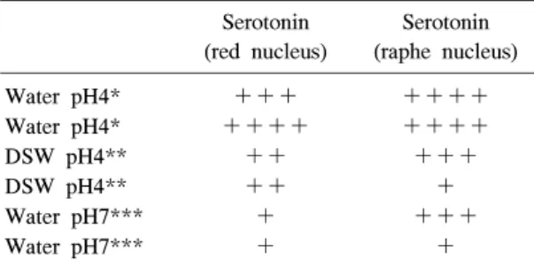
표 1은 적색핵과 솔기핵 두 부위에서 각 군(n=2) 에 대한 세로토닌 면역조직화학 염색의 평균을 반정 량적으로 나타낸 것이다(표 1).
다른 섬유근통 증후군과 관련된 신경전달물질인 glutamate receptor 2&3, substance P 및 tyrosine hydro- xylase (카테콜라민 지표)에 대한 항체들을 이용하여 시행한 면역 염색법에서 유발군과 대조군 및 심층수 음용군과 맹물 음용군 간의 발현 차이는 없었다.
Table 1. Semiquantitative expression of serotonin in red and raphe nucleus
Serotonin (red nucleus)
Serotonin (raphe nucleus)
Water pH4* +++ ++++
Water pH4* ++++ ++++
DSW pH4** ++ +++
DSW pH4** ++ +
Water pH7*** + +++
Water pH7*** + +
DSW: Deep-Sea Water, *: water treatment after pH4 saline injection, **: DSW treatment after pH4 saline injection, ***: water treatment after pH7 saline injection (control group)
대천문 부위로부터 차례대로 조사한 뇌실주위핵 (paraventricular nucleus) 및 창백핵(globus pallidus)을 비롯한 10 여군데 관심영역들에선 유발군과 대조군 간 및 심층수 음용군과 맹물 음용군 간의 발현 차이 는 없었다.
고 찰
저자들은 문헌상 최초로 산성 식염수 모델의 뇌단 면에서 신경전달물질들의 변화를 면역조직화학법을 통해서 보았고, 또한 심층수 음용 전후의 변화를 보 았다. 이 연구에서 산성 식염수 모델은 중성 식염수 를 처치한 대조군에 비해 적색핵과 솔기핵에서 세로 토닌에 강한 면역반응을 나타내는 세포가 매우 증가 되어 있었고, 산성 식염수 모델에서 심층수를 음용 시키면 세로토닌에 의한 면역반응이 현저히 감소되 었다.
산성 식염수 모델은 지속적이고 광범위한 근골격계 통증 및 변화된 중추 신경계 처리에 대한 증거를 비 롯한 섬유근통 증후군의 특성들을 나타낸다 (14,15).
산성 식염수 투여로 일차 구심성 C섬유에 위치한 acid sensing ion channels이 활성화되어 기계적 과통 각이 유발되며, 중추성 변화로 인해 반대측 과통각 도 일어난다고 생각된다 (14). 이 모델의 뇌 활성 패 턴의 특성을 기술하기 위해 신경세포 활성화의 지수 로서 국소적 대뇌 혈류량을 측정했다. 생리적으로 중성인 식염수를 주사한 대조군 쥐들에 비해, 산성
식염수를 주사한 쥐들에서 양측의 기계적 과통증이 나타났으며 더불어 통각입력의 처리와 조절에 관여 되는 신경 구조들에서 기저활성이 감소되었다. 특히 이런 효과들이 처음 산성 식염수 근주후 거의 2주후 관찰되었다 (15). 앞에서 언급했듯이 자극받지 않은 섬유근통 증후군 환자들에서 SPECT 영상을 사용해 서 비슷하게 국소적 대뇌 혈류량이 감소한 것을 증 명한 보고들이 있다 (11-13). 이런 동물과 사람 연구 사이의 일치되는 결과들을 볼 때, 이 산성 식염수 모델이 섬유근통 증후군이나 만성 광범위 통증에 대 해 적절하며, 이런 질환들의 신경 병리학적인 기전 에 대한 우리들의 이해를 높일 수 있는 잠재력을 지 닌 모델임을 나타낸다.
적색핵은 중간뇌의 구조로 주로 운동 조절에 관여 한다 (20). 하지만 많은 연구들에서 다양한 감각 자극 에 적색핵의 신경세포들이 반응한다고 밝혀져 있다 (20-24). 감각 정보를 받고 운동 반응을 유도하는 적색 핵의 이중 능력으로 인해 감각운동 통합에서 이 핵의 잠재적인 중요성이 제시되어 왔다 (20). 적색핵이 진통 에 관여할 수 있음을 제시하는 연구들이 있다 (25-27).
고양이의 피부를 꼬집을 때 전기생리학적 방법으로 적색핵 신경세포가 반응하는 것을 알게 되었다 (25).
더구나 쥐의 적색핵에 전기적 자극을 주면 peria- queductal gray (PAG)나 흑색질(substantia nigra)로부터 유도되는 것보다 더 강력하고 긴 기간의 진통(무통증) 이 발생한다 (26). 적색핵의 신경세포들은 들어오는 통 각에 반응하지만, 행동적 실험에 의하면 적색핵은 감 각 전달을 조절하는데는 관여하지 않는 것 같다 (27).
적색핵에서 많은 양의 세로토닌이 발견되는데 (28), 이 핵에 전반적으로 분포하고 있는 세로토닌계 신경섬유 들이 주로 방출하는 것같다 (29). 등쪽 솔기핵으로부터 오는 일련의 세로토닌계 신경섬유들은 적색핵에 이른 다 (30). 세로토닌계 경로는 전형적으로 수면-각성 주 기 (31,32), 통각 (33), 우울증 (34,35) 등에서 의미를 지 닌다.
청솔기핵 역시 중간뇌의 구조로 중추 신경계의 대 부분 전뇌 구조들에 연결되는 대다수의 세로토닌계 신경섬유들의 기원이다 (36). 청솔기핵 내의 세로토 닌 신경세포들이 자극되면, 등쪽의 PAG에 이르는 projections을 통하여 fight/flight 방어 행동이 줄어들 고 편도체에 이르는 projections을 통하여 fear/anxiety
가 증폭되는 경향이 있다는 것은 이미 알려져 있다 (37). 청솔기핵의 세로토닌 신경세포들은 축삭 곁 (axon collaterals) 및 세포체와 가지돌기로부터 청솔 기핵 자기 안에 세로토닌을 방출한다 (38). 통제되지 않는 스트레스나 피할 수 없는 스트레스는 통제되는 스트레스에 비해 상대적으로 청솔기핵내의 세로토닌 신경세포들을 더 활성화시킨다 (39). 청솔기핵의 세 로토닌 활성도를 억제하면 동물 모델에서 불안이 줄 고 세로토닌 활성도를 높여 주면 불안과 연관된 행 동이 증가된다는 보고들이 많다 (40).
척수의 NMDA에 의해 유발된 교감신경 반응들이 세로토닌에 의해 강화되는 등 세로토닌과 NMDA의 연관성은 많이 보고되고 있다 (41). 저자들은 마그네 슘과 칼슘이 풍부한 심층수를 음용한 산성 식염수 모델에서 NMDA 신경연접에 어떤 변화가 일어났다 고 생각한다. 그것이 심층수로 인한 NMDA 신경연 접의 주요 이온인 마그네슘과 칼슘의 회복 및 연이 은 NMDA와 연관된 세로토닌계의 변화로 이어졌을 것이라고 추정한다. 이 부분은 향후의 또 다른 연구 의 좋은 주제라고 생각한다.
이 연구에서도 역시 산성 식염수 모델에서는 통증 과 불안 등으로 인해 적색핵이나 청솔기핵에서 세로 토닌 신경세포들이 활성화된 것으로 생각된다. 심층 수의 음용으로 각 핵에서 세로토닌에 대한 염색 반 응이 현저히 줄어 든 것이 실제로 동물들의 통증 및 불안의 감소로 이어졌는지는 알 수 없으나 향후 행 동 테스트들을 포함한 대규모 실험으로 증명된다면 사람에서도 심층수를 이용해 진통 및 항불안 효과를 연구할 수 있을 것이다.
결 론
산성 식염수를 처치한 쥐의 뇌에서 적색핵과 솔기 핵 부위 세로토닌의 발현이 증가되었다. 심층수 음용 후 이 부위들의 세로토닌이 현저하게 감소한 것은 심 층수가 통증과 불안 등의 증상 치료에 도움이 될 수 도 있음을 제시한다. 이 연구의 예비적인 결과를 볼 때, 산성 식염수 모델은 섬유근통의 동물 모델로서 타당하고 향후 섬유근통에 기인하는 뇌의 여러 영역 들에 관한 연구에 기초를 제공할 것으로 기대된다.
감사의 글
본 연구는 산업자원부 지역연고산업진흥사업(06- RIS-02)의 지원에 의하여 수행되었으며, 이에 감사드 립니다.
참고문헌
1) Wolfe F, Smythe HA, Yunus MB, Bennett RM, Bombardier C, Goldenberg DL, et al. The American College of Rheumatology criteria for the classification of fibromyalgia: report of the multicenter criteria committee. Arthritis Rheum 1990;33:160-72.
2) Okifuji A, Turk DC, Marcus DA. Comparison of generalized and localized hyperalgesia in patients with recurrent headache and fibromyalgia. Psychosom Med 1999;61:771-80.
3) Staud R, Vierck CJ, Cannon RL, Mauderli AP, Price DD. Abnormal sensitization and temporal summation of second pain (wind-up) in patients with fibro- myalgia syndrome. Pain 2001;91:165-75.
4) Staud R. The neurobiology of chronic musculoskeletal pain. In: Wallace DJ, Clauw DJ, eds. Fibromyalgia &
other central pain syndromes. 1st ed. p. 45-62, Phila- delphia, Lippincott Williams & Wilkins, 2005.
5) Schwarz MJ, Spath M, Muller-Bardorff H, Pongratz DE, Bondy B, Ackenheil M. Relationship of sub- stance P, 5-hydroxyindole acetic acid and tryptophan in serum of fibromyalgia patients. Neurosci Lett 1999;
259:196-8.
6) Offenbaecher M, Bondy B, de-Jonge S, Glatzeder K, Krger M, Schoeps P, et al. Possible association of fibromyalgia with a polymorphism in the serotonin transporter gene regulatory region. Arthritis Rheum 1999;42:2482-8.
7) Russell IJ. Advances in fibromyalgia: possible role for central neurochemicals. Am J Med Sci 1998;315:
377-84.
8) Heils A, Teufel A, Petri S, Stober G, Riederer P, Bengel D, et al. Allelic variation of human serotonin transporter gene expression. J Neurochem 1996;66:
2621-4.
9) Moldofsky H. Rheumatic pain modulation syndrome:
the interrelationships between sleep, central nervous system serotonin, and pain. Adv Neurol 1982;33:51-7.
10) Harris RE, Clauw DJ. How do we know that the pain
in fibromyalgia is "real"? Curr Pain Headache Rep 2006;10:403-7.
11) Guedj E, Taieb D, Cammilleri S, Lussato D, de Laforte C, Niboyet J, et al. (99m)Tc-ECD brain perfusion SPECT in hyperalgesic fibromyalgia. Eur J Nucl Med Mol I 2007;34:130-4.
12) Kwiatek R, Barnden L, Tedman R, Jarrett R, Chew J, Rowe C, et al. Regional cerebral blood flow in fibromyalgia: single-photon-emission computed tomo- graphy evidence of reduction in the pontine teg- mentum and thalami. Arthritis Rheum 2000;43:2823- 33.
13) Mountz JM, Bradley LA, Modell JG, Alexander RW, Triana-Alexander M, Aaron LA, et al. Fibromyalgia in women. Abnormalities of regional cerebral blood flow in the thalamus and the caudate nucleus are associated with low pain threshold levels. Arthritis Rheum 1995;38:926-38.
14) Sluka KA, Kalra A, Moore SA. Unilateral intramu- scular injections of acidic saline produce a bilateral, long-lasting hyperalgesia. Muscle Nerve 2001;24:37- 46.
15) Harte SE, Kim SH, Clauw DJ, Morrow TJ. Altered regional cerebral blood flow at rest in an animal model of fibromyalgia. Arthritis Rheum 2007;56(Suppl):S92.
16) Hataguchi Y, Tai H, Nakajima H, Kimata H. Drinking deep-sea water restores mineral imbalance in atopic eczema/dermatitis syndrome. Eur J Clin Nutr 2005;
59:1093-6.
17) Yoshikawa S, Hamada A, Gue T, Yokota J, Yamamoto S, Kusunose M, et al. Pharmacological activity of deep-sea water: examination of hyperli- pemia prevention and medical treatment effect. Biol Pharm Bull 2003;26:1552-9.
18) Miyamura M, Yoshioka S, Hamada A, Takuma D, Yokota J, Kusunose M, et al. Difference between deep seawater and surface seawater in the preventive effect of atherosclerosis. Biol Pharm Bull 2004;27:
1784-7.
19) Katsuda SI, Yasukawa T, Nakagawa K, Miyake M, Yamasaki M, Katahira K, et al. Deep-sea water improves cardiovascular hemodynamics in Kurosawa and Kusanagi-Hypercholesterolemic (KHC) rabbits.
Biol Pharm Bull 2008;31:38-44.
20) Massion J. The mammalian red nucleus. Physiol Rev 1967;47:383-436.
21) Eccles JC, Scheid P. Taborikova H. Responses of red nucleus neurones to cutaneous afferent inputs. Brain Res 1973;53:440-4.
22) Larsen KD, Yumiya H. The red nucleus of the monkey. Topographical localization of somatosensory input and motor output. Exp Brain Res 1980;40:
393-404.
23) Padel Y, Sybirska E, Bourbannais D, Vinay L.
Electrophysiological identification of a somaesthetic pathway of the red nucleus. Behav Brain Res 1988;
8:139-51.
24) Vinay L, Padel Y. Spatio-temporal organization of the somaesthetic projections in the red nucleus trans- mitted through the spino-rubral pathway in the cat.
Exp Brain Res 1990;79:412-26.
25) Nishioka S, Nakahama H. Peripheral somatic activation of neurons in the cat red nucleus. J Neuro- physiol 1973;36:296-307.
26) Prado WA, Raghubir R, Roberts MHT. Long duration antinociception induced by red nucleus stimulation in the rat. Pain 1984;2 Suppl:329S.
27) Matsumoto RR, Walker JM. Inhibition of rubral neurons by noxious and non-noxious pressure. Brain Res 1991;556:78-84.
28) Palkovitz M, Brownstein M, Saavedra JM. Serotonin content of the brainstem nuclei in the rat. Brain Res 1974;80:237-49.
29) Steinbusch HWM. Serotonin-immunoreactive neurons and their projection in the CNS. In: Björklund A, Hokfelt T, Kuhar MJ, eds. Handbook of chemical neuroanatomy. Vol III. p. 68-125, Amsterdam, Elsevier, 1984.
30) Pierce ET, Foote WE, Hobson JA. The efferent connection of the nucleus raphe dorsalis. Brain Res 1976;107:137-44.
31) Hobson JA, Lydic R, Baghdoyan HA. Evolving concepts of sleep cycle generation: from brain centers to neuronal populations. Behav Brain Sci 1986;9:
371-448.
32) Jouvet M. Biogenic amines and states of sleep.
Science 1969;163:32-41.
33) Basbaum AI, Fields HL. Endogenous pain control systems: brainstem spinal pathways and endorphin circuitry. Annu Rev Neurosci 1984;7:309-38.
34) Kahn RS, van Praag HM, Wetzler S, Asnis GM, Barr G. Serotonin and anxiety revisited. Biol Psychiatry 1988;23:189-208.
35) Miller LE, van Kan PL, Sinkjaer T, Andersen T, Harris GD, Houk JC. Correlation of primate red nucleus discharge with muscle activity during free-form arm movements. J Physiol 1993;469:13-43.
36) Adell A, Celada P, Abellan MT, Artigas F. Origin and
functional role of the extracellular serotonin in the midbrain raphe nuclei. Brain Res Rev 2002;39:54-80.
37) Graeff FG, Guimaress FS, De Andrade TGCS, Deakin JFW. Role of S-NT in stress, anxiety and depression.
Pharmacol Biochem Behav 1996;54:129-41.
38) Matos FF, Urban C, Vocca FD. Serotonin release in the dorsal raphe and ventral hippocampus: raphe control of somatodendritis and terminal 5-HT release.
J Neural Transm 1996;103:173-90.
39) Maier SF, Watkins LR. Stressor controllability and learned helplessness: the roles of the dorsal raphe nucleus, serotonin, and corticotropin-releasing factor.
Neurosci Biobehav R 2005;29:829-41.
40) Millan MJ. The neurobiology and control of anxious states. Prog Neurobiol 2003;70:83-244.
41) Madden CJ, Morrison SF. Serotonin potentiates sympathetic responses evoked by spinal NMDA. J Physiol 2006;577:525-37.