Brief Report
644 Ann Dermatol
Received August 5, 2016, Revised September 7, 2016, Accepted for publication September 9, 2016
Corresponding author: Moon-Bum Kim, Department of Dermatology, Pusan National University Hospital, 179 Gudeok-ro, Seo-gu, Busan 49241, Korea. Tel:
82-51-240-7338, Fax: 82-51-245-9467, E-mail: drkmp@hanmail.net
This is an Open Access article distributed under the terms of the Creative Commons Attribution Non-Commercial License (http://creativecommons.org/
licenses/by-nc/4.0) which permits unrestricted non-commercial use, distribution, and reproduction in any medium, provided the original work is properly cited.
Copyright © The Korean Dermatological Association and The Korean Society for Investigative Dermatology cause Hailey-Hailey disease. Nat Genet 2000;24:61-65.
2. Dobson-Stone C, Fairclough R, Dunne E, Brown J, Dissanayake M, Munro CS, et al. Hailey-Hailey disease: molecular and clinical characterization of novel mutations in the ATP2C1 gene. J Invest Dermatol 2002;118:338-343.
3. Matsuda M, Hamada T, Numata S, Teye K, Okazawa H, Imafuku S, et al. Mutation-dependent effects on mRNA and protein expressions in cultured keratinocytes of Hailey-Hailey disease. Exp Dermatol 2014;23:514-516.
4. Btadini W, Abou Hassan OK, Saadeh D, Abbas O, Ballout F, Kibbi AG, et al. Identification of several mutations in ATP2C1 in Lebanese families: insight into the pathogenesis of Hailey- Hailey disease. PLoS One 2015;10:e0115530.
5. Kellermayer R, Szigeti R, Keeling KM, Bedekovics T, Bedwell DM. Aminoglycosides as potential pharmacogenetic agents in the treatment of Hailey-Hailey disease. J Invest Dermatol 2006;126:229-231.
https://doi.org/10.5021/ad.2017.29.5.644
Eruptive Melanocytic Nevi without Any Trigger in a 5-Year-Old Healthy Girl
Won-Ku Lee
1, Hyunju Jin
1, Hyang-Suk You
1, Woo-Haing Shim
3, Jeong-Min Kim
3, Gun-Wook Kim
1, Hoon-Soo Kim
1, Hyun-Chang Ko
3, Byung-Soo Kim
1,2, Moon-Bum Kim
1,21Department of Dermatology, Pusan National University School of Medicine, 2Biomedical Research Institute, Pusan National University Hospital, Busan, 3Department of Dermatology, Pusan National University Yangsan Hospital, Yangsan, Korea
Dear Editor:
Eruptive melanocytic nevi (EMN) are rare skin manifes- tations characterized by the simultaneous and abrupt de- velopment of numerous melanocytic nevi on the skin1. Although the exact mechanism of EMN development is not well understood, it has been associated with various triggers including light exposure, cutaneous injury such as the Koebner phenomenon, bullous dermatoses, systemic immunosuppression, biologic chemotherapeutics, increased hormone levels, and others including atopic dermatitis in children, postoperative fever, and seizures2. However, EMN without any trigger, especially in a healthy girl, are rather rare.
A 5-year-old Korean girl presented with multiple hyper- pigmented maculopapules over the whole body. The le- sions first appeared on her chest when she was 1 year old,
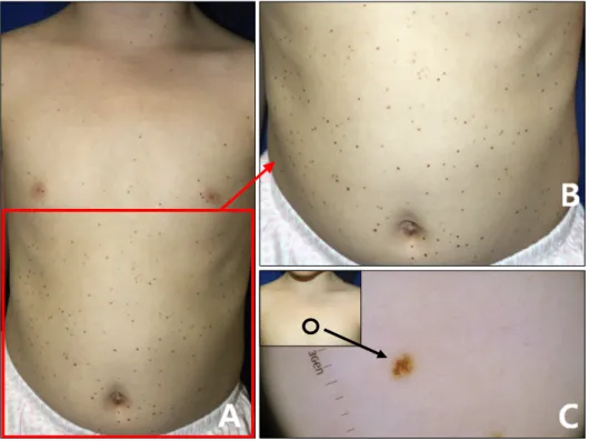
and then hundreds of similar lesions covering her entire skin surface developed continuously during the next 2 years. The girl was of Fitzpatrick skin type IV and had no specific medical and family histories including multiple nevi. On physical examination, there were no systemic abnormalities except for the skin lesions that appeared as multiple small (0.5∼3 mm diameter) brown to black pig- mented maculopapules with a globular pattern on dermo- scopy (Fig. 1). The histopathologic finding was compatible with compound nevus. Findings of routine laboratory ex- aminations including complete blood counts, peripheral blood smear, liver/renal function test, venereal disease re- search laboratory test, antinuclear antibody, and urine analysis were either negative or within the normal limits.
The test for BRAF V600E mutation was negative.
There have been very limited data about the changes in
Brief Report
Vol. 29, No. 5, 2017 645 Table 1. Eruptive melanocytic nevi in healthy children without any triggering events
Coskey5 (1975) Zalaudek et al.1 (2013) Our case (2016)
Sex (patient no.) M M (6), F (1) F
Age (yr) 5 Mean (range): 8.1 (4∼12) 5
Fitzpatrick skin type NM II∼III IV
Country USA Italy Korea
Onset age (yr) 5 NM 1
Number of nevus 24 Lower compared with previous EMN >200
Location Face, trunk, extremities NM Face, trunk, extremities
Color Dark brown Pink to skin colored Brown to black
Hispathology Junctional nevus Compound nevus Compound nevus
M: male, F: female, NM: not mentioned, EMN: eruptive melanocytic nevi.
Fig. 1. (A, B) Clinical photographs of eruptive melanocytic nevi in a 5-year-old healthy girl and its mag- nified image. (C) Dermoscopic fin- ding of brownish macules on the chest (marked by the circle) showing a globular pattern.
the number of melanocytic nevi with aging. In a Scottish study, there were 2∼3 nevi in the first decade, 22∼33 nevi in the third decade, and 4∼6 nevi in the seventh decade3. Considering this age-related change in nevus number, EMN seem to be a rather rare condition.
Recently, studies on molecular nevogenesis have been a hot topic and revealed significant mutations of NRAS in congenital nevi, GNAQ in blue nevi, and BRAF in ac- quired nevi. Although these mutations were not always detected, they were discovered with various frequencies of positivity. For example, BRAF mutation in acquired ne- vi was found in 67.2% of intradermal nevi, 57.5% of com- pound nevi, 37.8% of junctional nevi, and 43.3% of dys- plastic nevi4. In the present patient, no BRAF mutations were found. This could be due to the mutation hetero-
geneity of BRAF in the nevi. Furthermore, there is a possi- bility that the patient may have other mutations.
There were two previous reports on EMN in healthy chil- dren without any triggering events (Table 1)1,5. Our case differs from these reports in terms of ethnicity and the ne- vus number. The nevus counts in our child were much higher (>200 nevi) than those of Coskey5 (one boy with 24 nevi) and Zalaudek et al.1 (seven children with much lower nevus counts than those of previous EMN cases).
To our knowledge, this is a very rare case of EMN in a healthy Asian girl without any triggering factors. This case could highlight the complicated aspects of nevo- genesis and provide clues for further understanding of nevogenesis.
Brief Report
646 Ann Dermatol
Received August 1, 2016, Revised August 22, 2016, Accepted for publication September 13, 2016
Corresponding author: Yang Wang, Department of Dermatology and Venerology, Peking University First Hospital, No.8 Xishiku Street, Xi Cheng District, Beijing 100034, China. Tel: 86-13811232795, Fax: 86-10- 66551216, E-mail: yangwang_dr@bjmu.edu.cn
This is an Open Access article distributed under the terms of the Creative Commons Attribution Non-Commercial License (http://creativecommons.org/
licenses/by-nc/4.0) which permits unrestricted non-commercial use, distribution, and reproduction in any medium, provided the original work is properly cited.
Copyright © The Korean Dermatological Association and The Korean Society for Investigative Dermatology
ACKNOWLEDGMENT
This work was supported by clinical research grant from Pusan National University Hospital in 2017.
CONFLICTS OF INTEREST
The authors have nothing to disclose.
REFERENCES
1. Zalaudek I, Moscarella E, Sturm RA, Argenziano G, Longo C, Misciali C, et al. 'Eruptive' amelanotic compound nevi in children with facial freckles and pale skin colour: more than
an occasion? J Eur Acad Dermatol Venereol 2013;27:1583- 1585.
2. Navarini AA, Kolm I, Calvo X, Kamarashev J, Kerl K, Conrad C, et al. Trauma as triggering factor for development of melanocytic nevi. Dermatology 2010;220:291-296.
3. English JS, Swerdlow AJ, Mackie RM, O'Doherty CJ, Hunter JA, Clark J, et al. Site-specific melanocytic naevus counts as predictors of whole body naevi. Br J Dermatol 1988;118:
641-644.
4. Marghoob AA. Nevogenesis. 1st ed. New York: Springer, 2012:104-106.
5. Coskey RJ. Letter: eruptive nevi. Arch Dermatol 1975;111:
1658.
https://doi.org/10.5021/ad.2017.29.5.646
Type I Lepra Reaction as the Presenting Sign of Histoid Leprosy
Jingru Sun, Ping Tu, Shengguo Yi, Wenjing Fu, Yang Wang
Department of Dermatology and Venerology, Peking University First Hospital, Beijing, China
Dear Editor:
A 47-year-old woman presented with a two-week history of multiple asymptomatic erythematous eruptions over the face, trunk and extremities following a transient fever. She was otherwise healthy. Physical examination revealed dis- seminated erythematous to violaceous plaques and nod- ules with tumidity and sharp margination over her face, trunk and extremities. The lesions were neither painful nor tender (Fig. 1A, B). Additionally, one asymptomatic skin-col- ored nodule over her right arm was noted (Fig. 1C). It last- ed for 2 years and was previously misdiagnosed as dermatofibroma. Neither anesthesia nor enlarged periph-
eral nerves was presented. Laboratory tests revealed slight- ly increased C-reactive protein (11 mg/L; normal range, 0∼
10 mg/L), marked increased erythrocyte sedimentation rate (42 mm/h; normal range 0∼15 mm/h) and elevated serum IgM (3.52 g/L; normal range, 0.63∼2.77 g/L).
Skin biopsies were taken from a plaque on the face as well as the persistent nodule on the arm. Histologically, the plaque lesion showed marked dermal edema with loose lymphocyte and histiocyte infiltration (Fig. 2A, B), and the nodular lesion demonstrated dense infiltration of foamy histiocytes (Fig. 2D, E) with abundant acid-fast ba- cilli (Fig. 2F) which were confirmed as Mycobacterium