www.kjlm.org 201
Subcutaneous Phaeohyphomycosis Caused by Phaeoacremonium Species in a Kidney Transplant Patient: The First Case in Korea
Jonghyeon Choi, M.D.1, Yangsoon Lee, M.D.1, Hae-Sun Chung, M.D.1, Ja-Seung Koo, M.D.3, Dongeun Yong, M.D.1, Yu Sun Kim, M.D.2, Kyungwon Lee, M.D.1, and Yunsop Chong, Ph.D.1
Departments of Laboratory Medicine and Research Institute of Bacterial Resistance1, General Surgery2, and Pathology3, Yonsei University College of Medicine, Seoul, Korea
Phaeohyphomycosis is a subcutaneous infection caused by dark pigmented fungi, including fungi of the species Phaeoacremoni- um, Alternaria, Exophiala, and Pyrenochaeta. In August 2005, a 54-yr-old man who had received a renal transplant 5 yr ago was ad- mitted to our hospital with a subcutaneous mass on the third finger of the right hand; the mass had been present for several months. He had been receiving immunosuppressive agents for several years. He underwent excision of the mass, which was fol- lowed by aspiration of the wound for bacterial and fungal cultures. Many fungal hyphae were observed on the histology slide treated with periodic acid-Schiff stain. A few white waxy colonies with a woolly texture grew on the Sabouraud dextrose agar at 30˚C and changed to dark brown in color. Nucleotide sequencing of internal transcribed spacer regions revealed 100% homology to the Phaeoacremonium aleophilum anamorph and Togninia minima teleomorph (514 bp/514 bp). The patient completely recov- ered after wide surgical excision. Here, we report the first case of phaeohyphomycosis caused by Phaeoacremonium species in a kidney transplant patient in Korea.
Key Words: Subcutaneous phaeohyphomycosis, Phaeoacremonium species, Renal transplantation, Immunosuppressive agents
Received: December 13, 2010 Manuscript No: KJLM-10-169 Revision received: January 18, 2011
Accepted: March 4, 2011
Corresponding author: Dongeun Yong, M.D.
Department of Laboratory Medicine, Yonsei University College of Medicine, 250 Seongsan-ro, Seodaemun-gu, Seoul 120-752, Korea
Tel: +82-2-2228-2442, Fax: +82-2-313-0956, E-mail: [email protected] ISSN 1598-6535 © The Korean Society for Laboratory Medicine.
This is an Open Access article distributed under the terms of the Creative Commons Attribution Non-Commercial License (http://creativecommons.org/licenses/by-nc/3.0) which permits unrestricted non-commercial use, distribution, and reproduction in any medium, provided the original work is properly cited.
Korean J Lab Med 2011;31:201-204 DOI 10.3343/kjlm.2011.31.3.201
Case Report
Clinical Microbiology
KJLM
INTRODUCTION
The fungal genus Phaeoacremonium was described by Crous et al. [1]. Fungi of this genus have been isolated from a wide range of hosts such as humans, woody plants, and the larvae of bark beetles, but only some species of this ge- nus have been reported as pathogens of humans [2, 3]. Pres- ently, the Phaeoacremonium genus, formerly known as Phi- alophora, includes more than 20 species [4, 5]. The occur- rence of Phaeoacremonium species varies, with Phaeoacre- monium aleophilum being the most frequently isolated spe-
cies in esca diseased grapevines [2].
The term phaeohyphomycosis was first introduced in 1974 to describe local infections caused by darkly pigment- ed molds (black mold) [6, 7]. However, only a few human phaeohyphomycosis cases have been reported in the litera- ture worldwide [8]. We report the first case of primary pha- eohyphomycosis by Phaeoacremonium species in a kidney transplant patient in Korea.
CASE REPORT
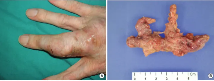
A 54-yr-old man with a large subcutaneous mass on the third finger of his right hand was admitted to our hospital in August 2005 (Fig. 1A). He had been working at a leather factory for 10 yr. He had chronic renal failure caused by dia- betes mellitus, and therefore, he underwent kidney trans- plantation in October 2000. After the operation, he was pre- scribed immunosuppressive agents such as tacrolimus, de- flazacort, and mycophenolate mofetil. He had no history of trauma, but had a small nodule on the third finger since 1995. The nodule grew gradually during several months prior to hospital admission. The patient also experienced
202 www.kjlm.org
Choi J, et al. • Subcutaneous Phaeohyphomycosis
DOI 10.3343/kjlm.2011.31.3.201
KJLM
mild pain and pruritus around the lesion. The mass was re- moved by an orthopedic surgeon. The patient had high se- rum glucose levels of 341 mg/dL, which suggested post-tra- nsplant diabetic mellitus; his serum blood urea nitrogen/
creatinine levels were 33.5/1.5 mg/dL. In complete blood count analysis, the white blood cell count and Hb and Hct levels were within reference ranges, although platelet count was slightly low at 117,000/µL. The patient underwent exci- sion of the mass, which was followed by culture of the wound aspirate collected in the operating room. The mass was removed entirely and had dimensions of 6.5×3×1.2 cm (Fig. 1B). In the bacterial culture, no colonies formed on the blood agar, MacConkey agar, or thioglycolate broth after incubation for 48 hr at 35˚C and ambient temperature.
Periodic acid-Schiff staining performed on the histology slide showed many fungal hyphae (Fig. 2). Few white waxy
colonies with a woolly texture were observed on Sabouraud dextrose agar at 30˚C, and the colonies changed into a dark- brown color. When the fungal hyphae were separated on the slide culture, the conidiophores were observed to be short and frequently unbranched. Furthermore, the apical cell of the conidiophores mainly represented 1 phialide. The phialides appeared to be elongate-ampulliform and were long or subcylindrical. This morphology further supported the presence of the genus Phaeoacremonium (Fig. 3). PCR using ITS1 (5´ TCC GTA GGT GAA CCT GCG G 3´) and ITS4 (5´ TCC TCC GCT TAT TGA TAT GC 3´) primers was performed for species identification [9]. The β-tubulin and actin genes were detected according to CLSI guidelines, 2008 [10]. Even though β-tubulin and actin were not ampli- fied, nucleotide sequencing of ITS1 and ITS4 revealed 100%
Fig. 1. (A) Subcutaneous lesion on the third finger of the right hand; (B) 6.5×3×1.2 cm-sized mass obtained after removal of the lesion.
Fig. 2. Periodic acid-Schiff stain showed many fungal hyphae under the mi- croscope (×400).
Fig. 3. On the slide culture of the specimen, many fungal hyphae were sep- tate, and conidiophores were observed to be short and usually unbranched (×400).
A B
Choi J, et al. • Subcutaneous Phaeohyphomycosis
www.kjlm.org 203
DOI 10.3343/kjlm.2011.31.3.201
KJLM
sequence homology to both P. aleophilum anamorph and Togninia minima teleomorph. As such, antifungal agents, including fluconazole and amphotericin B, were adminis- tered to the patient empirically for 2 days, and the patient recovered without any sequelae. Five years after the wide excision, the patient has had no further subcutaneous le- sions.
DISCUSSION
Phaeohyphomycosis is the term used to describe a subcu- taneous fungal infection caused by darkly pigmented molds (black mold), such as those from the species Phaeoacremo- nium, Phialophora, Alternaria, Exophiala, and Pyrenocha–
eta [6, 7]. This condition is rare in humans, but it can in- volve the skin, subcutis, paranasal sinuses, or the central nervous system [9]. The genus Phaeoacremonium has been isolated from patients who experience traumatic implanta- tion of fungi from contaminated plant thorns or soil, and was usually known as a causal agent of grapevine diseases such as Petri disease and esca, which can cause leaf drop and wood discoloration [7, 11]. Recently, cases of phaeohy- phomycosis caused by Phaeoacremonium species have been increasingly reported in humans [7, 8]. Notably, these dis- eases have been described in immunocompromised patie- nts with renal transplantation [12]. In our case, the patient was in an immunosuppressed state. Although he lived in an urban area, he frequently climbed mountains as a hobby before the appearance of the subcutaneous mass. When we consider the natural ecological niches of the Phaeoacremo- nium species, the patient may have been exposed to this pathogen during his frequent mountain-climbing trips. It is difficult to explain how he acquired the infection because he did not report any injury. However, it is possible that mi- crotraumas to the skin and immunosuppressive therapy may have played a role.
Morphologically, the genus Phaeoacremonium is an inter- mediate between the genera Acremonium and Phialophora [8, 13]. Organisms of the genus Acremonium usually pro- duce delicate hyaline septate hyphae, which carry unbran- ched tapering conidiophores with elliptical-shaped unicel- lular conidia. The characteristic tapering phialides of these organisms are derived from septate hyphae with one-celled ellipsoidal conidia [14]. In contrast, organisms of genus Phi- alophora show melanized hyphae and slimy one-celled co- nidia at their apex with distinct collarettes [15]. Phaeoacre- monium show medium brown hyphae, which become pale brown to hyaline and verruculose. The phialides have a fun- nel-shaped collarette and show a wide variety of diverse
forms, including ellipsoidal, obovate, cylindrical, or allan- toid (sausage-like) [5]. The morphological distinctions from a number of other relatively similar Phaeoacremonium spe- cies have been summarized by Mostert et al. [5]. Although it is difficult to identify the species in our isolate on the basis of morphological criteria, it could be distinguished from species of brown colonies, such as Phaeoacremonium para- siticum and Phaeoacremonium inflatipes, by its short and usually unbranched conidiophores. However, molecular- based tools must be used in order to confirm the species.
Although the additional PCRs for β-tubulin and actin were unsuccessfully repeated several times, sequence analysis of internal transcribed spacer (ITS) revealed 100% sequence homology to P. aleophilum and T. minima. T. minima is a te- leomorph of P. aleophilum [11]. CLSI guidelines indicate that the ITS region in Phaeoacremonium only provides lim- ited resolution for species identification [10]. Thus, we are able to conclude that the pathogen was of the genus Phaeo- acremonium. Optimal treatment of phaeohyphomycosis usually involves surgical wound excision combined with an- tifungal agents for several months [16]. For this patient, em- pirical antifungal agents were administered for 2 days and the wound excision was successfully completed. Total exci- sion of the nodule was important for uncomplicated healing in the patient. After 5 yr, no subcutaneous lesion has been recognized in the patient. Herein, we have reported the first case of phaeohyphomycosis caused by the Phaeoacremo- nium species in a kidney transplant patient in Korea.
Authors’ Disclosures of Potential Conflicts of Interest No potential conflict of interest relevant to this article was reported.
REFERENCES
1. Crous PW, Gams W, Wingfield MJ, Van Wyk PS. Phaeoacremo- nium gen. nov. associated with wilt and decline diseases of woody hosts and human infections. Mycologia 1996;88:786-96.
2. Aroca A, Raposo R, Lunello P. A biomarker for the identification of four Phaeoacremonium species using the beta-tubulin gene as the target sequence. Appl Microbiol Biotechnol 2008;80:1131-40.
3. Aguilar-Donis A, Torres-Guerrero E, Arenas-Guzman R, Hernan- dez-Hernandez F, Lopez-Garcia L, Criales-Vera S, et al. Mycetoma caused by Phaeoacremonium parasiticum- a case confirmed with B-tubulin sequence analysis. Mycoses Epub 2010 Aug 4. (http://
www.ncbi.nlm.nih.gov/pubmed/20701685).
4. Essakhi S, Mugnai L, Crous PW, Groenewald JZ, Surico G. Molec- ular and phenotypic characterisation of novel Phaeoacremonium species isolated from esca diseased grapevines. Persoonia 2008;21:
119-34.
5. Mostert L, Groenewald JZ, Summerbell RC, Robert V, Sutton DA,
204 www.kjlm.org
Choi J, et al. • Subcutaneous Phaeohyphomycosis
DOI 10.3343/kjlm.2011.31.3.201
KJLM
Padhye AA, et al. Species of Phaeoacremonium associated with in- fections in humans and environmental reservoirs in infected woody plants. J Clin Microbiol 2005;43:1752-67.
6. Naggie S, Perfect JR. Molds: hyalohyphomycosis, phaeohyphomy- cosis, and zygomycosis. Clin Chest Med 2009;30:337-53, vii-viii.
7. Guarro J, Alves SH, Gene J, Grazziotin NA, Mazzuco R, Dalmagro C, et al. Two cases of subcutaneous infection due to Phaeoacremo- nium spp. J Clin Microbiol 2003;41:1332-6.
8. Baradkar VP, Mathur M, Kumar S. Phaeohyphomycosis of subcu- taneous tissue caused by Phaeoacremonium parasiticum. Indian J Med Microbiol 2009;27:66-9.
9. Kumar KK, Hallikeri K. Phaeohyphomycosis. Indian J Pathol Mi- crobiol 2008;51:556-8.
10. Clinical and Laboratory Standerds Institute. Interpretive criteria for identification of bacteria and fungi by DNA target sequencing (MM18-A) 2008.
11. Damm U, Mostert L, Crous PW, Fourie PH. Novel Phaeoacremo- nium species associated with necrotic wood of Prunus trees. Per-
soonia 2008;20:87-102.
12. Farina C, Gotti E, Mouniee D, Boiron P, Goglio A. Phaeoacremo- nium parasiticum subcutaneous infection in a kidney-transplanted patient successfully treated by surgery. Transpl Infect Dis 2007;9:
253-5.
13. Reblova M, Seifert KA. A new fungal genus, Teracosphaeria, with a phialophora-like anamorph (Sordariomycetes, Ascomycota). Mycol Res 2007;111:287-98.
14. Das S, Saha R, Dar SA, Ramachandran VG. Acremonium species: a review of the etiological agents of emerging hyalohyphomycosis.
Mycopathologia 2010;170:361-75.
15. de Hoog GS, Mayser P, Haase G, Horre R, Horrevorts AM. A new species, Phialophora europaea, causing superficial infections in humans. Mycoses 2000;43:409-16.
16. Larsen CG, Arendrup MC, Krarup E, Pedersen M, Thybo S, Larsen FG. Subcutaneous phaeohyphomycosis in a renal transplant recip- ient successfully treated with voriconazole. Acta Derm Venereol 2009;89:657-8.