ISSN 2234-3806 • eISSN 2234-3814
http://dx.doi.org/10.3343/alm.2014.34.5.337
HMOX1 Gene Promoter Polymorphism is Not
Associated with Coronary Artery Disease in Koreans
Seong Woo Han, M.D.1, Wonkeun Song, M.D.2, Han-Sung Kim, M.D.2, Kyu-Sung Shin, M.D.2, Heejung Kang, M.D.2, Hyoun Chan Cho, M.D.2, Chang-Seok Ki, M.D.3, and Min-Jeong Park, M.D.2
Departments of Cardiology1 and Laboratory Medicine2, Hallym University College of Medicine, Seoul; Department of Laboratory Medicine & Genetics3, Samsung Medical Center, Sungkyunkwan University School of Medicine, Seoul, Korea
Background: The heme oxygenase-1 gene (HMOX1) promoter polymorphisms modulate its transcription in response to oxidative stress. This study screened for HMOX1 polymor- phisms and investigated the association between HMOX1 polymorphisms and coronary artery disease (CAD) in the Korean population.
Methods: The study population consisted of patients with CAD with obstructive lesions (n=110), CAD with minimal or no lesions (n=40), and controls (n=107). Thirty-nine pa- tients with CAD with obstructive lesions underwent follow-up coronary angiography after six months for the presence of restenosis. The 5’-flanking region containing (GT)n repeats of the HMOX1 gene was analyzed by PCR.
Results: The numbers of (GT)n repeats in the HMOX1 promoter showed a bimodal distri- bution. The alleles were divided into two subclasses, S25 and L25, depending on whether there were less than or equal to and more than 25 (GT)n repeats, respectively. The allele and genotype frequencies among groups were statistically not different. More subjects in the S25-carrier group had the low risk levels of high sensitivity C-reactive protein (hsCRP) for the CAD than those in the non-S25 carrier group (P =0.034). Multivariate logistic re- gression analysis revealed that the genotypes of (GT)n repeats were not related to CAD status. The restenosis group in the coronary angiography follow-up did not show any sig- nificant difference in HMOX1 genotype frequency.
Conclusions: The HMOX1 genotypes were not found to be associated with CAD, but the short allele carrier group contained more individuals with hsCRP values reflecting low risk of cardiovascular disease in the Korean population.
Key Words: Coronary artery disease, Heme oxygenase-1, HMOX1 gene, Polymorphism
Received: September 19, 2013 Revision received: May 21, 2014 Accepted: July 28, 2014
Corresponding author: Min-Jeong Park Department of Laboratory Medicine, Hallym University College of Medicine, Kangnam Sacred Heart Hospital, 1 Singil-ro, Youngdeungpo-gu, Seoul 150-950, Korea Tel: +82-2-829-5258
Fax: +82-2-847-2403 E-mail: [email protected]
© The Korean Society for Laboratory Medicine This is an Open Access article distributed under the terms of the Creative Commons Attribution Non-Commercial License (http://creativecom- mons.org/licenses/by-nc/3.0) which permits unrestricted non-commercial use, distribution, and reproduction in any medium, provided the original work is properly cited.
INTRODUCTION
Coronary artery disease (CAD) is a complex and multifactorial human disease. Age, environment, lifestyle, and genetic factors are thought to contribute to the disease by exerting interactive effects, and research efforts have focused on various gene poly- morphisms in relation to the prediction of disease occurrence and severity as well as drug response. Oxidation and inflamma- tion have recently been suggested to be two important etiologi-
cal mechanisms underlying atherosclerotic vascular disease [1, 2]. Increase in the expression of endogenous stress proteins such as heme oxygenase (HO) is considered a physiological re- sponse that prevents further cell damage caused by oxidative stress [3, 4].
HO is an enzyme that controls the rate of enzymatic degrada- tion, heme to biliverdin. HO-1 is the only inducible form of HO isoenzymes and has potent anti-inflammatory, anti-oxidant, and anti-proliferative effects [5, 6]. Among the polymorphisms of the
gene for HO-1 (HMOX1), the (GT)n repeat polymorphism in the promoter position is considered to have important functional implications [7, 8].
A number of studies have examined the association between cardiovascular diseases and HMOX1 polymorphisms. The risk of coronary heart disease was low in carriers of short (GT)n re- peats, whereas carriers of long (GT)n repeats had an increased risk [9]. Furthermore, it was reported that patients with short (GT)n repeats had a lower risk of in-stent restenosis by decreas- ing inflammation [10, 11], whereas those with long (GT)n re- peats had a higher risk [12].
The purpose of this study was to investigate the HMOX1 poly- morphisms in the Koreans and to elucidate the association be- tween HMOX1 polymorphisms and the occurrence of CAD as well as the risk of restenosis after coronary angioplasty or stent insertion.
METHODS 1. Study population
We examined 150 cardiac patients who underwent coronary angiography at a university hospital and were suspected of hav- ing heart disease because of the symptoms, such as chest pain at rest or on exertion and the electrocardiographic findings from November 2001 to October 2002.
According to the coronary angiography findings, patients with obstructive lesions of over 50% in one or more coronary arteries were classified as a group of CAD with obstructive lesions (n=
110), and those who had been found to be normal or to have less than 50% stenosis were classified as the CAD with minimal or no lesions group (n=40). Patients in the group of CAD with obstructive lesions were further divided into three groups de- pending on the number of vessels with obstructive lesions: 1 vessel disease (VD) (n=48), 2VD (n=36), and 3VD (n=26).
Patients with obstructive lesions also underwent coronary an- gioplasty and stent insertion and received follow-up coronary angiography after six months in order to verify the presence of restenosis. Thirty-nine patients completed the follow-ups.
Healthy adults who visited the health examination center were randomly included in the control group (n=107).
Exclusion criteria were as follows: 1) subjects who had any abnormal results in the complete blood count, chemistry tests (including liver enzymes), total protein and albumin, total and direct bilirubin, blood urea nitrogen (BUN) and creatinine, thy- roid hormones, and various tumor markers; 2) history of hyper- tension or diabetes; and 3) family history of cardiovascular dis-
ease. We gathered information regarding whether the subject had hypertension or diabetes, their smoking status, and family history of cardiovascular disease from their medical records.
Hypertension was defined when blood pressure exceeded 140/90 mmHg in repeated measurements or anti-hypertensive agents were administered. Diabetes was defined when fasting plasma glucose exceeded 126 mg/dL or anti-diabetic agents were administered. Hyperlipidemia was defined as fasting cho- lesterol level exceeding 200 mg/dL or LDL cholesterol (LDL-C) exceeding 130 mg/dL. The entire study population was classi- fied according to the risk factors for CAD: hypertension (n=70), diabetes (n=47), and hyperlipidemia (n=57). Restenosis was defined as 50% or greater coronary lumen stenosis that was confirmed in the 6-month follow-up coronary angiography.
Informed consent was obtained from all the subjects. This study was approved by the hospital’s institutional review board.
2. Specimens
Venipunctures were performed after 12-hr overnight fasting be- fore coronary angiography or regular health check-up. The ve- nous blood was collected in a serum-separating vacutainer and in an EDTA-containing vacutainer. The sera were stored in a deep freezer at -70°C until analysis, and DNA was extracted from whole blood by using Wizard Genomic DNA Purification kit (Promega, Madison, WI, USA) and stored in a deep freezer in -70°C until genotyping.
3. Biochemical parameters
The sera were used to analyze the levels of cholesterol, triglycer- ide, HDL cholesterol (HDL-C), bilirubin (Wako, Osaka, Japan), LDL-C, and high sensitivity C-reactive protein (hsCRP) (Denka Seiken, Tokyo, Japan) with a chemistry autoanalyzer (Hitachi 7600; Hitachi Chemical Co., Tokyo, Japan). The hsCRP had a lower detection limit of 0.1 mg/L and reference intervals of equal to or less than 3.0 mg/L.
4. HMOX1 promoter polymorphism analysis
HMOX1 polymorphism was analyzed by the number of (GT)n repeats in the promoter region by PCR with Gene Amp PCR system 9600 (Applied Biosystems, Foster City, CA, USA). The HMOX1 promoter polymorphisms were classified as (GT)n re- peats less than 25 were termed “S25 (S for short)” alleles and those with 25 repeats or more were termed “L25 (L for long)”
alleles, yielding S25/S25, S25/L25, and L25/L25 genotypes, and the carriers of S25 alleles comprised homozygous and heterozy- gous S groups. The following pair of primers was used: sense,
5’-AGAGCCTGCAGCTTCTCAGA-3’ and anti-sense, 5’-ACAAAG TCTGGCCATAGGAC-3’. The PCR products were analyzed using ABI 3130 (Applied Biosystems), and their sizes were determined with GeneScan software (Applied Biosystems).
5. Statistical analysis
All biochemical indicators except triglycerides and hsCRP were expressed as mean±standard deviation. Triglyceride and hsCRP values were expressed as median and interquartile range. In ad- dition, the positive rates of hsCRP were compared, and 1.0 mg/
L or more was considered positive according to the recommen- dations for clinical and public health practice, in which the pa- tients with less than 1 mg/L of hsCRP were categorized as low risk for cardiovascular disease [13].
Total cholesterol, LDL-C, and HDL-C were compared by using an ANOVA with Bonferroni post-hoc tests; however, triglyceride and hsCRP were compared by non-parametric Mann-Whitney test. Sex, hypertension, diabetes, smoking, frequency of positive hsCRP, and genotype and allele frequencies were compared by Chi-square test. The associations between CAD and risk factors were analyzed using multivariate logistic regression. Multivariate logistic regression analysis was applied to assess the association of HMOX1 genotype, group classification, and to adjust for po- tentially confounding variables. We compared all CAD patients to the control group. In the first step, we performed univariate analysis using Chi-square test. Next, for variables that showed suggestive evidence of association (P <0.2), multivariate logistic
regression were performed with adjustments for covariates. P values of <0.05 were considered statistically significant. SPSS software (ver. 18, SPSS Inc., Chicago, IL, USA) was used for statistical analyses.
The Hardy-Weinberg equilibrium test was performed on gen- otype and allele frequencies, and the sample size adequacy of this study was tested with a power analysis (Epi Info software, Centers for Disease Control and Prevention, Atlanta, GA, USA).
RESULTS
1. Clinical and biochemical characteristics of study populations
The average age of patients in both groups was significantly higher than that of the controls (63±11 and 60±10 yr vs. 52±
12 yr; P <0.001). There were no intergroup gender differences.
Moreover, while there were no differences between the CAD with obstructive lesions and CAD with minimal or no lesions groups with respect to the frequencies of hypertension, diabetes and smoking history; however, there were higher frequencies than the control group (hypertension, diabetes; P <0.001, smoking;
P =0.012). Among the biochemical parameters, total choles- terol, LDL-C, and hsCRP levels were significantly higher in both CAD groups than in the control group, and total bilirubin level was higher in CAD with obstructive lesions than in the control group (Table 1).
Regarding the subgroups of the number of coronary arteries Table 1. Demographic and biochemical data and comparisons of all study populations according to HMOX1
CAD with obstructive lesions (N=110)
CAD with minimal or no lesions (N=40)
Control group
(N=107) P Class S25 allele carrier (N=144)
Class S25 allele noncarrier (N=113) P
Age (yr) 63±11* 60±10† 52±12*,† <0.001 57±12 59±12 0.383
Male (%) 71 (64.5) 19 (47.5) 56 (52.3) 0.084 85 (59) 61 (54) 0.247
Hypertension (%) 50 (45.5)* 20 (50.0)† 0 (0)*,† <0.001 38 (26.4) 32 (28.3) 0.418
DM (%) 37 (33.6)* 10 (25.0)† 0 (0)*,† <0.001 27 (18.8) 20 (17.7) 0.480
Smoking (%) 41 (37.3)* 14 (35.0)† 21 (19.6)*,† 0.012 47 (43.9) 30 (26.5) 0.145
Total cholesterol (mg/dL) 188±41* 185±45† 163±26*,† <0.001 174±39 181±37 0.151
Triglycerides (mg/dL) 112 (84-175) 118 (70-179) 101 (70-136) 0.079 109 (77-156) 104 (68-166) 0.416
LDL-C (mg/dL) 119±37* 117±40† 95±22*,† <0.001 105±35 113±34 0.110
HDL-C (mg/dL) 44±10 42±10 46±10 0.113 43±9 46±11 0.081
Total bilirubin (mg/dL) 0.57±0.28* 0.61±0.31 0.69±0.30* 0.006 0.61±0.40 0.65±0.35 0.189 hsCRP (mg/L) 2.5 (1.5-6.4)* 2.2 (0.9-5.5)† 0.7 (0.5-1.5)*,† <0.001 1.1 (0.6-4.4) 1.7 (0.7-4.95) 0.707 hsCRP (N [%] of ≥1.0 mg/L) 86 (78.2)* 29 (72.5)† 39 (36.4)*,† <0.001 78 (54.2) 76 (67.3) 0.034 Data are given as counts and percentages, mean and standard deviation, or median and interquartile range. P <0.05 is considered statistically significant.
The class S25 allele carrier involves the genotypes of S25/S25 and S25/L25; class S25 allele noncarrier, genotype L25/L25.
*,†The P value was calculated between the groups marked with the same superscript.
Abbreviations: CAD, coronary artery disease; DM, diabetes mellitus; LDL-C, LDL cholesterol; HDL-C, HDL cholesterol; hsCRP, high sensitivity C-reactive protein.
with lesions, the intergroup comparisons revealed no significant differences in all parameters except for age, whereby the aver- age age of the 3VD group was significantly higher than that of the 1VD and 2VD groups (69±10 vs. 61±10 and 62±12 yr, re- spectively; P <0.007).
2. HMOX1 promoter polymorphism
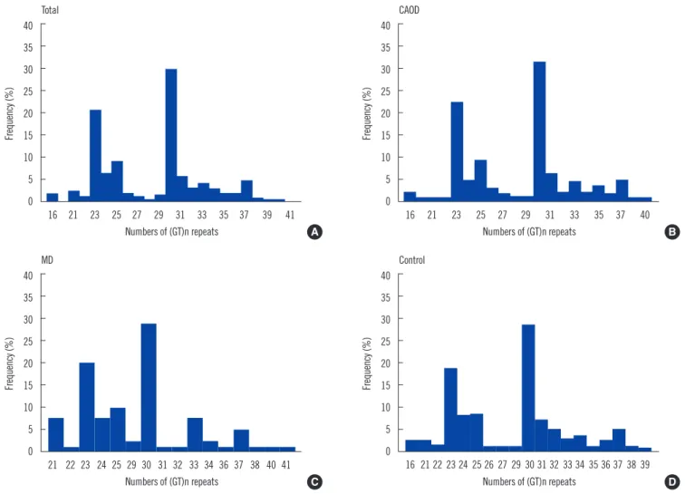
Analysis of the HMOX1 gene promoter polymorphism revealed that the number of (GT)n repeats ranged from 16 to 41, show- ing a bimodal distribution, in which the highest frequencies were shown at 23 and 30 repeats (Fig. 1).
There were no significant differences in allele and genotype frequencies between patient and control groups (Table 2).
Furthermore, we divided the entire study population into two groups: an S25-carrier group with the alleles of less than 25 (GT)n repeats and a non-S25 carrier group that lacked the S25 allele. We then performed comparative analyses with respect to
all parameters. The S25-carrier group included S25/L25 and S25/S25 genotypes, and the non-S25 group included only the L25/L25 genotype. Among age, sex, hypertension, diabetes, and biochemical parameters, only the positive rate of hsCRP was significantly higher in the non-S25 carrier group than in the S25 carrier group (P =0.034) (Table 1).
3. Restenosis following coronary stenting at the 6-month follow-up
In the CAD group with obstructive lesions, 39 patients com- pleted 6-month follow-ups and subsequent coronary angiogra- phies. Four patients underwent percutaneous transluminal cor- onary angioplasty, 34 patients had coronary angioplasty with a stent insertion, and 1 patient underwent coronary artery bypass graft. Among these 39 patients, 12 had vascular restenosis, whereas 27 were restenosis-free. Comparative analyses re- vealed no significant intergroup differences in any of the clinical
Fig. 1. The frequency distributions of the numbers of (GT)n repeats of the HMOX1 promoter. (A) Total study populations (N=257), (B) CAD with obstructive lesions group (N=110), (C) MD (N=40), and (D) controls (N=107).
Abbreviations: CAD, coronary artery disease, CAOD, coronary artery disease with obstructive lesions; MD, coronary artery disease with minimal or no lesions.
Frequency (%)
Total
Numbers of (GT)n repeats 40
35 30 25 20 15 10 5 0
16 21 23 25 27 29 31 33 35 37 39 41
Frequency (%)
Control
Numbers of (GT)n repeats 40
35 30 25 20 15 10 5 0
16 21 22 23 24 25 26 27 29 30 31 32 33 34 35 36 37 38 39
Frequency (%)
MD
Numbers of (GT)n repeats 40
35 30 25 20 15 10 5 0
21 22 23 24 25 29 30 31 32 33 34 36 37 38 40 41
Frequency (%)
CAOD
Numbers of (GT)n repeats 40
35 30 25 20 15 10 5 0
16 21 23 25 27 29 31 33 35 37 40
A B
C D
and biochemical parameters as well as allele and genotype fre- quencies (Table 3).
4. Multivariate logistic analysis of HMOX1 polymorphisms and other coronary risk factors
Analysis of the genotypes and CAD for each risk factor group did not yield any significant differences.
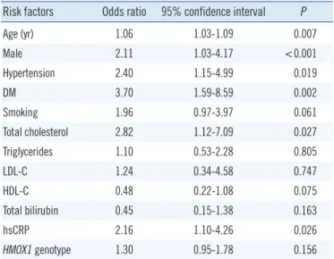
Multivariate regression analysis revealed a significantly high odds ratio for sex, age, hypertension, diabetes, cholesterol, and hsCRP, whereas HMOX1 genotype and bilirubin levels did not yield a significant odds ratio (Table 4). Most traditional cardio- vascular risk factors were significantly associated with CAD;
however, there was no significant association between HMOX1 genotype and CAD in these patients.
DISCUSSION
Inflammatory responses play a crucial role in various atheroscle- rosis-related diseases such as CAD. The fact that atherosclero- sis, which is the key cause of CAD, is an inflammatory disease
indicates that its relevant immune mechanism interacts with metabolic risk factors, thereby developing and activating arterial lesions [14]. Recently, oxidative stress has also been considered a pathogenic mechanism of a variety of diseases, such as ath- erosclerosis, myocardial ischemia, and neurodegenerative dis- eases [8, 15]. Increase in the expression of endogenous stress proteins such as HO can be a physiological response to prevent further cell damage by oxidative stress [1, 2].
The heme-HO system is a regulator of the integrity of endo- thelial cells and oxidative stress [3]. The anti-oxidant activity of HO-1 generates biliverdin and bilirubin by degrading heme dur- ing the process of releasing carbon monoxide. Carbon monox- ide plays an important role in controlling the vascular tone [16].
Induction of HO-1 activity has a protective effect for atheroscle- rosis, sepsis, diabetes, lung injury, vascular obstructive disease, and ischemic heart [6, 17]. Because bilirubin reduces oxidative Table 2. Distributions of HMOX1 promoter genotypes and allele frequencies of the study population
CAD with obstructive lesions (N=110) CAD with minimal or no lesions (N=40) Control (N=107) P Alleles, N (%)
S25 66 (30.0) 29 (36.2) 70 (32.7) 0.573
L25 154 (70.0) 51 (63.8) 144 (67.3)
Genotypes, N (%)
S25/S25 10 (9.1) 4 (10.0) 7 (6.6) 0.505
S25/L25 46 (41.8) 21 (52.5) 56 (52.3)
L25/L25 54 (49.1) 15 (37.5) 44 (41.1)
Abbreviations: CAD, coronary artery disease; S25, short (<25 [GT]n repeats); L25, long (≥25 [GT]n repeats).
Table 3. Distributions of HMOX1 promoter genotypes and allele fre- quencies of the CAD patients with and without restenosis at the 6-month follow-up
No restenosis
(N=27) Restenosis
(N=12) P
Alleles, N (%)
S25 14 (25.9) 7 (29.2) 0.787
L25 40 (74.1) 17 (70.8)
Genotypes, N (%)
S25/S25 0 (0) 1 (8.3) 0.296
S25/L25 14 (51.9) 5 (41.7)
L25/L25 13 (48.1) 12 (50.0)
Abbreviations: CAD, coronary artery disease; S25, short ( <25 [GT]n re- peats); L25, long (≥25 [GT]n repeats).
Table 4. Multivariate logistic regression analysis in CAD patients and controls
Risk factors Odds ratio 95% confidence interval P
Age (yr) 1.06 1.03-1.09 0.007
Male 2.11 1.03-4.17 <0.001
Hypertension 2.40 1.15-4.99 0.019
DM 3.70 1.59-8.59 0.002
Smoking 1.96 0.97-3.97 0.061
Total cholesterol 2.82 1.12-7.09 0.027
Triglycerides 1.10 0.53-2.28 0.805
LDL-C 1.24 0.34-4.58 0.747
HDL-C 0.48 0.22-1.08 0.075
Total bilirubin 0.45 0.15-1.38 0.163
hsCRP 2.16 1.10-4.26 0.026
HMOX1 genotype 1.30 0.95-1.78 0.156
Abbreviations: CAD, coronary artery disease; DM, diabetes mellitus; LDL-C, LDL cholesterol; HDL-C, HDL cholesterol; hsCRP, high sensitivity C-reactive protein.
stress, high bilirubin levels are related to a lower risk of LDL oxi- dation and hinders CAD [18, 19]. The increase in HMOX1 tran- scription mediated by statins is related to the increase in HO-1 proteins and decrease in free radicals [20].
The capacity to induce HO-1 differs from individual to indi- vidual, and there are 3 polymorphisms at the HMOX1 promoter, which regulates the quantitative activities of HO-1. Among them, (GT)n repeat polymorphism and the single nucleotide polymor- phism (SNP) at the -413 position are believed to have functional importance [8]. The (GT)n repeat polymorphism is a purine-py- rimidine sequence with a Z-conformation structure and is known to have negative effects on transcription activity [9].
With respect to the distribution of (GT)n repeats, several stud- ies found bimodal distributions with 23 and 30 repeats as the (GT)n alleles, which is consistent with the findings observed in our study. From this, we inferred that this distribution pattern is common to humans, regardless of racial differences. The previ- ous studies divided the alleles with this bimodal distribution pat- tern into either two groups of short- and long-allele groups or three groups by adding a medium-allele group. When classify- ing these groups, they used the reference sequences to be 22, 23, and 25 for the short alleles and 27, 29, and 30 for the long alleles [10-12, 21-24]. For the analyses in the present study, we classified the alleles into S and L groups, setting 25 repeats as a cut-point; 29 repeats were also used as a cut-point, but did not find any differences in genotypic and allelic frequencies be- tween the patient and control groups (data not shown).
There are a number of studies regarding the association be- tween HMOX1 polymorphisms and various diseases, such as chronic pulmonary emphysema [23], lung disease [6, 25], can- cer [24], hyperbilirubinemia [18, 26], renal disease [27], and stroke [16, 28, 29]. The consensus from these studies is that the ability of upregulation of HO-1 exerts an important protective role against specific diseases.
Research on the HMOX1 polymorphism is especially focused on CAD. These are based on the findings of studies that showed that HO-1 expression played a crucial role in the formation of atherosclerotic lesions in the aorta of LDL receptor knockout mice [5]. A previous study stated that short (GT)n repeats were associated with a reduced risk of CAD under high oxidative stress [15]. In a study on Chinese people with type 2 diabetes, the patients with long (GT)n repeats had a 4.7-fold higher risk for CAD [12]. A similar study conducted in Japanese people re- vealed that, among the high-risk groups of people with hyperlip- idemia or diabetes as well as smokers, the carriers of short (GT) n repeats had a reduced risk for CAD [9]. On the other hand, in
another study, similar distributions of polymorphic (GT)n repeats were found in patients with myocardial infarction and stable an- gina versus controls, and that only the group with short repeat sequences showed significantly high concentrations of bilirubin and high-density lipoprotein [30].
In the present study, there were no significant differences in genotype and allele frequencies between the CAD patients and control groups, but the non-S carrier group showed a signifi- cantly higher hsCRP positive rate than the S-carrier group. This result is similar to the one by Li et al. [31] that revealed the baseline CRP of the patients with CADs differed by genotype by showing that those carrying the long allele had higher CRP than those who did not have a long allele. These results seem to be consistent with other reports that carriers of the short allele showed fewer incidences of inflammation and thus they had a lower risk of restenosis [7, 11, 32].
The restenosis rate in this study was about 30%, which is similar to previous reports. Many of the factors inhibited by HO-1 are involved in restenosis occurrence by suppressing in- flammation of the vascular wall, vascular spasm and remodel- ing, and formation of neointimal thickening through smooth muscle cell proliferation. With regard to HMOX1 promoter geno- type, short (GT)n repeats are thought to decrease the risk for re- stenosis after coronary angioplasty by reducing inflammatory re- sponses compared with long repeats [11]. These results were verified in two prospective studies, in which the risk for resteno- sis in patients with short (<25) GT repeats who underwent per- cutaneous angioplasty on peripheral arteries was reduced to 0.24 and 0.43, respectively [7, 11].
The long allele has been reported to be a risk factor for the oc- currence of restenosis in several studies [22, 31]. As such, these findings are similar to other studies conducted on people of dif- ferent ethnic backgrounds, and therefore provide strong evi- dence that the mechanism of vascular restenosis is associated with the HMOX1 (GT)n repeat polymorphism. This can also be explained by its implications for predicting significant adverse ef- fects or outcomes [12]. In our study, however, no significant dif- ferences in the allelic and genotypic frequencies were exhibited by the restenosis and restenosis-free groups after coronary an- gioplasty or stent placement. This discrepancy between our re- sults and those of previous studies is presumably attributable to the small sample size of the patients in our study (n=39) who completed the follow-up coronary angiography to determine the risk factors for restenosis.
The clinical conditions which showed associations with the HMOX1 genotype are very different across studies, thus leaving
room for debate regarding how they are related with known risk factors in various clinical settings. In the present study, the com- parisons of disease frequencies according to genotype distribu- tion in the presence of various risk factors did not yield any sig- nificant differences.
The limitations of this study are that there was a small sample size and the gene-gene interaction and association with other genetic polymorphisms could not be taken into account be- cause only a specific gene was studied.
In conclusion, a microsatellite polymorphism in the HMOX1 gene promoter was observed in the Korean population with a bi- modal distribution, which is similar to that of previous studies.
The HMOX1 genotypes were not associated with CAD in the Ko- rean population, but the short allele carrier group contained more individuals with hsCRP values that place them at low risk for cardiovascular disease.
Authors’ Disclosures of Potential Conflicts of Interest
No potential conflicts of interest relevant to this article were re- ported.
REFERENCES
1. Berliner JA, Navab M, Fogelman AM, Frank JS, Demer LL, Edwards PA, et al. Atherosclerosis: basic mechanism.Oxidation, inflammation, and genetics. Circulation 1995;91:2488-96.
2. Lusis AJ. Atherosclerosis. Nature 2000;407:233-41.
3. Jornot L and Junod AF. Variable glutathione levels and expression of an- tioxidant enzymes in human endothelial cells. Am J Physiol 1993;264:
L482-9.
4. Maines MD. Hemeoxygenase: function, multiplicity, regulatory mecha- nisms, and clinical applications. FASEB J 1988;2:2557-68.
5. Ishikawa K, Sugawara D, Wang XP, Suzuki K, Itabe H, Maruyama Y, et al. Heme oxygenase-1 inhibits atherosclerotic lesion formation in LDL- receptor knockout mice. Circ Res 2001;88:506-12.
6. Ryter SW, Kim HP, Nakahira K, Zuckerbraun BS, Morse D, Choi AM.
Protective functions of heme oxygenase-1 and carbon monoxide in the respiratory system. Antioxid Redox Signal 2007;9:2157-73.
7. Exner M, Schillinger M, Minar E, Mlekusch W, Schlerka G, Haumer M, et al. Heme oxygenase-1 gene promoter microsatellite polymorphism is associated with restenosis after percutaneous transluminal angioplasty.
J Endovasc Ther 2001;8:433-40.
8. Ono K, Goto Y, Takagi S, Baba S, Tago N, Nonogi H, et al. A promoter variant of the heme oxygenase-1 gene may reduce the incidence of ischemic heart disease in Japanese. Atherosclerosis 2004;173:315-9.
9. Kaneda H, Ohno M, Taguchi J, Togo M, Hashimoto H, Ogasawara K, et al. Heme oxygenase-1 gene promoter polymorphism is associated with coronary artery disease in Japanese patients with coronary risk factors.
Arterioscler Thromb Vasc Biol 2002;22:1680-5.
10. Schillinger M, Exner M, Mlekusch W, Ahmadi R, Rumpold H, Mannhal-
ter C, et al. Heme oxygenase-1 genotype is a vascular anti-inflammatory factor following balloon angioplasty. J Endovasc Ther 2002;9:385-94.
11. Schillinger M, Exner M, Minar E, Mlekusch W, Müllner M, Mannhalter C, et al. Heme oxygenase-1 genotype and restenosis after balloon angioplas- ty: a novel vascular protective factor. J Am Coll Cardiol 2004;43:950-7.
12. Chen YH, Chau LY, Lin MW, Chen LC, Yo MH, Chen JW, et al. Heme oxygenase-1 gene promotor microsatellite polymorphism is associated with angiographic restenosis after coronary stenting. Eur Heart J 2004;
25:39-47.
13. Pearson TA, Mensah GA, Alexander RW, Anderson JL, Cannon RO 3rd, Criqui M, et al. Markers of inflammation and cardiovascular disease:
application to clinical and public health practice: A statement for health- care professionals from the Centers for Disease Control and Prevention and the American Heart Association. Circulation 2003;107:499-511.
14. Hansson GK. Inflammation, atherosclerosis, and coronary artery dis- ease. N Engl J Med 2005;352:1685-95.
15. Chen M, Zhou L, Ding H, Huang S, He M, Zhang X, et al. Short (GT) (n) repeats in heme oxygenase-1 gene promoter are associated with lower risk of coronary heart disease in subjects with high levels of oxidative stress. Cell Stress Chaperones 2012;17:329-38.
16. Chen YH, Yet SF, Perrella MA. Role of heme oxygenase-1 in the regula- tion of blood pressure and cardiac function. Exp Biol Med 2003;228:
447-53.
17. Bai CH, Chen JR, Chiu HC, Chou CC, Chau LY, Pan WH. Shorter GT re- peat polymorphism in the heme oxygenase-1 gene promoter has pro- tective effect on ischemic stroke in dyslipidemia patients. J Biomed Sci 2010;17:12.
18. Abraham NG and Kappas A. Hemeoxygenase and the cardiovascular- renal system. Free Radic Biol Med 2005;39:1-25.
19. Bozkaya OG, Kumral A, Yesilirmak DC, Ulgenalp A, Duman N, Ercal D, et al. Prolonged unconjugated hyperbilirubinaemia associated with the haem oxygenase-1 gene promoter polymorphism. Acta Paediatr 2010;
99:679-83.
20. Lee TS, Chang CC, Zhu Y, Shyy JY. Simvastatin induces heme oxygen- ase-1: a novel mechanism of vessel protection. Circulation 2004;110:
1296-302.
21. Chen YH, Lin SJ, Lin MW, Tsai HL, Kuo SS, Chen JW, et al. Microsatel- lite polymorphism in promoter of heme oxygenase-1 gene is associated with susceptibility to coronary artery disease in type 2 diabetic patients.
Hum Genet 2002;111:1-8.
22. Gulesserian T, Wenzel C, Endler G, Sunder-Plassmann R, Marsik C, Mannhalter C, et al. Clinical restenosis after coronary stent implantation is associated with the heme oxygenase-1 gene promoter polymorphism and the heme oxygenase-1 +99 G/C variant. Clin Chem 2005;51:1661-5.
23. Yamada N, Yamaya M, Okinaga S, Nakayama K, Sekizawa K, Shibahara S, et al. Microsatellite polymorphism in the heme oxygenase-1 gene pro- moter is associated with susceptibility to emphysema. Am J Hum Genet 2000;66:187-95.
24. Was H, Dulak J, Jozkowicz A. Hemeoxygenase-1 in tumor biology and therapy. Curr Drug Targets 2010;11:1551-70.
25. Raval CM and Lee PJ. Heme oxygenase-1 in lung disease. Curr Drug Targets 2010;11:1532-40.
26. Kanai M, Akaba K, Sasaki A, Sato M, Harano T, Shibahara S, et al. Neo- natal hyperbilirubinemia in Japanese neonates: analysis of the heme oxygenase-1 gene and fetal hemoglobin composition in cord blood. Pe- diatr Res 2003;54:165-71.
27. Chin HJ, Cho HJ, Lee TW, Na KY, Yoon HJ, Chae DW, et al. The heme oxygenase-1 genotype is a risk factor to renal impairment of IgA ne- phropathy at diagnosis, which is a strong predictor of mortality. J Korean Med Sci 2009;24(S):S30-7.
28. Funk M, Endler G, Schillinger M, Mustafa S, Hsieh K, Exner M, et al.
The effect of a promoter polymorphism in the heme oxygenase-1 gene on the risk of ischaemic cerebrovascular events: the influence of other vascular risk factors.Thromb Res 2004;113:217-23.
29. Kimpara T, Takeda A, Watanabe K, Itoyama Y, Ikawa S, Watanabe M, et al. Microsatellite polymorphism in the human heme oxygenase-1 gene promoter and its application in association studies with Alzheimer and Parkinson disease. Hum Genet 1997;100:145-7.
30. Endler G, Exner M, Schillinger M, Marculescu R, Sunder-Plassmann R, Raith M, et al. A microsatellite polymorphism in the heme oxygenase-1
gene promoter is associated with increased bilirubin and HDL levels but not with coronary artery disease. Thromb Haemost 2004;91:155-61.
31. Li P, Sanders J, Hawe E, Brull D, Montgomery H, Humphries S. Inflam- matory response to coronary artery bypass surgery: does the heme-oxy- genase-1 gene microsatellite polymorphism play a role? Chin Med J 2005;118:1285-90.
32. Tiroch K, Koch W, von Beckerath N, Kastrati A, Schömig A. Heme oxy- genase-1 gene promoter polymorphism and restenosis following coro- nary stenting. Eur Heart J 2007;28:968-73.