J Korean Soc Surg Hand 2015;20(1):8-14.
http://dx.doi.org/10.12790/jkssh.2015.20.1.8
THE HAND
INTRODUCTION
The fingers are immobilized in full extension for finger sta- bility in skin graft or flap operation on the volar aspect of the finger or the palm. A common method for finger immobi-
lization is Kirschner wire (K-wire) pinning from the distal phalangeal bone to the proximal phalangeal or metacarpal bone through the medullar cavity. However, this method requires a drilling device to insert a pin into the bone, and minor joint or tendon injury by the high-speed rotating wire
Manual Kirschner-Wire Insertion through the Soft Tissue for Finger Immobilization after Scar
Contracture Release
Jun Hee Lee, Kang Woo Lee, Jin Sik Burm, Won Yong Yang, Sang Yoon Kang
Department of Plastic and Reconstructive Surgery, Kyung Hee University School of Medicine, Seoul, Korea
Received:December 26, 2014 Revised:[1] February 13, 2015,
[2] March 10, 2015 Accepted:March 15, 2015 Correspondence to:Jin Sik Burm Department of Plastic and Reconstructive Surgery, Kyung Hee University School of Medicine, 23 Kyungheedae-ro, Dongdaemun- gu, Seoul 130-872, Korea
TEL:+82-2-958-8431 FAX:+82-2-963-5638 E-mail:[email protected]
Purpose: Finger immobilization by Kirschner-wire (K-wire) insertion may be used for postoperative stability after release of scar contracture. K-wire inser- tion through the phalangeal bone requires drilling and can result in joint and/or tendon injury or pain during wire removal. To prevent these problems, we inserted the K-wire through the soft tissue.
Methods:Seventy-five fingers of 45 patients who underwent reconstruction of scar contracture of the fingers were immobilized by K-wire. After contracture release, just before skin grafting and/or local flap surgery, in full extension of the finger, a K-wire was inserted manually from the fingertip to the proximal phalanx or metacarpal bone through the soft tissue under the phalangeal bone, along the longitudinal axis on the volar side. If the graft site did not have enough soft tissue or the K-wire was felt on the recipient bed, the K-wire was inserted on the dorsal side of the finger. K-wires were manually removed two weeks after surgery.
Results:In most cases, the time to insert the K-wire was 2-3 minutes per fin- ger, and immobilization and stability was maintained for two weeks. In two fin- gers, the K-wire came out prematurely during wound care; this did not affect the overall outcome. There were no complications due to K-wire insertion or pain during removal.
Conclusion:Finger immobilization by K-wire insertion through soft tissue is simple to perform, leads to stable immobilization, has no adding procedure.
This method is useful for temporary finger immobilization in full extension.
Keywords:Finger, Immobilization, Kirschner wire, Soft tissue, Contracture release
This is an Open Access article distributed under the terms of the Creative Commons Attribution Non-Commercial License (http://creativecommons.org/ licenses/by- nc/3.0/) which permits unrestricted noncommercial use, distribution, and reproduction in any medium, provided the original work is properly cited.
is not uncommon. In addition, wire removal is painful and requires special instruments. To avoid these problems, we have performed finger immobilization by manually insert- ing a K-wire through the soft tissue without the use of a drilling machine. The purpose of this study was to report the effectiveness of finger immobilization by manual K-wire splinting through the soft tissue.
MATERIALS AND METHODS
The study protocol confirmed to the ethical guidelines of the Declaration of Helsinki. Local ethics committee approval was obtained. No external funding was received for this study. This study included 75 fingers of 45 patients (11 female, 34 male) who underwent skin graft and/or mul- tiple Z-plasty to treat scar contracture of the fingers. The age of the patients ranged from 2 to 52 years (mean, 17 years).
The cause of contracture deformity was burn (31), crushing injuries (7), rotating belt injury (4), laceration (2), and surgi- cal incision1. Of the patients, 21 underwent operation for a single finger, and 24, for multiple fingers. Of the 75 fingers operated, 7 were the thumb; 25, the index finger; 17, the middle finger; 11, the ring finger; and 15, the little finger (Table 1).
The finger or volar scar contractures were excised and sufficiently relaxed before the K-wire was inserted for finger immobilization. While one of the surgeon’s hands extend- ed the finger completely, the other hand held a K-wire (diameter, 0.85 mm) and inserted it from the fingertip
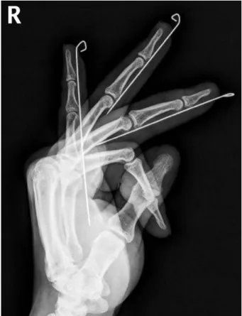
through the soft tissue of the volar pulp. The wire was inserted just beneath the phalangeal bone through the soft tissue and moved toward the volar side parallel to the longi- tudinal axis of the finger between the phalangeal bone and tendon (Fig. 1). If the graft site was located at a distal or mid- dle phalanx, the end of the wire was placed at the proximal third of the finger to allow motion in the metacarpopha- langeal joint. If the graft site was located at the proximal phalanx or metacarpal bone, the end of the wire was placed between the middle and the proximal third of the metacarpal bone.
The wire was usually inserted from the volar side. If the graft bed had little soft tissue or the wire was felt from the volar side, it was inserted into the lateral side of the finger near the skin graft site. If the wire could still be felt, it was inserted from the dorsal side (as in the fifth finger of Fig. 2).
The exposed K-wire was cut off after ensuring joint immobi-
Fig. 1. A postoperative X-ray after Kirschner wire insertion through the soft tissue. The wires placed on the volar side of the right third, fourth, and fifth fingers are immobilized in full exten- sion and are inserted up to the proximal third of the third and fourth fingers to allow motion of the metacarpophalangeal joint.
Table 1.Summary of patients
Thumb 1 (1)
Index 9 (9)
Middle 3 (3)
Little 8 (8)
Thumb, index 2 (4)
Thumb, index, middle 3 (9)
Thumb, index, ring 1 (3)
Index, middle 08 (16)
Index, middle, ring 2 (6)
Middle, ring 1 (2)
Ring, little 07 (14)
Total 45 (75)
Involved finger No. of patient (finger)
lization in full extension, leaving a small portion from the finger tip. The end of the K-wire was bent in a loop shape to prevent it from being buried into the skin and causing a pressure injury at the insertion site. At two weeks after surgery, the K-wire was removed by lightly pulling it out with the fingers or forceps in the out-patient office. The patients were required to apply antibiotic ointment and simple gauze dressing until the small hole due to the K-wire insertion was closed, and active finger movement was immediately allowed.
RESULTS
In most cases, the time to insert the K-wire was two to three minutes per finger, and the finger was usually immobilized in the extended state at once. Most patients maintained a stable finger position for 2 weeks (Figs. 2-4). In two of the 75 fingers, the K-wire came out accidentally because it became caught on the gauze during wound care. However, this did not affect the survival of the graft or the overall out- come. Two patients complained of pain at the proximal end of the K-wire on the palm when the finger was hyper- extended during treatment of the grafted skin but otherwise had no pain. No patients experienced complications, such as infection or ulcer at the wire insertion site, joint or ten- don injuries, limitation of joint motion, pain or difficulty
during K-wire removal.
1. Case 1
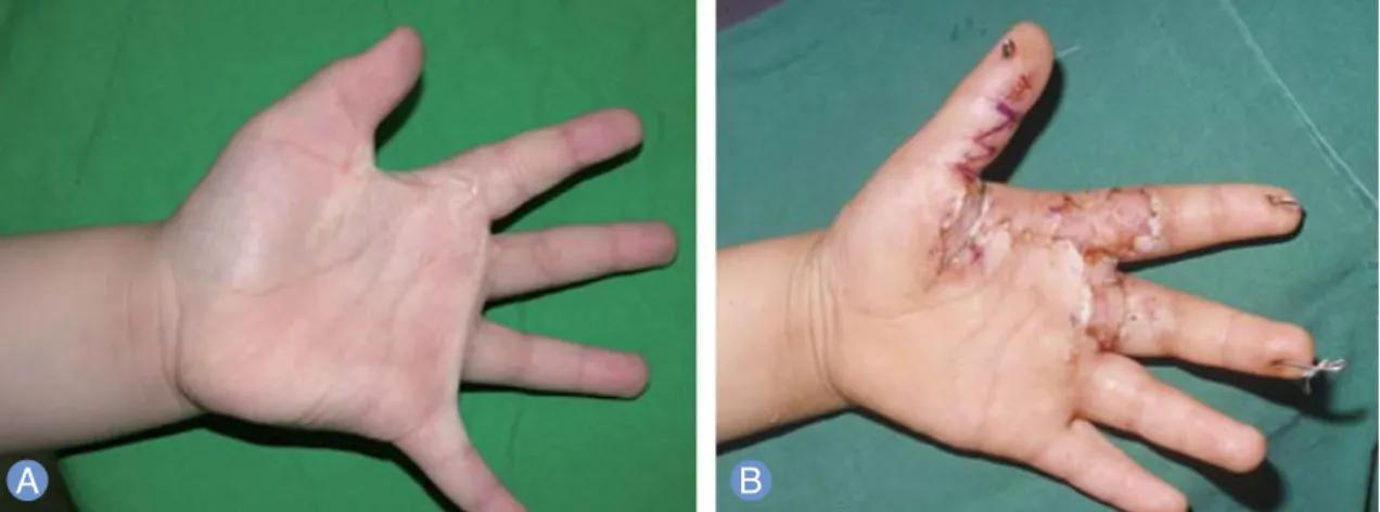
A 10-year-old male patient complained chiefly of scar con- tractures in the left ring and little fingers due to steam burns from a pressure rice cooker when he was 3 years old. After excisional release of the scar contractures, a K-wire was inserted to immobilize the finger during skin grafting. As the fourth finger had insufficient soft tissues at the skin graft site, the K-wire was inserted on the dorsal side, and the end of the wire was positioned at the proximal phalanx. For the fifth finger, the wire was inserted on the volar side, and the end of the wire was positioned at the proximal phalanx.
After the finger was immobilized, the defect was covered with a full-thickness graft. The skin graft survived without complications, and the K-wire was removed at 2 weeks after surgery (Fig. 2).
2. Case 2
A 6-year-old male patient had scar contractures of the left hand at an interdigital web space between the first and sec- ond fingers, at the proximal phalanxes of the first, second, and third fingers, and at the metacarpophalangeal joints, caused by iron burns at the age of 6 months. First, the scar contractures of the fingers were removed simultaneously using Z-plasty, and the fingers were completely relaxed.
Fig. 2.Case 1.(A) A10-year-old child with burn scar contracture on the volar side of the ring and little fingers.(B) Postoperative view after full-thickness skin grafting. As the soft tissue at the skin graft site was insufficient, the K-wires were inserted on the dorsal side of the fourth finger and on the lateral side of the fifth finger.
Then, the K-wire was inserted through the soft tissues on the volar side for finger immobilization. As the soft tissues were insufficient at the bed of the graft site on the third fin- ger, the wire was inserted along the lateral side of the finger, and the end of the wire was located at the middle of the metacarpal bone. For the second finger, the wire was insert- ed on the volar side. The defect was covered with a full- thickness graft, and Z-plasty was performed on the space between the first and second fingers. The skin graft survived without complications, and the K-wire was removed at 2 weeks after surgery (Fig. 3).
3. Case 3
A 9-year-old male patient had a scar contracture deformity of the right little finger due to deep abrasion one year previ-
ously. The scar contracture was relaxed by multiple Z-plasty so that the finger could be sufficiently extended. While care was taken to prevent the K-wire from being felt at the bed of the graft site, the wire was inserted into the soft tissues on the volar side for finger immobilization. After closure of the Z-plasty, the proximal defect was covered with a full-thick- ness graft. The skin graft and multiple flaps survived with- out complications, and the K-wire was removed at 2 weeks after surgery (Fig. 4).
DISCUSSION
After skin grafting to the volar surface of the finger, the fin- ger is immobilized in full extension for 2 or 3 weeks to stabi- lize the grafted skin effectively1-3. Finger immobilization
Fig. 3.Case 2.(A)A 6-year-old child with burn scar contracture on the first, second, and third web spaces and fingers of the left hand.(B)Ten days after full-thickness skin grafting and Z-plasty. The K-wires were inserted on the lateral side of the first and third fingers and on the volar side of the second finger.
Fig. 4.Case 3.(A)A 9-year-old child with scar contracture on the right little finger.(B)Five days after full-thickness skin graft and multiple Z-plasty. The K-wire was inserted through the soft tissue on the volar side of the finger.
using K-wires is a representative technique for stabilization of skin grafts after scar removal and to achieve sufficient relaxation in patients with finger scar contractures. It also plays a role in stretching the tissues surrounding the con- tracted articular capsule4-6. K-wire insertion achieves finger immobilization in full extension without much difficulty, even in children who tend not to cooperate during the heal- ing period necessary for the survival of the skin graft7.
There are some disadvantages to the traditional method of K-wire insertion through the phalangeal bone with a drill machine. This procedure always requires a drilling machine for insertion of the K-wire and an additional instrument for removing it. In addition, it is difficult to insert a K-wire exactly through the multiple medullary cavities of the pha- langeal or metaphalangeal bones along a longitudinal axis, especially in younger children with small phalangeal bones.
Therefore, there is a strong possibility of multiple trials due to inaccurate wire insertion. When the K-wire penetrates outside of the wall of the phalangeal bone, the rotating wire tip can injure the soft tissue, including adjacent ligaments or tendons. Moreover, it always penetrates multiple pha- langeal joints, and this may result in growth plate injury8, articular cartilage injury, or hemoarthrosis, although some researchers have reported that there is no permanent artic- ular damage on magnetic resonance imaging3. Finally, removal of an intramedullary K-wire may cause problems such as pain, bleeding, or hemoarthrosis.
Our technique of manual K-wire insertion through the soft tissues has some advantages. It can be performed easily without additional instruments. When the wire is not inserted properly on the first attempt, it can simply be removed and reinserted. Since no rotatory force is generat- ed during wire insertion, there is no risk of soft tissue injury.
Furthermore, the wire is easily removed without an addi- tional instrument, and there are no complications such as pain or bleeding during or after wire removal.
However, this technique does have some disadvantages.
K-wire splinting through the soft tissue allowed little flexi- bility of the finger joints. When the patients actively flexed their fingers, the metacarpophalangeal joints were flexed in the range of 5�to 10�. However, this minimal flexibility did not affect the skin graft outcome. In addition, it is possible
to remove a K-wire accidently by pulling out the gauge hanging on the K-wire tip during a dressing change. In the early period of this series, accidental wire removal during dressing change occurred in two cases. Fortunately, it did not affect the graft survival or the overall outcome. After that, the tip of the K-wire was bent in a loop shape, and no more cases of accidental removal occurred.
With this technique, the K-wire is always inserted parallel to the longitudinal axis of the finger, and the insertion point varies according to the state of the soft tissues at the bed of the skin graft. It is usually inserted from the fingertip pulp along the center line of the volar side. If the wire is felt at the bed of the graft site or if it is thought highly likely that the soft tissues are insufficient, the wire is inserted to the lateral side or the dorsal side. In any case, the wire is moved for- ward along the wall of the phalanx, and the K-wire is posi- tioned close to the bone and ligament to prevent protrusion and act as a splint. When mild resistance is felt during wire insertion, forceps are used to rotate the wire to the left or right. When strong resistance is felt, the wire is moved back and inserted again.
CONCLUSION
Finger immobilization by K-wire insertion through the soft tissue has several advantages: it is simple to perform, achieves stable immobilization without adding procedure, and allows easy and painless wire removal, resulting in high compliance in patients. This method may be a useful option for effective finger immobilization in full extension.
REFERENCES
1. Achauer BM, Bartlett RH, Furnas DW, Allyn PA, Wingerson EL. Internal fixation in the management of the burned hand. Arch Surg. 1974;108:814-20.
2. Green DP, Hotchkiss RN, Pederson WC. Green’s opera- tive hand surgery. New York: Churchill Livingstone;
1999.
3. Sungur N, Ulusoy MG, Boyacgil S, et al. Kirschner-wire fixation for postburn flexion contracture deformity and consequences on articular surface. Ann Plast Surg.
2006;56:128-32.
4. Larson DL, Evans EB, Abston S, Lewis SR. Skeletal sus- pension and traction in the treatment of burns. Ann Surg. 1968;168:981-5.
5. Kalliainen LK, Drake DB, Edgerton MT, Grzeskiewicz JL, Morgan RF. Surgical management of the hand in Freeman-Sheldon syndrome. Ann Plast Surg. 2003;50:
456-62.
6. Jang YC, Kwon OK, Lee JW, Oh SJ. The optimal manage- ment of pediatric steam burn from electric rice-cooker:
STSG or FTSG? J Burn Care Rehabil. 2001;22:15-20.
7. Stern PJ, Neale HW, Graham TJ, Warden GD. Classification and treatment of postburn proximal interphalangeal joint flexion contractures in children. J Hand Surg Am.
1987;12:450-7.
8. Galanakis I, Aligizakis A, Katonis P, Papadokostakis G, Stergiopoulos K, Hadjipavlou A. Treatment of closed unstable metacarpal fractures using percutaneous transverse fixation with Kirschner wires. J Trauma.
2003;55:509-13.
반흔구축이완술 후 연부조직내
K-강선 삽입을 이용한 손가락 고정술
이준희∙이강우∙범진식∙양원용∙강상윤
경희대학교 의과대학 성형외과학교실
목적: 손가락의 반흔구축이완술 시 K-강선을 수지골의 골강 내로 삽입하여 신전 상태로 고정하는데 관절 관통상, 힘줄
손상, 제거 시 통증 등의 문제점이 있어 K-강선을 연부 조직 내에 삽입하여 고정하고자 하였다.
방법:45명의 환자, 75예의 손가락을 대상으로 하였고 손가락을 완전 신전시킨 상태에서 K-강선을 연부 조직 내로 삽
입하여 수지골의 벽을 따라 장축에 평행하게 손바닥 쪽으로 진행시켰고 손가락 관절이 신전된 상태로 고정된 것을 확인 한 후 이식피부가 안정된 2주 경에 K-강선을 가볍게 잡아 뽑아 제거하였다.
결과:손가락이 신전된 상태로 2주간 안정된 상태가 유지되었고 2예에 있어 치료 도중 K-강선이 뽑혔으나 수술 결과에
는 큰 영향을 주지 않았다. K-강선 삽입에 의한 연부 조직의 감염, 관절 및 힘줄 손상, 제거 시 통증 등의 합병증은 전 례에서 없었다.
결론:손가락의 연부조직 내 K-강선 삽입 고정술은 조직 손상 없이 간단한 술기로 안정적인 고정 상태를 유지할 수 있
고 제거할 때 통증이 거의 없어 손가락의 고정이 필요한 경우 유용한 방법이라고 생각된다.
색인단어:손가락, 고정, K-강선, 연부 조직, 반흔구축이완
접수일2014년 12월 26일수정일1차 2015년 2월 13일, 2차 2015년 3월 10일 게재확정일2015년 3월 15일
교신저자범진식
서울시 동대문구 경희대로 23 경희대학교병원 성형외과학교실 TEL02-958-8431 FAX02-963-5638 [email protected]