2020 Korean Society for Surgery of the Hand, Ko- rean Society for Microsurgery, and Korean Society for Surgery of the Peripheral Nerve. All Rights re- served.
This is an open-access article distributed under the terms of the Creative Commons Attribution Non-Commercial license (http://creativecommons.
org/licenses/by-nc/4.0/), which permits unrestricted non-commercial use, distribution, and reproduction in any medium, provided the original work is prop- erly cited.
환자에서 원위요골 골절의 역학적 특성
김동희1, 곽상호2, 장효석3, 안성진4, 이광은1, 조윤재5, 이상현5
1성균관대학교 의과대학 삼성창원병원 정형외과학교실, 2부산대학교 의과대학 양산부산대학교병원 정형외과학교실, 3인제대학교 의과대학 인제대학교 해운대백병원 정형외과학교실, 4국군수도병원 정형외과, 5부산대학교 의과대학 부산대학교병원 정형외과학교실
Epidemiologic Features of Distal Radius Fractures in Severe Trauma Patients at the Busan Regional Trauma Center
Dong Hee Kim
1, Sang Ho Kwak
2, Hyo Seok Jang
3, Sung Jin An
4, Gwang Eun Lee
1, Yoon Jae Cho
5, Sang Hyun Lee
51Department of Orthopedic Surgery, Samsung Changwon Hospital, Sungkyunkwan University School of Medicine, Changwon, Korea
2Department of Orthopedic Surgery, Pusan National University Yangsan Hospital, Pusan National University School of Medicine, Yangsan, Korea
3Department of Orthopedic Surgery, Inje University Haeundae Paik Hospital, Inje University College of Medicine, Busan, Korea
4Department of Orthopedic Surgery, Armed Forces Capital Hospital, Seongnam, Korea
5Department of Orthopedic Surgery, Pusan National University Hospital, Pusan National University School of Medicine, Busan, Korea
Purpose: Distal radius fractures (DRFs) are often observed in simple trauma in older women with osteoporosis, and severe trauma caused by traffic or fall accidents. In this study, we aim to classify the DRFs according to injury mechanism, and statistically compare epidemiologic factors, radiological characteristics, and functional scores.
Methods: From 2013 to 2018, 112 cases of trauma in 104 patients (70 monotraumas and 42 severe traumas) diagnosed with DRFs were included. Patients were divided into the low-energy monotrauma (ML), high-energy monotrauma (MH), and severe trauma groups and analyzed for differences in sex, Injury Severity Score (ISS), accompanying ipsi- lateral injuries, radiologic indices, AO/OTA classification, and functional outcome scores (disabilities of the arm, shoulder, and hand [DASH] and Modified Mayo Wrist Score).
Results: Significant differences were observed in sex, age, ISS, and accompanying ipsi- lateral injury among three group (p<0.001). Distribution of AO/OTA classification was not significantly different among the groups. Especially, sex, age, and accompanying ipsilateral injury were significantly different between the ML and MH groups (p<0.001). Postoperative DASH and MMWS were significantly different between the monotrauma and severe trauma groups (p<0.001).
Conclusion: Severe trauma with DRFs was observed at a lower age and more fre- quently accompanied by ipsilateral injury and high ISS. Additionally, the functional outcomes were lower after severe trauma than after monotrauma. Therefore, for DRF patients with severe trauma, attention should be paid to the pattern of fracture as well as the accompanying injury and postoperative management and rehabilitation associated with it.
Keywords: Epidemiology, Distal radius fracture, Multiple trauma pISSN 2586-3290 · eISSN 2586-3533
Arch Hand Microsurg 2020;25(3):181-188 https://doi.org/10.12790/ahm.20.0030
Received: May 22, 2020 Revised: June 26, 2020 Accepted: July 8, 2020 Corresponding author:
Sang Hyun Lee
Department of Orthopedic Surgery, Pusan National University Hospital, 179 Gudeok-ro, Seo-gu, Pusan 49241, Korea Tel: +82-51-240-8718
Fax: +82-51-247-8395 E-mail: [email protected] ORCID:
https://orcid.org/0000-0002-2084-9824
Original Article
INTRODUCTION
Distal radius fractures (DRFs) are the most common frac- tures of the upper extremity, and various treatments for DRFs have been reported; however, options for DRFs due to trauma energy are seldom reported. DRFs may be sustained due to three types of trauma energy. First, it is often the result of a low-energy trauma, which is a simple injury encountered when a person lands on his/her wrist extended; in particular, elderly women with reduced bone density will have complex fractures even with simple trauma caused by lower-energy injuries [1-3].
Second, high-energy injuries occur only in the upper extremity due to events such as car accidents; this is simple trauma that is not accompanied by any severe trauma (ST) in other areas;
however, DRFs can occur as complex fractures due to their high-energy trauma [2,4]. Third, DRFs can occur after ST, which is accompanied by multiple organ injuries due to high-energy trauma. In this case, depending on the degree of high-energy injury affecting the upper extremity, both simple and complex fractures can occur. This ST is accompanied by damages to the brain, lung, liver, and intestines, which are re- lated to life support, or by open, pelvic, and multiple fractures that are related to orthopedically severe damage to soft tissues.
In particular, if these patients with ST miss appropriate surgery time or are unconscious, it may affect their future rehabilita- tion. In South Korea, trauma centers have been established to provide specialized treatment only to such ST patients.
This study aimed to evaluate and compare the epidemiologic features of ST patients with those of monotrauma patients with DRF. We categorized DRF patients according to the types of trauma. Subsequently, we compared the radiological features and functional results between the ST group and the mono- trauma group.
MATERIALS AND METHODS
Emergency medical centers in South Korea are subdivided into the regional emergency center, local emergency medical centers, and local emergency medical institutions. The regional trauma center is dedicated to ST. This study focused on patients who visited the Pusan National University Hospital, designated as a regional medical center and a regional trauma center. The study was approved by the Institutional Review Board of Pusan National University Hospital (No. H-1906-030-080).
Patients who visited the emergency center of Pusan National University Hospital from December 2013 to July 2018, were aged ≥20 years, were diagnosed with DRF and underwent sur-
gical treatment were initially included. Of the 109 enrolled pa- tients (119 cases), five patients (7 cases) were lost to follow-up.
Finally, 104 patients (112 cases) were included. Among the 104 patients, the evaluation of the disabilities of the arm, shoulder, and hand (DASH) score was not performed in five patients (5 cases) and a telephone survey was performed in five patients (5 cases).
We first classified patients into two groups according to the Injury Severity Score (ISS). The ISS is an established medical score that assesses trauma severity and used to define the term major trauma. It correlates with mortality, morbidity, and hos- pitalization time after trauma [5]. Multiple injuries are scored using the Abbreviated Injury Scale (AIS) according to six body regions. The ISS score is the sum of squares of the highest AIS grades in each of the three most severely injured body regions (Table 1).
ST patients were defined as those with DRFs and an ISS of
≥16. Of the 112 cases, 42 cases were of ST. Monotrauma pa- tients were defined as those with isolated DRFs or those with DRFs and fractures at other body parts and with an ISS of <16.
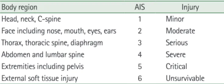
These patients were further categorized into low-energy mono- trauma (ML) and high-energy monotrauma (MH) according to the definition of MH [6]. The criteria for high-energy trau- ma are as follows [6]: a fall from >3 m, a car accident velocity of >60 km/hr, a motorcycle accident velocity of >30 km/hr, vehicle shortening of >50 cm, vehicle depression of the passen- ger side of >30 cm, vehicle rollover of passenger thrown from the vehicle, fatality in the same vehicle, and car or motorcycle versus pedestrian or bicyclist velocity of >10 km/hr. Addition- ally, it was classified as MH due to compression and crushing injury caused by a machine (Fig. 1).
Only one surgeon performed surgical treatment for all pa- tients. Surgical techniques used with a volar plate or Kirschner wire (K-wire) fixation with/without bone graft. K-wire fixation was used for simple and extra-articular fractures and bone graft
Table 1. Injury Severity Score
Body region AIS Injury
Head, neck, C-spine 1 Minor
Face including nose, mouth, eyes, ears 2 Moderate Thorax, thoracic spine, diaphragm 3 Serious Abdomen and lumbar spine 4 Severe Extremities including pelvis 5 Critical External soft tissue injury 6 Unsurvivable Calculated AIS for the most severely injured body part in each region.
Injury Severity Score is calculated as a sum of the square of AIS for each body region.
AIS, Abbreviated Injury Score.
was used for severe fragmented intra-articular fractures.
All patients were evaluated for differences in sex, ISS, accom- panying ipsilateral injury, and surgical method according to the medical chart. We assessed the radiological indices and AO/
OTA classification using radiological images at initial trauma and immediately after surgery. Radiologic indices included vo- lar tilt angle, radial inclination, and ulnar variance using the project-a-line technique. The ipsilateral injury was defined as the existence of a fracture at the ipsilateral extremity. To evalu- ate the functional results, we accessed the DASH questionnaire and calculated the Modified Mayo Wrist Score (MMWS) at 1 year after surgery.
For statistical analysis of radiological indices and functional outcomes, one-way analysis of variance and Pearson chi- squared test were used. The post-hoc test using the Bonferroni method was used to evaluate significant differences between the groups. The statistical significance level was set at p<0.05.
RESULTS
Of the total of 104 patients (112 cases) included in this study,
53 and 51 were females and males, respectively. The mode of injury was car accidents in 25 cases, fall accidents in 39 cases, and slip accidents in 45 cases. Furthermore, there was one case each of crush damage, rope wrapped accident during work, and stab wounds (Fig. 2).
Of the 112 cases, 51 were in the ML group, 19 in the MH group, and 42 in the ST group. By sex, males accounted for 29.4%, 84.2%, and 66.7% of all patients in the ML, MH, and ST groups, respectively (Table 2). The mean patient age was 64, 47, and 51 years in the ML, MH, and ST groups, respectively. Both sex and age were not significantly different between the MH and ST groups which included patients with injuries due to MH trauma (p=0.052, p=0.979), but they were significantly different between the ML and MH groups and between the ML and ST groups (p<0.001) (Table 3).
The ISS was 5.08 ±2.01, 7.74 ±3.66, and 26.79 ±9.8 in the ML, MH, and ST groups, respectively (Table 2). The difference in trauma energy between the monotrauma and ST groups was significant (p <0.0001), whereas within the monotrauma groups was not significant (p=0.365) (Table 3).
The time from injury to surgery was 8.9, 7.9, and 12.1 days in Enrolled patients with distal radius fractures who visited
the emergency center of Pusan National University Hospital from December 2013 to July 2018 (n=112)
· Fall >3 m or higher
· Car accident >60 km/hr
· Motorcycle accident >30 km/ hr
· Vehicle shortening >50 cm
· Vehicle depression passenger side >30 cm
· Vehicle rollover
· Passenger thrown from vehicle
· Fatality in same vehicle
· Car or motorcycle vs pedestrian or bicyclist >10 km/hr
· Motorcycle or bicycle vs motorcycle or bicycle or stationary object
ISS<16
Isolated distal radius fractures ISS≥16
No
ML group (n=51) MH group (n=19) ST group (n=42)
Yes
Fig. 1. Enrollment of patients with distal radius fractures. ML, low-energy monotrauma; MH, high-energy monotrauma; ST, severe trauma.
the ML, MH, and ST groups, respectively. The difference in the time from injury to surgery among the three groups was not significant (p=0.130).
Two patients in the ST group required revision surgery be- cause reduction loss was observed owing to patient incorpora- tion. One patient had a brain injury and another had a psycho- logical problem. All patients underwent bone union at the final follow-up.
1. Surgical methods
Patients who underwent only volar plate fixation after injury
accounted for 98.0%, 84.2%, and 97.6% in the ML, MH, and ST groups, respectively (Table 2), showing a significant difference (p=0.032) between the ML and ST groups (p=0.009), and be- tween the ML and MH groups (p=0.02) (Table 3). Whether using K-wire fixation or bone graft, the difference was not sig- nificant in the three groups (p≥0.05).
2. Ipsilateral combined injury
The incidence of ipsilateral injury was 5.9%, 42.1%, and 64.3% in the ML, MH, and ST groups, respectively. The post- hoc test revealed a significant difference between the ML and Slip down,
80%
Fall down
<3 m, 20%
Crushing injury,
10%
Motorcycle TA, 32%
Fall down
>3 m, 26%
Motorcycle TA, 10%
Stab injury, 2%
Rolling down
>3 m, 7%
Pedestrian TA, 7%
Car accident,
16%
Car accident, 12%
Fall down
>3 m, 62%
Pedestrian TA, 16%
Fig. 2. Distribution of patients in the three groups according to the AO/OTA classification. (A) Low-energy monotrauma group (n=51). (B) High-energy monotrauma group (n=19). (C) Severe trauma group (n=42). TA, traffic accident.
Table 2. Comparison of sex, age, ISS, AO/OTA classification, ipsilateral injury, and surgical methods among the study groups
Variable Group
p-value
ML (n= 51) MH (n= 19) ST (n= 42)
Sex < 0.001
Male 15 (29.4) 16 (84.2) 28 (66.7)
Female 36 (70.6) 3 (15.8) 14 (33.3)
Age (yr) 64.61± 12.74 47.37± 18.75 51.33± 14.35 < 0.001
ISS 5.08± 2.01 7.74± 3.66 26.79± 9.80 < 0.001
Time from injury to surgery (day) 8.88 7.89 12.14 0.130
AO/OTA classification 0.250
A 13 (25.5) 3 (15.8) 6 (14.3)
B 20 (39.2) 5 (26.3) 2 (4.8)
C 18 (35.3) 11 (57.9) 34 (81.0)
Surgical methods
Plate 50 (98.0) 16 (84.2) 41 (97.6) 0.032
Kirschner wire 5 (9.8) 4 (21.1) 6 (14.3) 0.515
Bone graft 1 (2.0) 2 (10.5) 5 (11.9) 0.114
Ipsilateral injury 0 (0) 8 (42.1) 27 (64.3) < 0.001
ISS, Injury Severity Score; ML, low-energy monotrauma; MH, high-energy monotrauma; ST, severe trauma.
p<0.05 indicates statistical significance.
A B C
MH groups and between the ML and ST groups (p<0.0001), but not between the MH and ST groups, which included pa- tients with injuries due to MH (p=0.105).
3. Radiologic indices
The results of the radiological examination conducted before and after surgery showed no significant differences, except the fracture site gap that was significantly different between the monotrauma and ST groups (p<0.001), but not between the ML and MH groups (p>0.999).
All patients underwent bone union at the last follow-up.
None of the patients required revision surgery.
4. Functional outcomes
The results of DASH and MMWS to evaluate functional out-
comes showed significant differences in all groups (p<0.001).
As the energy of the mechanism of injury increased, functional outcomes worsened (Tables 3, 4). The distribution of AO/OTA Classification did not show significant differences (p=0.25), but the predominance of type C was identified in the MH and ST groups (Table 2).
DISCUSSION
There are several classification systems for DRFs, but it is dif- ficult to classify fractures according to trauma energy [6]. Few papers have compared and analyzed the results by classifying DRFs according to the degree of trauma energy [2,4,6]. We can expect easily that patients with high-energy trauma will have a higher rate of complex fractures than those with low-energy trauma, which subsequently results in worse functional out- comes. However, previous studies have reported no correlation between the extent of trauma and AO/OTA classification [7].
This is due to the involvement of various factors, including os- teoporosis, concomitant injuries, the degree of external trauma, and the mechanism [1-3]. Therefore, we classified the clinical characteristics of each group by dividing them into three groups based on the injury mechanism. Subsequently, we ana- lyzed the radiological findings, types of fractures, and function- al outcomes in patients after surgery.
The ST and MH groups mostly included young and male pa- tients among the three groups. According to the injury mecha- nism, high-energy trauma such as fall or traffic accidents was the main injury mechanism in the ST and MH groups (Fig. 2).
Relatively, slip down is the most common injury mechanism, and most of the patients included in the ML group were post- Table 3. Results of the post-hoc test for evaluating significant
differences among the study groups
Variable ML vs. MH ML vs. ST MH vs. ST
Sex < 0.001 < 0.001 0.052
Age < 0.001 < 0.001 0.979
ISS 0.382 < 0.001 < 0.001
Time from injury to surgery > 0.999 0.259 0.276
Plate 0.020 0.333 0.009
Ipsilateral injury < 0.001 < 0.001 0.054 DASH score 0.546 < 0.001 < 0.001
MMWS 0.066 < 0.001 < 0.001
ML, low-energy monotrauma; MH, high-energy monotrauma; ST, severe trauma; ISS, Injury Severity Score; DASH, disabilities of the arm, shoulder, and hand; MMWS, Modified Mayo Wrist Score.
Bonferroni correction method was used, and p<0.05 indicates statistical significance.
Table 4. Statistical analysis results of radiological indices and functional outcomes
Variable Group
p-value
ML (n= 51) MH (n= 19) ST (n= 42)
Volar tilt
Preoperative 1.28± 14.48 –3.95± 15.16 1.29± 18.67 0.447
Postoperativer 7.64± 7.41 4.47± 7.28 6.48± 8.23 0.311
Radial inclination
Preoperative 16.24± 6.21 19.89± 7.32 16.76± 7.98 0.158
Postoperativer 20.08± 5.79 20.37± 7.00 20.95± 4.27 0.749
Ulnar variance
Preoperative 4.01± 13.50 2.26± 4.43 3.17± 4.87 0.788
Postoperative –0.10± 2.43 –1.11± 2.54 –0.12± 2.35 0.094
Functional outcome
DASH score 13.44± 7.49 17.96± 13.87 30.30± 16.10 < 0.001
MMWS 87.20± 8.34 81.05± 12.54 63.81± 15.69 < 0.001
ML, low-energy monotrauma; MH, high-energy monotrauma; ST, severe trauma; DASH, disabilities of the arm, shoulder, and hand; MMWS, Modified Mayo Wrist Score.
menopausal women with decreasing bone density. Moreover, there might be a selection bias because our study was conduct- ed in a tertiary university hospital where patients with simple fractures do not usually visit. In this study, although the statisti- cal significance of simple trauma was not high, there were sev- eral cases of complex fractures in simple trauma. This is con- sidered a feature of the ML group that comprised several elder- ly patients with osteoporosis. In previous studies assessing DRFs in patients with high-energy trauma [6,8], there were several complex articular fracture types and high ipsilateral damages in the high-energy group. In this study, the same re- sult was observed, that is the accompanying ipsilateral injury was high, but there was no statistical significance for the type of fracture. Additionally, the accompanying rate of hand injuries among ST patients was 3.6%, and the concomitant injury was a significant factor affecting the functional outcomes after treat- ment. This result is probably due to the high-energy injury in the ST group, which likely causes other injuries to the affected area.
We expected that the MH and ST groups would have poor results in terms of radiological indices than the ML group;
however, the actual statistical analysis results showed no signif- icant difference. The preoperative and postoperative radiologi- cal indices did not show any difference between the three groups. Most of the patients in the MH and ST groups are young men; thus, performing union in the fracture site is con- sidered advantageous in these young patients, which might af- fect statistical results. Additionally, due to the recent develop- ment of surgical techniques and tools and equipment, types of fracture do not seem to affect radiological parameters [9].
There was no significant difference in the preoperative radio- logical parameters among the three groups. This indicates that simple slip down might cause a complex fracture because sev- eral elderly women were likely to have osteoporosis in the ML group.
In a previous study that compared patients with isolated frac- tures and those with polytrauma, there were no significant dif- ferences between the groups in terms of union, but the overall results were significantly lower in patients with polytrauma than in patients with isolated fractures in terms of functional outcomes [4,10]. Although no statistical difference in radiolog- ical indices was observed among the groups, poor functional outcomes were observed in the MH and ST groups. We hy- pothesized that this is due to the high ISS and accompanying injuries. In our experience, patients in the ST group have poor cooperation due to polytrauma, which lead to difficulty in di- agnosing and treating these patients. In this study, the time
from injury to surgery was longer in the ST group than that in the ML group. In the ML group, 90.2% of patients underwent surgery within 10 days from injury. Moreover, 36.8% and 45.2% of patients in the MH and ST groups, respectively, un- derwent surgery after >10 days from the injury date. In partic- ular, all four patients in the ML group who underwent surgery after 3 weeks were all operated due to reduction loss during conservative treatment. In ST patients with accompanying pol- ytrauma, surgery related to vital signs, such as thoracic surgery, general surgery, or neurosurgery, is prioritized, except in the case of vascular injury. Therefore, orthopedic problems are usually low in surgical priority. Accordingly, it is difficult to perform orthopedic surgery at the appropriate time, and sur- gery is often delayed. Furthermore, ST patients have difficulties in managing soft tissue damage or swelling, and rehabilitation may not be performed or delayed due to unconsciousness or damage to other areas, which in turn affects functional out- comes after surgery.
This study has the following limitations. First, the sample size was small. Second, the study included only patients who visited the emergency center after trauma. In our hospital where the study was conducted, DRF patients are not allowed to visit through the outpatient hospital unless a medical problem or trauma to the other body areas is observed. Therefore, the number of patients with ML trauma to whom ambulatory care can be easily provided would be relatively lower. Third, the ST group had an ISS of ≥16, but this did not mean that whether their high-energy trauma had directly affected the fracture could be distinguished.
CONCLUSION
DRFs accompanying high-energy trauma were observed more frequently in younger age patients and male than DRFs accompanying simple trauma. We found that fracture severity and radiological indices after surgery based on the AO/OTA classification were not different between patients who visited the emergency center with high-energy. Furthermore, patients with high-energy DRFs showed worse functional outcomes af- ter surgery and required extra care considering the type of frac- tures, damages to soft tissues, and condition of the patient’s whole body as well so that they could receive appropriate post- operative management and rehabilitation after fracture treat- ment.
CONFLICTS OF INTEREST
The authors have nothing to disclose.
ACKNOWLEDGEMENTS
This work was supported by a clinical research grant from Pusan National University Hospital in 2020.
REFERENCES
1. Chung KC, Spilson SV. The frequency and epidemiology of hand and forearm fractures in the United States. J Hand Surg Am. 2001;26:908-15.
2. MacIntyre NJ, Dewan N. Epidemiology of distal radius frac- tures and factors predicting risk and prognosis. J Hand Ther.
2016;29:136-45.
3. Vogt MT, Cauley JA, Tomaino MM, Stone K, Williams JR, Herndon JH. Distal radius fractures in older women: a 10- year follow-up study of descriptive characteristics and risk factors. The study of osteoporotic fractures. J Am Geriatr Soc.
2002;50:97-103.
4. Hodel S, Schraner C, Oehme F, et al. Factors predicting ad- verse outcome in complete intra-articular distal radius frac-
tures. Eur J Trauma Emerg Surg. 2019 Feb 28 [Epub]. https://
dx.doi.org/10.1007/s00068-019-01102-8.
5. Baker SP, O’Neill B, Haddon W Jr, Long WB. The injury se- verity score: a method for describing patients with multiple injuries and evaluating emergency care. J Trauma. 1974;
14:187-96.
6. Ferree S, van der Vliet QM, Nawijn F, et al. Epidemiology of distal radius fractures in polytrauma patients and the influ- ence of high traumatic energy transfer. Injury. 2018;49:630-5.
7. Wæver D, Madsen ML, Rölfing JHD, et al. Distal radius frac- tures are difficult to classify. Injury. 2018;49 Suppl 1:S29-32.
8. Ferree S, van der Vliet QM, van Heijl M, Houwert RM, Leenen LP, Hietbrink F. Fractures and dislocations of the hand in polytrauma patients: Incidence, injury pattern and func- tional outcome. Injury. 2017;48:930-5.
9. Alluri RK, Hill JR, Ghiassi A. Distal radius fractures: ap- proaches, indications, and techniques. J Hand Surg Am.
2016;41:845-54.
10. Ferree S, Hietbrink F, van der Meijden OAJ, Verleisdonk EJ, Leenen LP, Houwert RM. Comparing fracture healing disor- ders and long-term functional outcome of polytrauma pa- tients and patients with an isolated displaced midshaft clavicle fracture. J Shoulder Elbow Surg. 2017;26:42-8.
부산 권역 외상센터에 내원한 중증외상 환자에서 원위요골 골절의 역학적 특성
김동희1, 곽상호2, 장효석3, 안성진4, 이광은1, 조윤재5, 이상현5
1성균관대학교 의과대학 삼성창원병원 정형외과학교실, 2부산대학교 의과대학 양산부산대학교병원 정형외과학교실,
3인제대학교 의과대학 인제대학교 해운대백병원 정형외과학교실, 4국군수도병원 정형외과, 5부산대학교 의과대학 부산대학교병원 정형외과학교실
목적: 원위요골 골절은 골밀도가 감소된 나이든 여성에서는 단순외상뿐만 아니라 교통사고나 추락 등으로 인한 중증외상에서도 많이 발생한다. 저자들은 본 연구에서 이러한 손상 에너지 차이에 의한 원위요골 골절을 분류하여 중증외상 환자에 동반된 원위요골 골절의 역학적 인자 및 방사선학적 특징, 기능적 수술 결과를 일반 외상 환자와 통계적으로 비교 분석하여 보고하고자 한다.
방법: 환자군은 2013년부터 2018년까지 본원을 방문하였던 104명의 환자에서 112례의 원위요골 골절 환자들을 대상으로 하였다.
중증외상이 동반되지 않아서 응급의료센터로 방문한 손상 중증도 점수(Injury Severity Score, ISS) 15 미만의 70례를 단순 외상(monotrauma)군으로 보고, 이를 다시 손상 기전에 의해서 단순 외상-저에너지 손상군(low-energy monotrauma, ML) 51례와, 단순외상-고에너지 손상군(high-energy monotrauma, MH) 19례로 분류하였다. ISS 점수가 15 이상으로 중증외상센터로 내원한 중증외상 동반 42례는 중증 외상군(severe trauma, ST)으로 구분하여 세 군으로 나누어 분석하였다. 각 그룹을 성별, ISS 점수, 동측 동반 손상 여부, 방사선학적 지표, AO/OTA 분류, 그리고 기능적 결과 평가를 위한 disabilities of the arm, shoulder, and hand (DASH) 점수와 Modified Mayo Wrist Score (MMWS)의 결과에 유의한 차이가 있는지를 분석하였다.
결과: 분석 결과에 따르면 환자의 성별, 연령, ISS 및 동측 손상 항목에서 유의한 차이가 나타났다(p<0.001). AO/OTA 분류의 분포는 MH군과 ST군에서 type C가 우세하였으나 세 그룹에서 통계적으로 유의한 차이를 보이지 않았다. 특히, ML군과 MH군 사이에서 성별, 연령 및 동측부 동반 손상 항목에서 유의한 차이를 보였다(p<0.001). 술 후 DASH 점수와 MMWS는 외상군과 ST군 사이에서 유의한 차이를 보였다(p<0.001).
결론: 중증 외상에 의한 원위요골 골절 환자의 경우 젊은 연령대에 호발하고 동측 손상이 같이 동반되는 경우가 많으며 높은 ISS 점수를 보였다. 또한 술 후의 기능적 결과가 중증외상 환자에서 단순외상에 비해 더 떨어졌다. 그러므로 중증외상을 동반한 원위요골 골절 환자의 경우 골절 및 그에 관련한 수술적 치료뿐만 아니라 동반된 손상 그리고 이에 관련한 수술 치료 시기 및 술 후의 재활 등에도 주의가 필요하다.
색인단어: 역학, 원위요골 골절, 다발성 외상
접수일 2020년 5월 22일 수정일 2020년 6월 26일 게재확정일 2020년 7월 8일 교신저자 이상현
49241, 부산광역시 서구 구덕로 179, 부산대학교병원 정형외과
TEL +82-51-240-8718 FAX +82-51-247-8395 E-mail [email protected] ORCID https://orcid.org/0000-0002-2084-9824