The Clinical Accuracy of Endoscopic Ultrasonography and White Light Imaging in Gastric Endoscopic
Submucosal Dissection
Soon Hong Park, Sang Hun Sung, Seung Jun Lee, Min Kyu Jung, Sung Kook Kim, and Seong Woo Jeon Division of Gastroenterology and Hepatology, Department of Internal Medicine, Kyungpook National University Hospital,
Kyungpook National University School of Medicine, Daegu, Korea
Purpose: Gastric mucosal neoplastic lesions should have characteristic endoscopic features for successful endoscopic submucosal dis- section.
Materials and Methods: Out of the 1,010 endoscopic submucosal dissection, we enrolled 62 patients that had the procedure cancelled.
Retrospectively, whether the reasons for cancelling the endoscopic submucosal dissection were consistent with the indications for an en- doscopic submucosal dissection were assessed by analyzing the clinical outcomes of the patients that had the surgery.
Results: The cases were divided into two groups; the under-diagnosed group (30 cases; unable to perform an endoscopic submucosal dissection) and the over-diagnosed group (32 cases; unnecessary to perform an endoscopic submucosal dissection), according to the second endoscopic findings, compared with the index conventional white light image. There were six cases in the under-diagnosed group with advanced gastric cancer on the second conventional white light image endoscopy, 17 cases with submucosal invasion on endoscopic ultrasonography findings, 5 cases with a size greater than 3 cm and ulcer, 1 case with diffuse infiltrative endoscopic fea- tures, and 1 case with lymph node involvement on computed tomography. A total of 25 patients underwent a gastrectomy to remove a gastric adenocarcinoma. The overall accuracy of the decision to cancel the endoscopic submucosal dissection was 40% (10/25) in the subgroup that had the surgery.
Conclusions: The accuracy of the decision to cancel the endoscopic submucosal dissection, after conventional white light image and endoscopic ultrasonography, was low in this study. Other diagnostic options are needed to arrive at an accurate decision on whether to perform a gastric endoscopic submucosal dissection.
Key Words: Endoscopic submucosal dissection; Accuracy; Low grade dysplasia; Early gastric cancer
Correspondence to: Seong Woo Jeon
Division of Gastroenterology and Hepatology, Department of Internal Medicine, Kyungpook National University Hospital, Kyungpook National University School of Medicine, 130, Dongdeok-ro, Jung-gu, Daegu 700-721, Korea
Tel: +82-53-420-5514, Fax: +82-53-426-8773 E-mail: swjeon@knu.ac.kr
Received February 21, 2012 Revised April 3, 2012 Accepted May 2, 2012
Copyrights © 2012 by The Korean Gastric Cancer Association www.jgc-online.org
This is an open-access article distributed under the terms of the Creative Commons Attribution Non-Commercial License (http://creativecommons.org/
licenses/by-nc/3.0) which permits unrestricted noncommercial use, distribution, and reproduction in any medium, provided the original work is properly cited.
Introduction
Gastric cancer is one of the leading causes of cancer related deaths in the world, and it is especially common in East Asian
countries.(1) One of the major factors associated with improved survival of patients with gastric cancer is early detection. As a result of expansion of both nationwide cancer-screening programs and of private health check-ups, premalignant gastric lesions and early gastric cancer (EGC) are now detected with increasing frequency in Korea.(2) The endoscopic mucosal resection (EMR) has become one of the established treatment procedures for small early gastric cancers without any lymph node involvement and precancerous gastric lesions.(3) EMR is an effective technique for the removal of early GI tract neoplasm. Complication rates associated with the EMR are low. However, lesions over 20 mm cannot be resected in a single piece and piecemeal resection leads to local recurrence
rates of up to 15%.(4,5)
A novel technique, the endoscopic submucosal dissection (ESD) has been proposed to guarantee en bloc resection.(6-8) This tech- nique has several advantages: the resected size and shape can be controlled, en bloc resection is possible even with large tumors, and tumors with ulcerative findings are also resectable. In addition, the ESD can provide a precise histological diagnosis and has been as- sociated with a reduced recurrence rate.(5) The ESD can be used for the resection of complex tumors, such as large tumors, ulcer- ative non-lifting tumors and recurrent tumors.
When performing the ESD, difficulty can arise because the fea- tures of the target mucosal lesion might be different in the second endoscopic examination, from the index endoscopy. Erroneous diagnosis by forceps biopsy is a reason to cancel an ESD in ad- dition to disease progression and spontaneous regression.(9,10) If we could enroll the patients with appropriate indication for gastric ESD, the benefit would be avoiding unnecessary surgery and also be avoding unnecessary ESD vice versa.
In this study, the clinical outcomes of cancelled ESD procedures were evaluated. The objective of this study was to determine the accuracy of the decision to cancel an ESD.
Materials and Methods
1. Patients
Out of 1,010 cases of gastric ESD performed from January 2007 to April 2010, 62 were included in this study. These patients had an ESD planned, but then cancelled. The mean follow-up period was 474.81±285.54 days. The patients with advanced gastric lesions on the initial conventional white light image (cWLI) were excluded.
The patients were divided into two groups: Under-diagnosed and over-diagnosed. For example, in cases where the initial diagnosis from a local clinic suggested the need for an ESD, the lesion might have been observed as more advanced than appropriate for an ESD on the second cWLI and endoscopic ultrasonography (EUS), and these cases were defined as under-diagnosed (unable or dif- ficult to perform an ESD). By contrast, if the lesion on the ESD was vague in appearance such as with indistinct margins or as too small or more consistent with gastritis on biopsy, during the second cWLI, these cases were defined as over-diagnosed (unnecessary to perform an ESD). The institutional review board of our center ap- proved this study.
2. Methods
When an ESD was planned, the expanded ESD indications
were adopted in cases with adenocarcinoma. The expanded indica- tions for EMR were suggested by Gotoda in 2000. Based on the report, at the National Cancer Center and other groups, the indica- tions for ESD are: (i) non-ulcerated, differentiated-type mucosal carcinomas, regardless of tumor size; and (ii) differentiated-type mucosal carcinoma with an ulcer scar ≤30 mm.(11) If the target lesion did not meet the criteria for the procedure during the second cWLI such as in cases with advanced disease (i.e. submucosal or muscularis propria invasion on EUS, size over 3 cm with ulcer- ation), the procedure was cancelled. In the case of dysplasia, the ESD was cancelled when the lesion to undergo ESD was vague in appearance with indistinct margins, too small, or was confirmed as
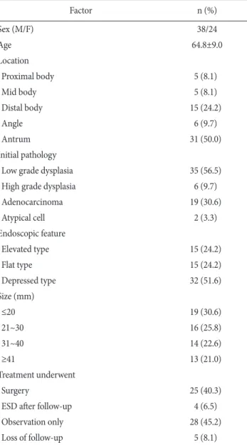
Table 1. Clinical characteristics of the patients when referred for EMR/ESD (n=62)
Factor n (%)
Sex (M/F) 38/24
Age 64.8±9.0
Location
Proximal body 5 (8.1)
Mid body 5 (8.1)
Distal body 15 (24.2)
Angle 6 (9.7)
Antrum 31 (50.0)
Initial pathology
Low grade dysplasia 35 (56.5)
High grade dysplasia 6 (9.7)
Adenocarcinoma 19 (30.6)
Atypical cell 2 (3.3)
Endoscopic feature
Elevated type 15 (24.2)
Flat type 15 (24.2)
Depressed type 32 (51.6)
Size (mm)
≤20 19 (30.6)
21~30 16 (25.8)
31~40 14 (22.6)
≥41 13 (21.0)
Treatment underwent
Surgery 25 (40.3)
ESD after follow-up 4 (6.5)
Observation only 28 (45.2)
Loss of follow-up 5 (8.1)
EMR = endoscopic mucosal resection; ESD = endoscopic submucosal dissection; M = male; F = female.
gastritis on the biopsy from the second endoscopy.
All 62 patients were referred for ESD from local clinics. The endoscopic features and pathology from the local clinics were reviewed. The mean time lag between the first and second endos- copy was 37.65±15.12 days. The second endoscopy for the ESD (GIF-H260, GIF-H180, Olympus, Tokyo, Japan) was performed at our clinic by a single expert (SW Jeon), who has performed more than 1,000 gastric ESDs.
A radial scanning, 20-MHz catheter, probe (UM3R, Olympus), was used by the same physicians in all patients except the patients with advanced gastric cancer (AGC) features on second endoscopy.
The probe was passed through the instrument channel of a two channel endoscope (GIF-2T200, Olympus). When the ESD was cancelled, a repeat biopsy (defined as the second diagnosis) and another description of the target lesions was performed.
The reasons for cancelling the ESD were categorized into four groups in the under-diagnosed group: gross AGC features, submu- cosal invasion in the EUS, size >3 cm with ulceration, and diffuse infiltrative lesion. The consistency between the second diagnosis and the final surgical pathology in the patients that went to surgery was evaluated. In addition, the clinical and endoscopic data were analyzed to assess the relationship between the accuracy and vari- ables that were assumed to be predictive of the accuracy associated with canceling the ESD. In the cases that underwent surgery, the
clinical outcomes were reviewed and the accuracy of cancelled cases assessed.
3. Statistical analysis
A statistical software package (SPSS ver. 14.0, SPSS Inc., Chi- cago, IL, USA) was used for the data analysis. For assessment of the association between the accuracy of the decision and the study variables (i.e. category of size, location, endoscopic features, and pathology), the c2 test was used, and the independent t-test for quantitative variables. P-values less than 0.05 were considered to be significant.
Results
1. Clinical characteristics of patients
The target lesions were more frequently located in the lower portion of the stomach. Macroscopic types were classified as el- evated, flat, and depressed. The depressed type was more common (32 [51.6%]) than the others (15 flat lesions [24.2%] and 15 elevated lesions [24.2%]). The tumor size was categorized into four groups:
(<20 mm, 19 [30.6%]; 21~30 mm, 16 [25.8%]; 31~40 mm 14 [22.6%], >40 mm 13 [21.0%]). The pathological diagnosis when referred was 35 low grade dysplasias (LGDs) (56.5%), six high grade dysplasias (HGDs) (9.7%), 19 adenocarcinomas (30.6%) and
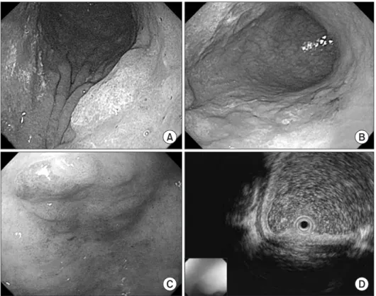
Fig. 1. Two centimeter elevated muco- sal lesion was revealed as HGD on the initial diagnosis of a 63-year-old male (A). However, ulceration and a size over 3 cm were observed on the second cWLI 59 days later (B), therefore surgi- cal treatment was performed. EGC was diagnosed on a 3 cm IIa lesion of the antrum in a 75-year-old female (C).
However, the lesion already involved over 1/3 of the upper submucosa on the EUS performed 9 days later com- pared to the initial endoscopy (D).
HGD = high grade dysplasia; cWLI = conventional white light image; EGC = early gastric cancer; EUS = endoscopic ultrasonography.
two atypical cells (3.3%). Twenty-five (40.3%) patients underwent surgical treatment and four (6.5%) patients underwent ESD later (Table 1).
2. Clinical outcomes of the patients
Among the 62 cancelled cases, 30 (19 adenocarcinoma, 5 HGD, 5 LGD, 1 atypical cell in initial diagnosis) were under-diagnosed when referred, and consisted of six cases of gross AGC on the sec- ond cWLI (Fig. 1A, B), 18 cases with submucosal invasion on the EUS (Fig. 1C, D), four cases with a size over 3 cm and ulceration, one case with diffuse infiltrative endoscopic features and one case with lymph node involvement on computed tomography (CT) (Fig. 2).
Thirty-two patients were over-diagnosed (1 atypical cells, 1 HGD, and 30 LGD on the initial diagnosis) and their pathology
at the time of the second endoscopy was one adenocarcinoma, 17 dysplasias, 11 cases of chronic gastritis and three cases with no sus- pected mucosal lesions for re-biopsy. Adenocarcinoma identified on the re-biopsied cases underwent ESD later and three cases with dysplasia had ESD later.
3. Clinical outcomes in the under-diagnosed group (unable to perform an ESD)
In the under-diagnosed group, 25 patients underwent a gas- trectomy (subtotal or total, according to the location) with D1-2 dissection. The clinical outcomes of these 25 operated patients are described in Table 2. There was no lymph node involvement on the final surgical pathology.
Fig. 2. Clinical outcomes of cancelled ESD cases. Among the 62 cancelled cases, 30 (19 adenocarcinoma, 5 HGD, 5 LGD, 1 atypical cell in initial diagnosis) were under-diagnosed when referred, and included six cases of gross AGC by endoscopic features, 17 cases with submucosal invasion on EUS findings, five cases over 3 cm with ulceration, one case with diffuse infiltrative endoscopic features and one case of lymph node involve- ment on CT. Twenty-five among the 30 under-diagnosed cases underwent subtotal gastrectomy and none had lymph node involvement. Thirty-two patients were over-diagnosed (1 atypical cell, 1 HGD, 30 LGD in initial diagnosis) and their pathology at the time of the second endoscopy was: 1 adenocarcinoma, 16 adenomas, 11 with chronic gastritis, and four cases with no suspected mucosal lesion for re-biopsy at the second endoscopy.
Adenocarcinoma found in re-biopsied cases had ESD later and there were three cases that underwent ESD later with adenomas. ESD = endoscopic submucosal dissection; Adenoca = adenocarcinoma; HGD = high grade dysplasia; LGD = low grade dysplasia; AGC = advanced gastric cancer;
EUS = endoscopic ultrasonography; LN = lymph node; CT = computed tomography; F/u = follow-up.
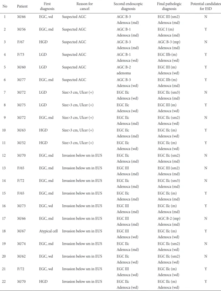
Table 2. Review of 25 cases in the under-diagnosed group that had surgery
No Patient First
diagnosis Reason for
cancel Second endoscopic
diagnosis Final pathologic
diagnosis Potential candidates for ESD
1 M/66 EGC, wd Suspected AGC AGC B-3
Adenoca (md)
EGC III (sm2) Adenoca (md)
N
2 M/56 EGC, md Suspected AGC AGC B-1
Adenoca (md)
EGC I (m) Adenoca (md)
Y
3 F/67 HGD Suspected AGC AGC B-3
Adenoca (md)
AGC B-3 (mp) Adenoca (md)
N
4 F/73 LGD Suspected AGC AGC B-1
Adenoca (wd)
EGC IIb (m) Adenoca (wd)
Y
5 M/60 LGD Suspected AGC AGC B-2
adenoma
EGC III (m) Adenoca (wd)
Y
6 M/77 EGC, md Suspected AGC AGC B-3
Adenoca (wd)
EGC IIb (m) Adenoca (md)
Y
7 M/72 LGD Size>3 cm, Ulcer (+) EGC IIc
Adenoca (wd)
EGC IIc (sm3) Adenoca (md)
N
8 M/75 LGD Size>3 cm, Ulcer (+) EGC IIc
Adenoca (wd)
EGC III (m) Adenoca (wd)
Y
9 M/72 EGC, md Size>3 cm, Ulcer (+) EGC IIc
Adenoca (md)
EGC IIc (sm2) Adenoca (wd)
N
10 M/63 HGD Size>3 cm, Ulcer (+) EGC IIc
Adenoca (md)
EGC IIc (m) Adenoca (wd)
Y
11 M/52 HGD Size>3 cm, Ulcer (+) EGC IIc
Adenoca (wd)
EGC IIc (m) Adenoca (wd)
Y
12 M/70 EGC, md Invasion below sm in EUS EGC IIc
Adenoca (md)
EGC IIc (sm2) Adenoca (md)
N
13 F/65 EGC, md Invasion below sm in EUS EGC III
Adenoca (md)
EGC III (sm2) Adenoca (md)
N
14 F/72 EGC, md Invasion below sm in EUS EGC IIc
Adenoca (md)
EGC IIc (sm3) Adenoca (md)
N
15 F/65 EGC, md Invasion below sm in EUS EGC IIc
Adenoca (md)
EGC IIc (m) Adenoca (md)
Y
16 M/73 EGC, wd Invasion below sm in EUS EGC III
Adenoca (md)
EGC IIc (m) Adenoca (md)
Y
17 M/66 EGC, md Invasion below sm in EUS EGC III
Adenoca (md)
AGC B-2 (mp) Adenoca (md)
N 18 M/67 Atypical cell Invasion below sm in EUS EGC III
Adenoca (wd)
EGC IIc (m) Adenoca (wd)
Y
19 M/74 EGC, md Invasion below sm in EUS EGC IIc
Adenoca (md)
EGC IIc (sm2) Adenoca (wd)
N
20 M/62 EGC, wd Invasion below sm in EUS EGC IIc
Adenoca (wd)
EGC IIc (sm2) Adenoca (wd)
N
21 F/72 EGC, wd Invasion below sm in EUS EGC III
Adenoca (wd)
EGC IIc (m) Adenoca (wd)
Y
22 M/70 HGD Invasion below sm in EUS EGC IIc
Adenoca (wd)
EGC IIc (m) Adenoca (wd)
Y
4. Clinical Outcomes in the over-diagnosed group (unnecessary to perform an ESD)
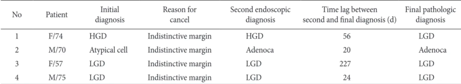
In the over-diagnosed group, four cases underwent an ESD later. All 4 cases were considered unnecessary to perform ESD due to indistinctive margins on the second endoscopy. The time lag between the second diagnosis and the ESD performed later varied from 20 days to 227 days (Table 3).
Clear margins were obtained by ESD in all four cases and the location of the final lesion on ESD was consistent with the initial location described at the local clinic.
5. Accuracy of the decision to cancel an ESD
The overall accuracy of the decision to cancel an ESD was 40%
(10/25); 33.3% (2/6) in the gross AGC subgroup, 40% (2/5) in the over 3 cm with ulceration subgroup, 46.2% (6/13) in the submucosal invasion by EUS subgroup and none in the one suspected lymph node involvement case on CT (Table 4). Lesion size, endoscopic features, pathology and location of the lesion were not associated with the decision accuracy in the statistical analysis.
Discussion
The worldwide clinical application of ESD, has allowed more cases of early stage gastric cancer to be treated by endoscopic resection. With the widespread use of endoscopic resection for the treatment of gastric neoplasms, precise staging has become mandatory in order to assess the appropriateness of the procedure for curative treatment.(12-14) With regard to the clinical decision Table 2. Continued
No Patient First
diagnosis Reason for
cancel Second endoscopic
diagnosis Final pathologic
diagnosis Potential candidates for ESD
23 M/61 LGD Invasion below sm in EUS EGC IIc
Adenoca (wd)
EGC III (m) Adenoca (wd)
Y
24 F/75 EGC, wd Invasion below sm in EUS EGC IIa
Adenoca (wd)
EGC I (m) Adenoca (md)
Y
25 M/68 EGC, wd Suspected LN
involvement in CT
EGC IIc Adenoca (wd)
EGC IIc (m) Adenoca (md)
Y
ESD = endoscopic submucosal dissection; M = male; F = female; EGC = early gastric cancer; wd = well differentiated; md = moderately differentiated; HGD = high grade dysplasia; LGD = low grade dysplasia; AGC = advanced gastric cancer; sm = submucosa; EUS = endoscopic ultrasonography; LN = lymph node; CT = computed tomography; Adenoca = adenocarcinoma; m = mucosa; mp = muscularis proper; N = no; Y
= yes.
Table 3. Review of 4 cases of ESD in the over-diagnosed group
No Patient Initial
diagnosis Reason for
cancel Second endoscopic
diagnosis Time lag between
second and final diagnosis (d) Final pathologic diagnosis
1 F/74 HGD Indistinctive margin HGD 56 LGD
2 M/70 Atypical cell Indistinctive margin Adenoca 20 Adenoca
3 F/57 LGD Indistinctive margin LGD 227 LGD
4 M/75 LGD Indistinctive margin LGD 24 LGD
ESD = endoscopic submucosal dissection; F = female; M = male; HGD = high grade dysplasia; LGD = low grade dysplasia; Adenoca = adeno- carcinoma.
Table 4. Accuracy of decision to cancel the gastric ESD in the under- diagnosed group that had surgery
Reason for cancellation at second diagnosis Accuracy
Grossly AGC (n=6) 33.3 (2/6)
Size >3 cm and ulcer (+) (n=5) 40.0 (2/5)
Sm invasion in EUS (n=13) 46.2 (6/13)
Suspected in LN involvement in CT (n=1) 0 (0/1)
Total (n=25) 40.0 (10/25)
Values are presented as % (n). ESD = endoscopic submucosal dissec- tion; AGC = advanced gastric cancer; Sm = submucosa; EUS = endo- scopic ultrasonography; LN = lymph node; CT = computed tomogra- phy.
to perform an ESD, it is impossible to assess the precise invasion depth of the forceps biopsy. Thus, EUS is the first-choice imaging modality for determining the depth of invasion.(12,14)
A meta-analysis of 22 studies showed that the accuracy of EUS for T staging in gastric cancer ranges from 65% to 92%.(15) These studies confirmed that EUS is the most accurate staging method for gastric cancer. However, when the studies are limited to early gastric cancer, the accuracy for T staging is only 70~76%.(16,17) A systematic review of 18 studies demonstrated that the sensitivity of EUS in differentiating mucosal cancer from cancer extension beyond the mucosa varied significantly, ranging from 18.2% to 100%.(12) The accuracy of EUS in differentiating between early and AGC is high. However, there are problems in distinguishing T1a from T1b cancer, which is critical in the selection of patients for endoscopic resection of early gastric cancer. The reasons for an incorrect diag- nosis with EUS include tumor microinvasion, peritumor inflamma- tion, a distinctly protruding lesion, and oblique scanning.(18) Mi- croinvasion may result in under-staging, as it is difficult to diagnose minimal submucosal invasion. Over-staging may be associated with ulceration, peritumor inflammatory changes, or fibrosis. About 10~30% of early gastric cancers have ulceration with accompany- ing fibrosis, which is seen as a hypoechoic lesion on EUS, similar to tumor invasion.(18)
In this study of cancelled ESD cases, only six out of 13 cases (operated cases due to cancer invasion suspected in over one third of submucosa by EUS) were accurate according to the final surgical pathology reports. Another 53.8% of cases had intramucosal cancer on the surgical pathology reports, which suggests they underwent avoidable surgery. In this study, large lesions and a high frequency of depressed lesions may have been responsible for lower EUS ac- curacy in comparison to other studies.
To distinguish cancer invasion from ulcer fibrosis, a method of pattern analysis was introduced. This pattern analysis was based on the fact that ulcer fibrosis always has a fan-shaped spread, while cancer invades in an arched-shaped spread. However, micro-inva- sion into the ulcer fibrosis does not change the contours of the fan- shaped ulcer fibrosis, so micro-invasion is not detectable by EUS.
By using this pattern analysis, it was reported that the diagnostic accuracy for depressed-type EGC with ulceration was 76.1%.(19) Contrast-enhanced EUS was recommended to be another method to improve the accuracy of EUS for lesions with ulcerous changes.
If the area of the carcinoma cells was selectively enhanced by intravenous contrast, it might contribute to distinguishing tumor invasions from fibrosis and lead to an accurate diagnosis for lesions
with ulcerations.(20) Kida et al.(21) reported that three-dimensional EUS (3D-EUS) provided a practical way to diagnose small inva- sion of tumors larger than 500 mm with an accuracy of 78.7% when EGC had no ulcerous changes, suggesting that 3D-EUS may be more useful and more accurate for diagnosis. However, even with the use of 3D-EUS, differentiating the minute gastric cancer inva- sion of ulcer fibrosis from ulcer fibrosis alone has been difficult.
The accuracy of the decision to cancel the procedure due to gross AGC features and a size larger than 3 cm with ulcerations was also low (50%). Perhaps due to the small sample size; however, the endoscopist must be cautious with regard to the decisions on how to manage the AGC lesions. Performing EUS is acceptable in cases that appear to be AGC. Fifty percent of cases with EGC that were large (>3 cm) with ulceration had the potential for complete resection by ESD, in this study. Although a larger study with a randomized controlled design is needed, EUS examination can be carefully performed to assess the possibility of complete resection by ESD in patients with EGC; especially for those where more in- vasive procedures are contraindicated.
Thirty-two out of 62 cancelled cases were considered to be over-diagnosed on the first endoscopy. For the second diagnosis, well demarcated, definite mucosal lesions for ESD were not ob- served as in the first diagnosis. In previous studies, the proportion of spontaneous regression of gastric adenomas has been reported to be from 11% up to 74%.(9,22) This wide variability was associated with a diverse proportion of LGD and HGD in the enrolled cases.
In 28 cases, lesions that needed endoscopic resection did not reap- pear during the follow up period. However, demarcation was clearly observed in four cases on the follow up endoscopic examination, and ESD was performed later. One case had an adenocarcinoma on the final ESD pathology.
Narrow band imaging (NBI) has been used as a tool for well demarcation of the lesion and is a video endoscopic imaging tech- nique that enhances the visualization of microstructures and capil- laries in the superficial mucosal layer by the use of narrow band filters that change the spectral features of the observed light relative to that of the narrow band filters.(23) Predictions of the histological characteristics of gastric cancer lesions can also be made using NBI and magnifying endoscopy (NBI-ME), which yield very clear im- ages of microvessels on mucosal surfaces.(24,25) One study dem- onstrated that determining the border of the lesion recognized by differences in capillary structures with NBI-ME is useful and helps with the en bloc EMR of EGC.(26) In addition, autofluorescence imaging (AFI) is one of the newly developed technologies that
produce real-time pseudocolor images by detecting natural tissue fluorescence from endogenous fluorophores that are emitted by the excitation of light.(27,28) Several studies have reported that the AFI can reveal minute lesions of the stomach that were not detected by cWLI.(28-31) Therefore, the AFI might also be helpful for the de- termination of the extent of small gastric lesions before treatments such as EMR/ESD. In this study, determination of the depth of the invasion was the main problem in the under-diagnosed group, whereas a definite demarcation line was an important factor in the over-diagnosed group. These two factors are essential in the deter- mination of the endoscopic resectability of gastric neoplasms. By using only cWLI, it is very difficult to confirm these factors before obtaining surgical pathology. The clinical usefulness of NBI-ME and AFI should be further validated.
The limitations of this study include the following. The study used a retrospective design. Also, 1st endoscopy was performed by various endoscopist in local clinics, but data base on ESD was maintained prospectively by a single endoscopist, thus bias was minimized. Because of the low ESD cancellation rate in this study (6.1%), there was a small sample size (n=62). The third limitation was the use of multiple endoscopy results from multiple local clin- ics; different imaging procedures might have been performed in the first and second endoscopic procedures with different equip- ment. The variation in the time interval between the index and second endoscopy might be another limitation of this study. A large prospective randomly controlled trial is needed to further assess the decision-making procedure for ESD.
There are some technical difficulties for achieving successful complete resections and very specific indications are needed to achieve successful complete resections. Other new diagnostic op- tions other than EUS and conventional white light endoscopy are needed to make more accurate clinical decision when considering an ESD.
References
1. Parkin DM, Bray FI, Devesa SS. Cancer burden in the year 2000. The global picture. Eur J Cancer 2001;37 Suppl 8:S4-66.
2. Nam SY, Choi IJ, Park KW, Kim CG, Lee JY, Kook MC, et al.
Effect of repeated endoscopic screening on the incidence and treatment of gastric cancer in health screenees. Eur J Gastroen- terol Hepatol 2009;21:855-860.
3. Soetikno RM, Gotoda T, Nakanishi Y, Soehendra N. Endo- scopic mucosal resection. Gastrointest Endosc 2003;57:567-
579.
4. Muto M, Miyamoto S, Hosokawa A, Doi T, Ohtsu A, Yoshida S, et al. Endoscopic mucosal resection in the stomach using the insulated-tip needle-knife. Endoscopy 2005;37:178-182.
5. Oka S, Tanaka S, Kaneko I, Mouri R, Hirata M, Kawamura T, et al. Advantage of endoscopic submucosal dissection com- pared with EMR for early gastric cancer. Gastrointest Endosc 2006;64:877-883.
6. Gotoda T, Yamamoto H, Soetikno RM. Endoscopic sub- mucosal dissection of early gastric cancer. J Gastroenterol 2006;41:929-942.
7. Cao Y, Liao C, Tan A, Gao Y, Mo Z, Gao F. Meta-analysis of endoscopic submucosal dissection versus endoscopic mucosal resection for tumors of the gastrointestinal tract. Endoscopy 2009;41:751-757.
8. Nakamoto S, Sakai Y, Kasanuki J, Kondo F, Ooka Y, Kato K, et al. Indications for the use of endoscopic mucosal resection for early gastric cancer in Japan: a comparative study with endo- scopic submucosal dissection. Endoscopy 2009;41:746-750.
9. Yamada H, Ikegami M, Shimoda T, Takagi N, Maruyama M.
Long-term follow-up study of gastric adenoma/dysplasia. En- doscopy 2004;36:390-396.
10. Kim YJ, Park JC, Kim JH, Shin SK, Lee SK, Lee YC, et al. His- tologic diagnosis based on forceps biopsy is not adequate for determining endoscopic treatment of gastric adenomatous le- sions. Endoscopy 2010;42:620-626.
11. Gotoda T, Yanagisawa A, Sasako M, Ono H, Nakanishi Y, Shi- moda T, et al. Incidence of lymph node metastasis from early gastric cancer: estimation with a large number of cases at two large centers. Gastric Cancer 2000;3:219-225.
12. Kwee RM, Kwee TC. The accuracy of endoscopic ultrasonogra- phy in differentiating mucosal from deeper gastric cancer. Am J Gastroenterol 2008;103:1801-1809.
13. Ichikawa T, Kudo M, Matsui S, Okada M, Kitano M. Endo- scopic ultrasonography with three miniature probes of dif- ferent frequency is an accurate diagnostic tool for endoscopic submucosal dissection. Hepatogastroenterology 2007;54:325- 328.
14. Puli SR, Batapati Krishna Reddy J, Bechtold ML, Antillon MR, Ibdah JA. How good is endoscopic ultrasound for TNM stag- ing of gastric cancers? A meta-analysis and systematic review.
World J Gastroenterol 2008;14:4011-4019.
15. Kwee RM, Kwee TC. Imaging in local staging of gastric cancer:
a systematic review. J Clin Oncol 2007;25:2107-2116.
16. Kim JH, Song KS, Youn YH, Lee YC, Cheon JH, Song SY, et al.
Clinicopathologic factors influence accurate endosonographic assessment for early gastric cancer. Gastrointest Endosc 2007;66:901-908.
17. Hizawa K, Iwai K, Esaki M, Matsumoto T, Suekane H, Iida M.
Is endoscopic ultrasonography indispensable in assessing the appropriateness of endoscopic resection for gastric cancer? En- doscopy 2002;34:973-978.
18. Choi J, Kim SG, Im JP, Kim JS, Jung HC, Song IS. Comparison of endoscopic ultrasonography and conventional endoscopy for prediction of depth of tumor invasion in early gastric can- cer. Endoscopy 2010;42:705-713.
19. Kida M, Tanabe S, Watanabe M, Kokutou M, Kondou I, Yamada Y, et al. Staging of gastric cancer with endoscopic ul- trasonography and endoscopic mucosal resection. Endoscopy 1998;30 Suppl 1:A64-68.
20. Nomura N, Goto H, Niwa Y, Arisawa T, Hirooka Y, Hayakawa T. Usefulness of contrast-enhanced EUS in the diagnosis of up- per GI tract diseases. Gastrointest Endosc 1999;50:555-560.
21. Kida M, Kikuchi H, Ikeda H, Miyazawa S, Iwai T, Araki M, et al. Diagnostic of gastric cancer invasion with three-dimension- al endoscopic ultrasonography, especially in cases with ‘‘SM1’’
invasion. Stomach Intest 2007;42:88-98.
22. Di Gregorio C, Morandi P, Fante R, De Gaetani C. Gastric dys- plasia. A follow-up study. Am J Gastroenterol 1993;88:1714- 1719.
23. Gono K, Yamazaki K, Doguchi N, Nonami T, Obi T, Yamagu- chi M, et al. Endoscopic observation of tissue by narrow band illumination. Opt Rev 2003;10:211-215.
24. Nakayoshi T, Tajiri H, Matsuda K, Kaise M, Ikegami M, Sa- saki H. Magnifying endoscopy combined with narrow band imaging system for early gastric cancer: correlation of vascular
pattern with histopathology (including video). Endoscopy 2004;36:1080-1084.
25. Sumiyama K, Kaise M, Nakayoshi T, Kato M, Mashiko T, Uchi- yama Y, et al. Combined use of a magnifying endoscope with a narrow band imaging system and a multibending endoscope for en bloc EMR of early stage gastric cancer. Gastrointest En- dosc 2004;60:79-84.
26. Kadowaki S, Tanaka K, Toyoda H, Kosaka R, Imoto I, Hamada Y, et al. Ease of early gastric cancer demarcation recognition: a comparison of four magnifying endoscopy methods. J Gastro- enterol Hepatol 2009;24:1625-1630.
27. Ohkawa A, Miwa H, Namihisa A, Kobayashi O, Nakaniwa N, Ohkusa T, et al. Diagnostic performance of light-induced fluorescence endoscopy for gastric neoplasms. Endoscopy 2004;36:515-521.
28. Uedo N, Iishi H, Tatsuta M, Yamada T, Ogiyama H, Imanaka K, et al. A novel videoendoscopy system by using autofluores- cence and reflectance imaging for diagnosis of esophagogastric cancers. Gastrointest Endosc 2005;62:521-528.
29. Bhunchet E, Shibata T. Proposal for two strategies to prevent remnants of gastric cancers after endoscopic mucosal resec- tions: fluorescein electronic endoscopy and rapid stump diag- nosis based on pit patterns. Gastric Cancer 2004;7:221-232.
30. Haringsma J, Tytgat GN, Yano H, Iishi H, Tatsuta M, Ogihara T, et al. Autofluorescence endoscopy: feasibility of detection of GI neoplasms unapparent to white light endoscopy with an evolv- ing technology. Gastrointest Endosc 2001;53:642-650.
31. Bhunchet E, Hatakawa H, Sakai Y, Shibata T. Fluorescein elec- tronic endoscopy: a novel method for detection of early stage gastric cancer not evident to routine endoscopy. Gastrointest Endosc 2002;55:562-571.