http://dx.doi.org/10.3988/jcn.2014.10.2.140 J Clin Neurol 2014;10(2):140-147
Antioxidant Effects of Statins in Patients with Atherosclerotic Cerebrovascular Disease
Gyeong Joon Moon,a,b Suk Jae Kim,c Yeon Hee Cho,b Sookyung Ryoo,c Oh Young Bangc,d
aMedical Research Institute, Sungkyunkwan University School of Medicine, Suwon, Korea
bClinical Research Center, Samsung Biomedical Research Institute, Seoul, Korea
cDepartment of Neurology, Samsung Medical Center, Sungkyunkwan University School of Medicine, Seoul, Korea
dSamsung Advanced Institute for Health Sciences and Technology, Seoul, Korea
Received March 30, 2013 Revised November 8, 2013 Accepted November 8, 2013 Correspondence Oh Young Bang, MD, PhD Department of Neurology, Samsung Medical Center, Sungkyunkwan University School of Medicine, 81 Irwon-ro, Gangnam-gu, Seoul 135-710, Korea Tel +82-2-3410-3599 Fax +82-2-3410-0052 E-mail nmboy@unitel.co.kr
Background and PurposezzOxidative stress is involved in the pathophysiological mecha- nisms of stroke (e.g., atherosclerosis) and brain injury after ischemic stroke. Statins, which in- hibit 3-hydroxy-3-methylglutaryl coenzyme A (HMG-CoA) reductase, have both pleiotropic and low-density lipoprotein (LDL)-lowering properties. Recent trials have shown that high- dose statins reduce the risk of cerebrovascular events. However, there is a paucity of data re- garding the changes in the oxidative stress markers in patients with atherosclerotic stroke after statin use. This study evaluated changes in oxidative stress markers after short-term use of a high-dose statin in patients with atherosclerotic stroke.
MethodszzRosuvastatin was administered at a dose of 20 mg/day to 99 patients who had suf- fered an atherosclerotic stroke and no prior statin use. Blood samples were collected before and 1 month after dosing, and the serum levels of four oxidative stress markers–malondialdehyde (MDA), oxidized LDL (oxLDL), protein carbonyl content (PCO), and 8-hydroxy-2’-deoxy- guanosine (8-OHdG)–were evaluated to determine the oxidation of MDA and lipids, proteins, and DNA, respectively, at both of those time points.
ResultszzThe baseline levels and the degrees of reduction after statin use differed among the oxidative stress markers measured. MDA and PCO levels were associated with infarct volumes on diffusion-weighted imaging (r=0.551, p<0.05, and r=0.444, p=0.05, respectively). Statin use decreased MDA and oxLDL levels (both p<0.05) but not the PCO or 8-OHdG level. While the reduction in MDA levels after statin use was not associated with changes in cholesterol, that in oxLDL levels was proportional to the reductions in cholesterol (r=0.479, p<0.01), LDL (r=0.459, p<0.01), and apolipoprotein B (r=0.444, p<0.05).
ConclusionszzThe impact of individual oxidative stress markers differs with time after isch- emic stroke, suggesting that different oxidative markers reflect different aspects of oxidative stress. In addition, short-term use of a statin exerts antioxidant effects against lipid peroxidation via lipid-lowering-dependent and -independent mechanisms, but not against protein or DNA oxidation in atherosclerotic stroke patients. J Clin Neurol 2014;10(2):140-147 Key Wordszz atherosclerosis, ischemic stroke, statin, oxidative stress, cholesterol.
Open Access
cc This is an Open Access article distributed under the terms of the Cre- ative Commons Attribution Non-Commercial License (http://creative- commons.org/licenses/by-nc/3.0) which permits unrestricted non-com- mercial use, distribution, and reproduction in any medium, provided the ori- ginal work is properly cited.
Introduction
Oxidative stress is involved in the pathophysiological mech-
anisms of stroke (e.g., atherosclerosis) and brain injury after ischemic stroke (e.g., reperfusion injury).1 Antioxidant levels and activities have been found to decrease after stroke as a consequence of increased oxidative stress, and thereafter in- crease gradually over time.2 It has also been reported that low plasma antioxidant activity is associated with high lesion vol- ume and severe neurological impairment in stroke.3 Moreover,
the brain is at risk for oxidative damage because it has high ox- idative damage potential but a low antioxidant capacity.4
Statins, which inhibit HMG-CoA reductase, have both pleio- tropic and low-density lipoprotein (LDL)-lowering properties.5 Two randomized trials–Stroke Prevention by Aggressive Re- duction in Cholesterol Levels (SPARCL) and Justification for the Use of Statin in Prevention: an Intervention Trial Evalu- ating Rosuvastatin (JUPITER)–showed that medication with high-dose statins reduces the risk of cerebrovascular events.6,7 Based on the results of the SPARCL trial, the American Heart Association/American Stroke Association recently recom- mended the use of statins in patients who had suffered ischemic stroke or transient ischemic attack.8 In addition, the prior use of statins has been shown to reduce the severity of ischemic stroke and to be associated with better clinical stroke out- come.9,10 However, the precise mechanisms underlying the ef- fects of statins in this context remain unclear. The improved clinical outcomes observed with statin use may be due to fa- cilitated recanalization, collateral perfusion enhancement, or a direct neuroprotective effect.11 Statins have also been shown to exert plaque-stabilizing effects, with evidence of the occur- rence of atherosclerotic plaque regression and reverse remod- eling in medicated patients.12
There is a paucity of data regarding the effects of statins on oxidative stress in patients with atherosclerotic stroke. Under the assumption that the short-term use of statins exerts various antioxidant effects in patients with atherosclerotic stroke, a comprehensive study of the antioxidant effects of a statin was conducted using a variety of markers of oxidative stress, from lipids to DNA.
Methods
Patient selection and blood collection
Stroke patients with presumed atherosclerotic stroke and no use of statins prior to the onset of stroke were enrolled pro- spectively. All patients underwent electrocardiography, echo- cardiography, and brain magnetic resonance imaging (3.0 tes- la; Achieva, Philips Medical Systems, Best, The Netherlands) including diffusion-weighted imaging (DWI) and magnetic resonance angiography of the cervical and intracranial vessels.
Briefly, the semiautomatic threshold approach was used to measure the lesion volume on DWI using Medical Image Pro- cessing, Analysis and Visualization (MIPAV, version 6.0.1; Na- tional Institutes of Health, Bethesda, MD, USA). The catego- ries for the stroke mechanisms were assigned based on a modified Trial of Org 10172 in Acute Stroke Treatment classi- fication system.13
In addition to patients with acute ischemic stroke (within 7 days of symptom onset), chronic atherosclerotic stroke pa-
tients (after 3 months of symptom onset) were included in this study to preclude the effects of stroke at the level of oxidative stress markers. All human samples were used in accordance with procedures approved by the local institutional review boards. All patients provided written informed consent to par- ticipate in the study.
Rosuvastatin was administered at a dose of 20 mg/day after work-ups for stroke and blood sampling. Blood samples were collected using ethylenediaminetetraacetic acid-plasma col- lection tubes before and 1 month after treatment. Plasma was separated by centrifugation at 3000 rpm and 4ºC for 15 min, and then stored at -70°C until analysis.
Laboratory assays
All blood samples were subjected to comprehensive biochem- ical assays. Serum concentrations of lipids, high-sensitivity C- reactive protein (hs-CRP), apolipoprotein B (ApoB), and lipo- protein lipase (LPL) were measured by immunoturbidimetry.
Changes in the oxidative stress in the peripheral blood were determined using molecular targets of reactive oxygen species (ROS), which comprise proteins, lipids, and DNA. Four oxi- dative stress markers were evaluated, covering the oxidation of lipids [oxidized LDL (oxLDL) and malondialdehyde (MDA)], proteins [protein carbonyl content (PCO)], and DNA [8-hydroxy-2’-deoxyguanosine (8-OHdG)]. Plasma MDA, oxLDL, and 8-OHdG levels were determined using commer- cially available enzyme-linked immunosorbent assay kits ac- cording to the manufacturers’ protocols (Cell Biolabs, San Di- ego, CA, USA; Mercodia, Uppsala, Sweden; and Cayman Chemical, Ann Arbor, MI, USA). The level of PCO, a marker of protein oxidation, was determined by 2,4-dinitrophenyl- hydrazine spectrophotometry, as described previously.14 Statistical analysis
The Statistical Package for the Social Sciences (SPSS) 17.0 software (SPSS Inc., Chicago, IL, USA) was used for statistical analysis, and the level of statistical significance was set at p<
0.05. Except where indicated otherwise, data are expressed as mean±SD values. Statistical analysis consisted of the indepen- dent t-test for continuous variables, and the chi-squared or Fish- er’s exact test for categorical variables. In addition, a paired Student’s t-test was used to compare values between baseline and treatment samples. Pearson’s correlation coefficient was used to assess associations between measured parameters.
Results
Baseline characteristics of the patients
The patterns of oxidative stress markers were measured in the plasma from 99 patients who had suffered ischemic stroke (71
in the acute stroke group and 28 in the chronic stroke group) with intracranial (n=73) or extracranial (n=26) atherosclerotic stenosis. Rosuvastatin (20 mg/day) was typically administered for at least 1 month (39.1±15.1 days), starting at 5.4±3.7 days after stroke onset in the acute stroke group and 161.1±97.0 days in the chronic stroke group.
The patients’ baseline clinical characteristics and laboratory findings are given in Table 1. Age, gender, and risk-factor pro- files did not differ significantly between the groups, with the exception of current smoking, which was more prevalent in the acute stroke group (p=0.01). Furthermore, the initial lipid profile and ApoB levels did not differ significantly between the two groups.
Effects of rosuvastatin on lipid profile and inflammation
Table 2 lists the changes in the lipid profile and inflammatory markers before and after statin treatment. In both stroke groups, the statin dramatically reduced the serum levels of total choles- terol (mean changes from baseline: 34% in acute stroke and 33% in chronic stroke; p<0.01), triglycerides (27% and 22%, p<0.01 and <0.05, respectively), LDL-cholesterol (50% and 51%, p<0.01 for both), and ApoB (51% and 46%, p<0.01 for both). High-density lipoprotein levels significantly increased after treatment in both the acute (9%, p<0.05) and chronic (7%, p<0.05) stroke groups.
Rosuvastatin significantly reduced the LPL levels in both the acute (64%, p<0.01) and chronic (48%, p<0.01) stroke groups.
Table 1. Baseline characteristics of the patients
Variable Acute stroke (n=71) Chronic stroke (n=28) p
Age (years) 66.8±10.7 66.1±11.1 0.76
Male gender, n (%) 46 (64.8) 12 (42.9) 0.05
Risk factors, n (%)
Hypertension 39 (54.9) 15 (53.6) 0.90
Diabetes 21 (29.6) 4 (14.3) 0.30
Current smoking 25 (35.2) 1 (3.6) <0.01
Laboratory findings
Fasting glucose 124.8±41.5 115.2±26.4 0.28
Total cholesterol (mg/dL) 191.2±39.4 188.1±48.3 0.75
Triglyceride (mg/dL) 164.6±84.9 133.0±66.1 0.09
HDL-cholesterol (mg/dL) 43.9±12.4 46.1±8.6 0.41
LDL-cholesterol (mg/dL) 120.1±37.2 121.9±43.3 0.84
ApoB (μg/mL) 95.0±30.9 108.3±39.1 0.21
Inflammatory biomarkers
hs-CRP (mg/dL), mean (range) 0.09 (0.05–0.23) 0.07 (0.03–0.15) 0.24
LPL (mg/dL) 52.1±15.1 79.9±27.8 0.03
Except where indicated otherwise, data are mean±SD values.
ApoB: apolipoprotein B, HDL: high-density lipoprotein, hs-CRP: high-sensitivity C-reactive protein, LDL: low-density lipoprotein, LPL: lipo- protein lipase, N/A: not assessed.
Table 2. Changes in lipid profiles and inflammatory markers following statin use
Marker Acute stroke Chronic stroke
Pre-Tx Post-Tx p Pre-Tx Post-Tx p
Lipid profile (mg/dL)
Total cholesterol 191.2±39.4 127.1±25.4 <0.01 188.1±48.3 125.7±27.6 <0.01
Triglycerides 164.6±84.9 120.8±46.3 <0.01 133.0±66.1 103.5±29.5 0.02
HDL-cholesterol 43.9±12.4 43.9±12.5 0.04 46.1±8.6 49.5±13.4 0.02
LDL-cholesterol 120.1±37.2 59.6±20.2 <0.01 121.9±43.3 60.1±17.4 <0.01
ApoB (μg/mL) 95.0±30.9 47.0±14.1 <0.01 108.3±39.1 58.9±10.8 <0.01
Inflammatory markers
hs-CRP (mg/dL), mean (range) 0.09 (0.05–0.23) 0.07 (0.04–0.14) 0.05 0.07 (0.03–0.15) 0.05 (0.03–0.15) 0.61
LPL (ng/mL) 52.1±15.1 18.7±7.9 <0.01 79.9±27.8 41.3±19.1 <0.01
Except where indicated otherwise, data are mean±SD values.
ApoB: apolipoprotein B, HDL: high-density lipoprotein, hs-CRP: high-sensitivity C-reactive protein, LDL: low-density lipoprotein, LPL: lipo- protein lipase, Post-Tx: posttherapy, Pre-Tx: before therapy.
Although there was a trend toward a reduction in serum hs- CRP levels with statin use in the acute stroke group, it did not reach statistical significance.
Levels of oxidative stress markers in stroke patients
The increases in the serum levels of MDA and PCO were greater in the acute stroke patients than in the chronic stroke patients, while levels of oxLDL and 8-OHdG did not differ significantly between the two groups.
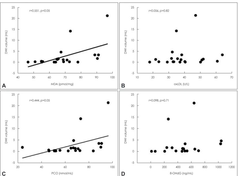
Serum MDA was significantly correlated with the initial DWI lesion volume in the acute stroke group (r=0.551, p<0.05) (Fig. 1A). Serum PCO also increased with increasing DWI lesion volume in the acute stroke patients (r=0.444, p=
0.05) (Fig. 1C). Conversely, oxLDL and 8-OHdG levels were not correlated with DWI lesion volume in the acute stroke group (Fig. 1B and D).
Effects of rosuvastatin on oxidative stress The degree of reduction in oxidative stress markers after statin treatment differed according to the specific marker (Fig. 2).
Specifically, levels of MDA decreased in the acute stroke pa- tients (p<0.05) but not in the chronic stroke patients (p=0.49) (Fig. 2A), whereas the levels of oxLDL were decreased in both of the patient groups (p<0.01 for both) (Fig. 2B). Rosuvastatin did not significantly affect PCO levels in either the acute or chronic stroke group (p=0.97 and 0.08, respectively) (Fig. 2C).
Plasma levels of 8-OHdG were significantly increased after statin treatment (p<0.01) in the acute stroke group, but tended to increase in the chronic stroke group, although that change was not statistically significant (p=0.89) (Fig. 2D).
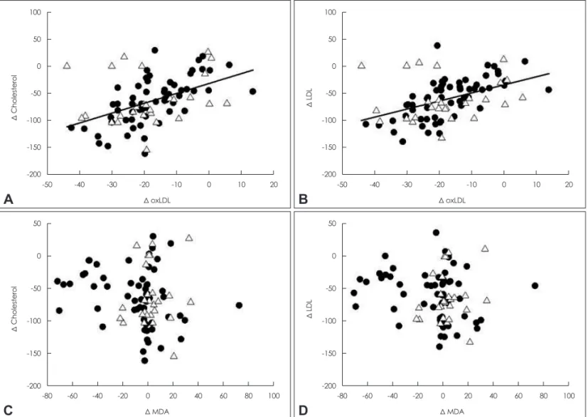
The mechanisms underlying the effects of rosuvastatin on oxidative stress markers were explored by assessing the corre- lations between changes in lipid and oxidative stress markers from the respective pretreatment levels to the posttreatment levels (Fig. 3). The posttreatment reduction of MDA levels was not associated with the observed changes in cholesterol levels
Fig. 1. Correlations between oxidative stress markers and lesion volume on initial DWI in acute stroke patients. Individual values and the linear regression line are displayed. DWI: diffusion-weighted imaging, MDA: malondialdehyde, oxLDL: oxidized low-density lipoprotein, PCO: protein carbonyl content, 8-OHdG: 8-hydroxy-2’-deoxyguanosine.
25 20 15 10 5 0 -5
25 20 15 10 5 0 -5
25 20 15 10 5 0 -5 25 20 15 10 5 0 -5
DWI volume (mL)DWI volume (mL) DWI volume (mL)DWI volume (mL)
40
20
10
50 60 20
40 0 200 400
30 MDA (pmol/mg)
PCO (nmol/mL) 8-OHdG (ng/mL)
oxLDL (U/L) 70
60 600
40 80
80 800
50
90 100 60
100 1000 1200
70 r=0.551, p<0.05
r=0.444, p=0.05 r=0.098, p=0.71
r=0.056, p=0.82
A
C D
B
(Fig. 3C and D). Conversely, significant positive correlations were observed between the changes in oxLDL levels and those in total cholesterol (r=0.479, p<0.01), LDL (r=0.459, p<0.01), and ApoB (r=0.444, p<0.05), suggesting a lipid-low- ering-dependent reduction in serum oxLDL (Fig. 3A and B).
Rosuvastatin had no effect on serum PCO and 8-OHdG lev- els (data not shown).
Discussion
The major findings of this study were as follows: 1) the levels of circulating oxidative stress markers appear to reflect differ- ent aspects of oxidative stress, 2) short-term use of a high-dose statin exerted antioxidant effects against lipid peroxidation but not protein oxidation or DNA damage, and 3) the antioxidant effects of statin against lipid peroxidation occurred via lipid- lowering-dependent and -independent mechanisms.
Reactive oxygen species might play critical roles in brain damage after ischemia and reperfusion by destroying the bal- ance of the redox potential in cells and triggering protein oxi- dation, lipid peroxidation, and DNA damage.15-17 ROS are also involved in the pathogenesis of atherosclerosis. Thus, measure- ment of oxidative stress markers in peripheral blood provides a useful tool for exploring the pathophysiological mechanisms and assessing the effects of antioxidants in stroke patients.
However, the findings regarding the circulating oxidative stress markers and the effects of statin on these markers in stroke pa-
tients are conflicting. The possible reasons for this are three- fold. First, stroke is a heterogeneous condition compared to coronary heart disease. Previous studies have encompassed all major stroke etiological subtypes, including cardioembolic, la- cunar, and even hemorrhagic stroke. Second, and more impor- tantly, different oxidative stress markers may be involved in different clinical settings in patients with ischemic stroke (Ta- ble 3). Few studies have evaluated the kinship between serum oxidative stress markers and the phase of ischemic stroke (i.e., acute and chronic) using a comprehensive approach.18 Finally, various types and doses of statin have been studied; the lipid- lowering and non-lipid-lowering (pleiotropic) effects may dif- fer among individual statins, which would explain the differing results between statin trials. Thus, in the present study the an- tioxidant effects of a high-dose statin were tested using vari- ous markers of oxidative stress in atherosclerotic stroke pa- tients.
The results of this study show that the baseline levels and the degrees of reduction observed after statin use differed among the oxidative stress markers measured. Interestingly, there was no correlation between the baseline levels of the four oxidative stress markers and the changes after statin treatment (data not shown). MDA levels were associated with the index of stroke severity (initial DWI lesion volume), whereas oxLDL was not.
These findings suggest that these two oxidative stress markers reflect different aspects of oxidative stress. Several studies have demonstrated that acute stroke patients possess high serum lev- Fig. 2. The changes in oxidative stress markers before and after statin use. A: Rosuvastatin significantly reduced serum MDA levels in the acute stroke group but not in the chronic stroke group. B: There was a marked reduction in serum oxLDL level in both the acute and chronic stroke groups. C: There was no significant change in PCO in either stroke group after statin use. D: Surprisingly, 8-OHdG levels increased more in the acute stroke group than in the chronic stroke group after statin use. *p<0.05, **p<0.01. MDA: malondialdehyde, oxLDL: oxidized low-density lipoprotein, PCO: protein carbonyl content, Post-Tx: posttherapy, Pre-Tx: before therapy, 8-OHdG: 8-hydroxy-2’-deoxyguanosine.
160 140 120 100 80 60 40 20 0
160 140 120 100 80 60 40 20 0
3500 3000 2500 2000 1500 1000 500 0 160 140 120 100 80 60 40 20 0 160
140 120 100 80 60 40 20 0
160 140 120 100 80 60 40 20 0
3500 3000 2500 2000 1500 1000 500 0 160 140 120 100 80 60 40 20 0
MDA (pmol/mg)PCO (nmol/mL) 8-OHdG (pg/mL)oxLDL (U/L)
MDA (pmol/mg)PCO (nmol/mL) 8-OHdG (pg/mL)oxLDL (U/L)
Pre-Tx
Pre-Tx Pre-Tx
Pre-Tx Pre-Tx
Pre-Tx Pre-Tx
Pre-Tx
* ** **
Post-Tx
Post-Tx Post-Tx
Post-Tx Post-Tx
Post-Tx Post-Tx
Post-Tx Acute
Acute Acute
Acute Chronic
Chronic Chronic
Chronic
* **
A
C D
B
els of MDA,19,20 but few studies have addressed the level of MDA in the chronic stage of stroke, and the obtained results have been controversial.18,21 On the contrary, oxLDL is report- edly increased in patients with atherosclerosis and is associat- ed with the progression and severity of that condition.22,23 Nev- ertheless, few studies have focused on the levels of oxLDL in acute stroke patients.24
The present findings also show that statin exerts an antioxi- dant action against lipid peroxidation via both lipid-lowering-
dependent (by reducing oxLDL) and -independent (by reduc- ing MDA) mechanisms. Several studies have indicated that therapy with statins could reduce lipoprotein oxidation and ameliorate free-radical injury. In general, plasma levels of ox- LDL are significantly correlated with the total levels of LDL.
As expected, statin therapy reduced the circulating oxLDL, an effect that was dependent upon the degree of LDL reduction.25,26 There was also no significant relationship between the chang- es in MDA and cholesterol levels. There have been conflicting Table 3. Changes in oxidative stress markers of ischemic cerebrovascular disease; a literature review
Marker Clinical setting
Subclinical (atherosclerosis) Acute stroke Chronic stroke
MDA Increased18-20,30,35,36 Increased18,21
No change36
oxLDL Increased22,37,38
Reduced with statin38
Increased24 Increased21
PCO Increased18
No change30
No change18
8-OHdG Increased15,* 31,* 32*
*Animal model of transient middle cerebral artery occlusion.
MDA: malondialdehyde, oxLDL: oxidized low-density lipoprotein, PCO: protein carbonyl content, 8-OHdG: 8-hydroxy-2’-deoxyguanosine.
Fig. 3. Correlation between changes in the levels of lipids and markers of oxidative stress. Black circle and white triangle signify acute and chronic stroke patients, respectively. Individual values and linear regression lines are displayed. LDL: low-density lipoprotein, MDA: malo- ndialdehyde, oxLDL: oxidized low-density lipoprotein.
100 50 0 -50 -100 -150 -200
50 0 -50 -100 -150 -200
100 50 0 -50 -100 -150 -200
50 0 -50 -100 -150 -200
Δ CholesterolΔ Cholesterol Δ LDLΔ LDL
-50
-80
-50
-80 -40
-60
-40
-60 -30
-40 -20
-30
-40 -20 Δ oxLDL
Δ MDA
Δ oxLDL
Δ MDA -20
0
-20
0 -10
20
-10
20 0
40 60
0
40 60
10
80
10
80 20
100
20
100
A
C
B
D
results regarding the concordant decrease of MDA and the de- crease in cholesterol levels. Specifically, one study showed that with statin therapy MDA levels reduced in parallel with reduc- tions in cholesterol levels,27 whereas in another study there was no significant decrease in MDA levels after statin use, despite a significant reduction in cholesterol levels.28 MDA is a highly reactive, three-carbon dialdehyde that is produced as a byprod- uct of polyunsaturated fatty acid peroxidation.29 It can thus be assumed that the MDA-lowering mechanism of statin is a by- product of peroxidation.
The present study found that markers of protein oxidation but not of DNA damage were changed in patients with isch- emic stroke. The most widely studied marker of protein oxida- tion is PCO. High levels of protein carbonyls have been report- ed in several neurological disorders.16,18,30 Plasma levels of 8-OHdG, which has been employed as a marker of oxidative DNA damage, were found to be increased in an animal model of ischemic stroke;15,31,32 however, there have been no studies of the plasma level of 8-OHdG in stroke patients.
Protein carbonyl content and 8-OHdG have been investigat- ed in only a small number of studies. Several preclinical stud- ies demonstrated that statin exerts antioxidant effects by pre- venting protein oxidation or DNA damage due to scavenging hydroxyl radicals.33 However, in the present study, statin ther- apy did not reduce either protein oxidation or oxidative DNA damage in the plasma of stroke patients. Several reports have suggested that proteins and lipids interact during the oxidation process. The presence of protein carbonyl is not necessarily an indication of direct oxidation of amino acid residues in pro- teins, and may occur via the Michael addition reaction with products of lipid peroxidation.29 In addition, lipid-dependent carbonylation is inhibited by antioxidants, while lipid-indepen- dent protein carbonylation is antioxidant-insensitive.34 Based on the data obtained in the present study, it appears that statin therapy does not reduce protein carbonylation by lipid-inde- pendent direct oxidation. Further studies are needed to estab- lish the antioxidant effects against protein oxidation and DNA damage after long-term statin use.
The present study was subject to some limitations. First, a relatively small cohort from a single center was studied. That being said, the cohort comprised a relatively homogeneous group of patients, all of whom had been diagnosed with ath- erosclerotic stroke after an extensive work-up for stroke mechanisms, for which the current guidelines recommend the use of a statin.8 Second, these results represent the short-term effects of statin therapy, and long-term follow-up data are needed. However, stroke recurrence and neuronal damage as a result of oxidative stress occur most frequently soon after an index stroke, suggesting the importance of short-term an- tioxidant therapy. Finally, a high dose of rosuvastatin was
used in the present study because it is well known that inten- sive statin therapy may further reduce the risk of stroke com- pared to less-intensive therapy. However, the antioxidant ef- fects may differ among the various statins, and the pleiotropic effects of rosuvastatin are less well known.
In conclusion, the results of this study suggest the need for a comprehensive approach in the study of oxidative stress mark- ers. Short-term use of a high-dose statin has not only lipid- lowering and LDL-dependent antioxidant effects, but also ex- erts antioxidant effects against lipid peroxidation in an acute setting (e.g., reperfusion injury or acute ischemic injury) via LDL-independent mechanisms. Further studies are needed to establish the long-term antioxidant effects of statins in patients with atherosclerotic stroke.
Conflicts of Interest
The authors have no financial conflicts of interest.
Acknowledgements
This work was supported by the Korea Healthcare Technology R&D Project, Ministry of Health & Welfare (A110208), the Research Fellow Program of Sungkyunkwan University (2013).
REFERENCES
1. Kelly PJ, Morrow JD, Ning M, Koroshetz W, Lo EH, Terry E, et al.
Oxidative stress and matrix metalloproteinase-9 in acute ischemic stroke: the Biomarker Evaluation for Antioxidant Therapies in Stroke (BEAT-Stroke) study. Stroke 2008;39:100-104.
2. Sánchez-Moreno C, Dashe JF, Scott T, Thaler D, Folstein MF, Martin A. Decreased levels of plasma vitamin C and increased concentrations of inflammatory and oxidative stress markers after stroke. Stroke 2004;
35:163-168.
3. Leinonen JS, Ahonen JP, Lönnrot K, Jehkonen M, Dastidar P, Molnár G, et al. Low plasma antioxidant activity is associated with high lesion volume and neurological impairment in stroke. Stroke 2000;31:33-39.
4. Floyd RA, Hensley K. Oxidative stress in brain aging. Implications for therapeutics of neurodegenerative diseases. Neurobiol Aging 2002;23:
795-807.
5. Tuñón J, Martín-Ventura JL, Blanco-Colio LM, Egido J. Mechanisms of action of statins in stroke. Expert Opin Ther Targets 2007;11:273- 6. Amarenco P, Bogousslavsky J, Callahan A 3rd, Goldstein LB, Henne-278.
rici M, Rudolph AE, et al. High-dose atorvastatin after stroke or tran- sient ischemic attack. N Engl J Med 2006;355:549-559.
7. Ridker PM, Danielson E, Fonseca FA, Genest J, Gotto AM Jr, Kastelein JJ, et al. Rosuvastatin to prevent vascular events in men and women with elevated C-reactive protein. N Engl J Med 2008;359:
2195-2207.
8. Adams RJ, Albers G, Alberts MJ, Benavente O, Furie K, Goldstein LB, et al. Update to the AHA/ASA recommendations for the preven- tion of stroke in patients with stroke and transient ischemic attack.
Stroke 2008;39:1647-1652.
9. Bang OY, Ovbiagele B, Liebeskind DS, Restrepo L, Yoon SR, Saver JL. Clinical determinants of infarct pattern subtypes in large vessel atherosclerotic stroke. J Neurol 2009;256:591-599.
10. Hennerici MG. Report of the 20th European Stroke Conference, Ham- burg, May 24-27, 2011. Cerebrovasc Dis 2011;32:589-613.
11. LaRosa JC. Pleiotropic effects of statins and their clinical significance.
Am J Cardiol 2001;88:291-293.
12. Lima JA, Desai MY, Steen H, Warren WP, Gautam S, Lai S. Statin-in- duced cholesterol lowering and plaque regression after 6 months of magnetic resonance imaging-monitored therapy. Circulation 2004;110:
2336-2341.
13. Ay H, Furie KL, Singhal A, Smith WS, Sorensen AG, Koroshetz WJ.
An evidence-based causative classification system for acute ischemic stroke. Ann Neurol 2005;58:688-697.
14. Levine RL, Williams JA, Stadtman ER, Shacter E. Carbonyl assays for determination of oxidatively modified proteins. Methods Enzymol 1994;233:346-357.
15. Nagayama T, Lan J, Henshall DC, Chen D, O’Horo C, Simon RP, et al. Induction of oxidative DNA damage in the peri-infarct region after permanent focal cerebral ischemia. J Neurochem 2000;75:1716-1728.
16. Moon GJ, Shin DH, Im DS, Bang OY, Nam HS, Lee JH, et al. Identi- fication of oxidized serum albumin in the cerebrospinal fluid of isch- aemic stroke patients. Eur J Neurol 2011;18:1151-1158.
17. de Nigris F, Lerman A, Ignarro LJ, Williams-Ignarro S, Sica V, Baker AH, et al. Oxidation-sensitive mechanisms, vascular apoptosis and atherosclerosis. Trends Mol Med 2003;9:351-359.
18. Corrêa Mde C, Maldonado P, da Rosa CS, Lunkes G, Lunkes DS, Kai- zer RR, et al. Oxidative stress and erythrocyte acetylcholinesterase (AChE) in hypertensive and ischemic patients of both acute and chronic stages. Biomed Pharmacother 2008;62:317-324.
19. Demirkaya S, Topcuoglu MA, Aydin A, Ulas UH, Isimer AI, Vural O.
Malondialdehyde, glutathione peroxidase and superoxide dismutase in peripheral blood erythrocytes of patients with acute cerebral ischemia.
Eur J Neurol 2001;8:43-51.
20. Polidori MC, Cherubini A, Stahl W, Senin U, Sies H, Mecocci P. Plas- ma carotenoid and malondialdehyde levels in ischemic stroke patients:
relationship to early outcome. Free Radic Res 2002;36:265-268.
21. Alexandrova ML, Bochev PG, Markova VI, Bechev BG, Popova MA, Danovska MP, et al. Oxidative stress in the chronic phase after stroke.
Redox Rep 2003;8:169-176.
22. Ishigaki Y, Katagiri H, Gao J, Yamada T, Imai J, Uno K, et al. Impact of plasma oxidized low-density lipoprotein removal on atherosclerosis.
Circulation 2008;118:75-83.
23. Gil-Núñez AC, Villanueva JA. Advantages of lipid-lowering therapy in cerebral ischemia: role of HMG-CoA reductase inhibitors. Cerebro- vasc Dis 2001;11 Suppl 1:85-95.
24. Uno M, Harada M, Takimoto O, Kitazato KT, Suzue A, Yoneda K, et al. Elevation of plasma oxidized LDL in acute stroke patients is associ- ated with ischemic lesions depicted by DWI and predictive of infarct enlargement. Neurol Res 2005;27:94-102.
25. Ky B, Burke A, Tsimikas S, Wolfe ML, Tadesse MG, Szapary PO, et al. The influence of pravastatin and atorvastatin on markers of oxida-
tive stress in hypercholesterolemic humans. J Am Coll Cardiol 2008;
51:1653-1662.
26. Mason RP, Walter MF, Jacob RF. Effects of HMG-CoA reductase in- hibitors on endothelial function: role of microdomains and oxidative stress. Circulation 2004;109(21 Suppl 1):II34-II41.
27. Skrha J, Stulc T, Hilgertová J, Weiserová H, Kvasnicka J, Ceska R. Ef- fect of simvastatin and fenofibrate on endothelium in Type 2 diabetes.
Eur J Pharmacol 2004;493:183-189.
28. Molcányiová A, Stancáková A, Javorský M, Tkác I. Beneficial effect of simvastatin treatment on LDL oxidation and antioxidant protection is more pronounced in combined hyperlipidemia than in hypercholes- terolemia. Pharmacol Res 2006;54:203-207.
29. Burcham PC, Kuhan YT. Introduction of carbonyl groups into proteins by the lipid peroxidation product, malondialdehyde. Biochem Biophys Res Commun 1996;220:996-1001.
30. Chang CY, Lai YC, Cheng TJ, Lau MT, Hu ML. Plasma levels of an- tioxidant vitamins, selenium, total sulfhydryl groups and oxidative products in ischemic-stroke patients as compared to matched controls in Taiwan. Free Radic Res 1998;28:15-24.
31. Liu H, Uno M, Kitazato KT, Suzue A, Manabe S, Yamasaki H, et al.
Peripheral oxidative biomarkers constitute a valuable indicator of the severity of oxidative brain damage in acute cerebral infarction. Brain Res 2004;1025:43-50.
32. Li S, Zheng J, Carmichael ST. Increased oxidative protein and DNA damage but decreased stress response in the aged brain following ex- perimental stroke. Neurobiol Dis 2005;18:432-440.
33. Aydin S, Uzun H, Sozer V, Altug T. Effects of atorvastatin therapy on protein oxidation and oxidative DNA damage in hypercholesterolemic rabbits. Pharmacol Res 2009;59:242-247.
34. Blakeman DP, Ryan TP, Jolly RA, Petry TW. Protein oxidation: exami- nation of potential lipid-independent mechanisms for protein carbonyl formation. J Biochem Mol Toxicol 1998;12:185-190.
35. Cano CP, Bermúdez VP, Atencio HE, Medina MT, Anilsa A, Souki A, et al. Increased serum malondialdehyde and decreased nitric oxide within 24 hours of thrombotic stroke onset. Am J Ther 2003;10:473- 36. Zimmermann C, Winnefeld K, Streck S, Roskos M, Haberl RL. Anti-476.
oxidant status in acute stroke patients and patients at stroke risk. Eur Neurol 2004;51:157-161.
37. Steinberg D, Parthasarathy S, Carew TE, Khoo JC, Witztum JL. Be- yond cholesterol. Modifications of low-density lipoprotein that in- crease its atherogenicity. N Engl J Med 1989;320:915-924.
38. Rosenson RS. Statins in atherosclerosis: lipid-lowering agents with an- tioxidant capabilities. Atherosclerosis 2004;173:1-12.