https://doi.org/10.9721/KJFST.2021.53.3.348
348
©The Korean Society of Food Science and Technology
Isolation and characterization of bacteriophage infecting
Lactobacillus plantarum KCCM 12116
Jiyoung Oh1 and Jong-Hyun Park1,*
1Department of Food Science and Biotechnology, Gachon University
Abstract Bacteriophages (phages) are known determinants of kimchi microbial ecology. Lactobacillus plantarum is related to kimchi over-acidification during the late stages of kimchi fermentation. A phage infecting Lac. plantarum was isolated from kimchi and characterized. The phage population for kimchi in a market was 2.3 log particles/mL, which corresponded to 32% of the bacterial population on a log scale. The isolated phage was designated as ΦLP12116. ΦLP12116 which belonged to the Siphoviridae family and has a very narrow host range, infecting only Lac. plantarum. The phage was stable at a lactic acid concentration of 1.0% and pH 4.0 at 4oC, indicating that it could survive in kimchi. In the kimchi extract broth treated by the phage, the growth of Lac. plantarum KCCM 12116 was inhibited by 2.2 log CFU/mL compared to the growth in non-phage-treated broth. Therefore, this study suggests that the growth of Lac. plantarum, which is known as an acid-producing strain during late fermentation in kimchi, may be controlled using the phage.
Keywords: kimchi, bacteriophage, Lactobacillus plantarum, biocontrol
Introduction
Bacteriophages (phages) are viruses that infect specifically bacteria and propagate on the host. Phages are widely present in all habitats where bacterial hosts exist. Phages exist as approximately 1031 particles on Earth, about 10 times more than the number of bacteria, which may be considered to be the most abundant microbiomes (Weitz et al., 2013). Of all characterized phages, most phages of 96% belong to the order Caudovirales, known as tailed phages. Based on the classification by the International Committee on Taxonomy of Viruses (Acker HW, 1999), the order Caudovirales consists of three families depending on tail morphology: Podoviridae have short non-contractile tails, Siphoviridae have long non-contractile tails, and Myoviridae have long contractile tails. Phages can be classified into two types for their life cycles: lytic and lysogenic cycles. For the lytic cycle, the phages attach to the host bacteria and inject their genetic materials into the cell. The injected genetic materials are replicated and progeny phages are produced, and released from host cells. For the lysogenic cycle, the phages exist as prophages that are inserted into the chromosome of the host bacteria, and the phage is replicated along with the genome of the host cell (Wang et al., 2019). However, when the prophage is exposed to stressful environmental conditions such as UV, high temperature, or high acidity, the lysogenic cycle is switched to the lytic cycle, and the lytic cycle proceeds (Lunde et al., 2005).
Various phages are present in foods fermented by lactic acid bacteria (LAB) and many studies have reported on phages isolated from fermented foods. Phages from cucumber pickles, fermented sausages, kimchi, and sauerkraut target Weissella, Leuconostoc, and Lactobacillus, respectively (Kleppen et al., 2012; Lu et al., 2003; Pringsulaka et al., 2011). Phage studies on sauerkraut and cucumber fermentation have demonstrated that the emergence of a new phage infecting LAB can affect LAB ecology (Jung et al., 2011; Lu et al., 2012). This indicates that the presence of LAB phages can influence the type and number of LAB during fermentation so that phages are closely correlated with a microbial succession of LAB (Lu et al., 2012).
Kimchi is one of the traditional fermented foods in Korea and known to be a health-promoting food. The components are cabbage, radish, ginger, green onion, and garlic, according to the regions and manufacturers, and fermented mainly by lactic acid bacteria. Among the various bacteria, LAB are the principal ones involved in kimchi fermentation. Most LAB strains are obligate or facultative heterolactic fermentative bacteria that can grow at low temperatures and in acidic environments. They make a better kimchi taste than homolactic fermentative LAB by producing acetate, lactate, ethanol, and CO2 (Kim et al., 2016). Previous studies have shown that the heterolactic genera Weissella and Leuconostoc are dominant at the initial and mid-stages of fermentation; since then, the homolactic genus Lactobacillus is shown to be predominant at the late stage of fermentation (Park et al., 2012). In particular, Lac. plantarum is responsible for the increased acidity and over-acidification of kimchi (Lee and Lee, 2010). Such progression results in sour taste, off-odor, and texture softening, influencing to the quality deterioration of kimchi (Lee and Byun, 2007).
It is important to prevent the deterioration of kimchi caused by over-acidification and homolactic fermentation in order to maintain *Corresponding author: Jong-Hyun Park, Department of Food
Sci-ence and Biotechnology, Gachon University, Seongnam 13120, Korea
Tel: +82-31-750-5523 Fax: +82-31-750-5238 E-mail: [email protected]
Received Marh 12, 2021; revised April 22, 2021; accepted April 23, 2021
the high quality of kimchi. The methods such as refrigeration, heat treatment, gamma irradiation, and food additives have been studied extensively (Han and Kang, 2004; Park et al., 2008; Shon and Lee, 1998). Furthermore, applications of Leu. mesenteroides, other lactic acid bacteria, nisin, and lytic enzymes have been used to kill Lac. plantarum (Chang and Chang, 2010; Lee and Lee, 2011). However, such treatments are not being commercialized because of the degradation of sensory quality and safety concerns associated with them. A biological trial using the bacterial enemy, phage, might be a way to control Lac. plantarum by growth inhibition for improving kimchi flavor.
Therefore, in this study, a phage infecting Lac. plantarum was isolated and characterized for the control of Lac. plantarum.
Materials and Methods
Kimchi samples and enumeration of microbes
Twenty kimchi samples, including cabbage kimchi, radish kimchi, green onion kimchi, Chonggak-kimchi with young radish, and Nabak-kimchi with cut radish in water were purchased from local markets in the Seoul area.
For total bacterial count, 25 g sample was mixed with 225 mL of 0.85% saline in a sampling bag (3M, St. Paul, MN, USA), homogenized using a stomacher (B&F Korea, Seoul, Korea), and diluted decimally in a saline solution. Appropriately diluted solutions were spread on plate count agar (PCA; Difco, Osi, Elancourt, France) and incubated overnight at 37oC, then counted as CFU/mL. To count lactic acid-producing bacteria, the homogenized sample was diluted and spread on bromocresol purple (BCP) late count agar (Eiken Chemical, Tokyo, Japan). The plates were incubated with a gas pack (BD, Franklin Lakes, New Jersey, USA) at 30oC for 24 h. After re-streaking on MRS agar plates (BD), yellowish colonies were counted.
To count bacteriophages in kimchi, the samples were filtered using a sampling bag (3M) and centrifuged at 8,000×g for 10 min. The supernatant was filtered through a 0.22-µm syringe filter (Sartorius, Goettingen, Germany). A 0.45-µm cellulose nitrate membrane filter (Whatman, GE, Germany) was placed on the aspirator and a 0.02 µm AnodiscTM 25 (Whatman) was placed on it. One milliliter of purified sample diluted properly was added to the Anodisc and filtered under vacuum. SYBRTM Gold nucleic acid gel stain (×1,000, Invitrogen, Carlsbad, CA, USA) was diluted to 1/10, and 2 μL of this solution was mixed with 78 μL of diethyl pyrocarbonate water (Bioneer, Daejeon, Korea). The filtered Anodisc was dyed with 80 μL of SYBR Gold for 15 min in a dark room. Subsequently, the Anodisc was exsiccated with Whatman paper until opaque forms were placed on the glass slide. To prevent the fluorescence from becoming blurred, ProlongTM Gold antifade reagent (Invitrogen) was added to the Anodisc, and cover glass was placed on top. Anodiscs were analyzed with a microscope (OPTIKA Srl, Ponteranica, Italy) at 1,000× magnification and at least 25 mesh out of 100 mesh were counted using a lens comprising 10×10 mesh. Three points of the Anodisc were randomly selected and counted. Phages per one mL were then calculated using the formula reported by Ortman and Suttle (2009).
Isolation of Lac. plantarum phage, spot assay, and efficiency of plating (EOP)
As a host for phage isolation, Lac. plantarum KCCM 12116 cells were cultured and harvested. MRS soft agar (0.7%) with a culture of 2% was overlaid on an MRS agar plate (Oxoid), and then the collected sample filtrate was applied. After overnight incubation, the plaques were collected as phage isolates.
For phage propagation, the phage filtrate on MRS and bacterial culture (1:1) were combined into a mixture. After 18 h of incubation, the culture was centrifuged, and the supernatant was filtered. MRS soft agar with 2% bacterial culture was overlaid on an MRS agar plate (Oxoid), and 10 μL of the filtrate was spotted. After overnight incubation, the appearance of plaques was considered to indicate the presence of phage (Manohar et al., 2018).
The host range for the isolated phage was tested against Weissella, Leuconostoc, and Lactobacillus. All the strains were incubated at 30oC for 18 h. One hundred microliters of each overnight culture was inoculated into 5 mL of MRS soft agar and the mixture was overlaid on MRS plate (Oxoid). After drying the plates for a few minutes, 10 μL of phage solution was spotted on the double-layer agar plate, and the plates were incubated overnight at 30oC. The host range of phages was determined on the basis of plaque formation, and plaque clarity was assessed according to the previous study.
The efficiency of plating (EOP) was defined as the ratio of plaque forming unit (PFU)/mL on the target bacteria to the number of host bacteria (Kutter, 2009).
Transmission electron microscopy and one-step growth curve assay
The morphological characteristics of the phage were analyzed using transmission electron microscopy (TEM). The phage solution was concentrated to 10-11 log PFU/mL by centrifuging at 26,000×g for 1 h. Briefly, 20 μL of phage suspension was attached to a 200 mesh carbon-coated copper grid (Ted Pella, Redding, CA, USA) for 2 min and negatively stained with 2% uranyl acetate for 1 min. The grid was then washed with sterilized distilled water and dried. The samples were examined using TEM (H-7600, Hitachi, Tokyo, Japan) at an operating voltage of 80 kV. Phages were classified according to the International Committee on the Taxonomy of Viruses.
A one-step growth curve assay was performed as described previously, with some modifications (Renata et al., 1993). Host bacteria were grown in MRS broth at 30oC for 18 h. One milliliter of the host was cultured overnight, and 1 mL of phage solution was mixed in 8 mL MRS broth at an appropriate MOI. The mixed solution was allowed to adsorb at 30oC for 10 min. After phage adsorption, the mixture was centrifuged at 8,000×g for 10 min. The supernatant containing free phages was discarded, and the pellet containing the adsorbed phage was re-suspended in 10 mL of fresh MRS broth and incubated at 30oC. Three sets of samples were obtained at 5 min intervals for up to 110 min and immediately titrated using the double-layer agar plate method. Through this assay, the latent period, burst size, and one cycle of the isolated
phage were calculated. All experiments were performed in triplicates.
Stability of ΦLP12116 after exposure to temperatures, salt concentrations, pH values, and organic acids
The effect of high temperature on the phage was assessed at various temperatures using a heat block (FINEPCR, Gunpo, Korea). The phage solution (9 log PFU/mL) was exposed to 50, 60, 70, and 80oC for 30 min and the samples were taken every 5 min. Aliquots (10 μL) of the phage solution were spotted on a lawn of MRS agar. After drying and incubating at 30oC for 18 h, the phage titer was determined.
To examine phage stability with regard to salinity, the phage was inoculated into MRS broth adjusted to the desired salinity. Phages of 9 log PFU/mL were suspended in 1, 5, and 10% salinity in MRS broth and incubated at 4 or 30oC for 48 h. The number of surviving phages was counted using the top agar overlay method (Manohar et al., 2018) and the results were represented as percentage survivability.
The phage (1%) was added to MRS broth adjusted to pH 3, 4, and 5, and incubated at 4 or 30oC for 48 h. After incubation, viable phages were immediately diluted and counted using the spotting method. The results are represented as the percentage viability.
For the organic acid stability test, the phage was exposed to various organic acid conditions. Lactic acid and acetic acid were selected and used to make an organic acid environment like kimchi with MRS broth. The phage lysate (1%) was suspended in MRS broth containing organic acid and incubated at 4 or 30oC for 48 h. After 0, 24, and 48 h of incubation, the solution was serially diluted and spotted on the double-layered MRS agar by the spotting method. The number of plaques was counted, and the surviving phages were expressed as a percentage of the initial phages.
Growth of Lac. plantarum with template ΦLP12116 for kimchi fermentation
To evaluate the bacterial control ability of ΦLP12116 for Lac. plantarum in the kimchi environment, ΦLP12116 and its host bacteria were inoculated into kimchi broth, and the number of bacteria was counted. Lac. plantarum KCCM 12116 were diluted to 4 log CFU/mL and mixed with ΦLP12116 at an appropriate MOI. MOI was determined based on the results of the growth inhibition assay as MOI 100. The host bacteria-phage mixture (1%) was inoculated into kimchi broth and incubated at 4oC, similar to the temperature of the refrigerator. To prepare the kimchi extract broth medium, Nabak-kimchi was manufactured, stomached, gauze-filtered, and sterilized (Kong et al., 2005). The number of bacteria and phages were counted by spreading on MRS agar and soft agar overlay plaque assay during the incubation. Non-phage-treated bacterial culture was used as a control. All experiments were performed in triplicate.
Results and Discussion
Microbe and bacteriophage populations of kimchi in a market
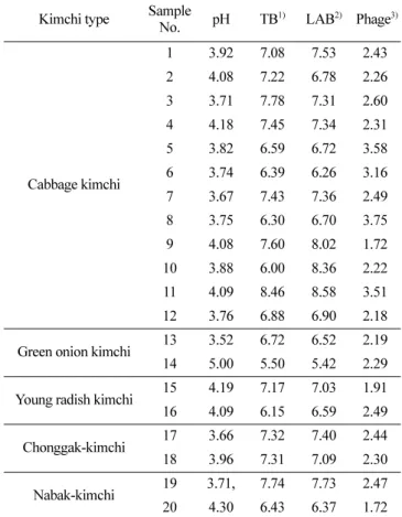
Twenty kimchi samples were collected from the local market and analyzed for total bacteria (TB), lactic acid bacteria (LAB), and bacteriophages (phages) (Table 1). The pH values ranged from 3.5 to 5.0, indicating a ripened state (Mheen and Kwon, 1984). The total bacterial counts of the samples were 5.5-8.5 log CFU/ mL, and the LAB counts were 5.4-8.6 log CFU/mL with a mean value of 7.1 log CFU/mL. The numbers of TB and LAB indicated that most of the bacteria present in kimchi seemed to be almost LAB. Total phages were 1.7-3.8 log particles/mL (mean 2.3 log particles/mL) and relatively lower in quantity than the total bacteria and LAB. The counts of phage amounted to 32% of LAB population in log scale. The total number of phages in the kimchi as assessed by epifluorescence microscopy showed no correlation with the number of bacteria regardless of the type of kimchi. Phages are reported to be a major population in the environment and greatly influence microbial ecology (Lu et al., 2012; Jung et al., 2011; Weitz et al., 2013). They have been detected in kimchi
Table 1. Distribution of total bacteria (TB), lactic acid bacteria (LAB), and phage
Kimchi type Sample
No. pH TB 1) LAB2) Phage3) Cabbage kimchi 1 3.92 7.08 7.53 2.43 2 4.08 7.22 6.78 2.26 3 3.71 7.78 7.31 2.60 4 4.18 7.45 7.34 2.31 5 3.82 6.59 6.72 3.58 6 3.74 6.39 6.26 3.16 7 3.67 7.43 7.36 2.49 8 3.75 6.30 6.70 3.75 9 4.08 7.60 8.02 1.72 10 3.88 6.00 8.36 2.22 11 4.09 8.46 8.58 3.51 12 3.76 6.88 6.90 2.18
Green onion kimchi 13 3.52 6.72 6.52 2.19
14 5.00 5.50 5.42 2.29
Young radish kimchi 15 4.19 7.17 7.03 1.91
16 4.09 6.15 6.59 2.49
Chonggak-kimchi 17 3.66 7.32 7.40 2.44
18 3.96 7.31 7.09 2.30
Nabak-kimchi 19 3.71, 7.74 7.73 2.47
20 4.30 6.43 6.37 1.72
Symbols: 1)TB and 2)LAB with conventional culture method by log CFU/mL. 3)Phage with epifluorescence microscopy by log particle/mL.
and fermented foods, mainly as LAB phages for Weissella, Pediococcus, Lactobacillus, and Leuconostoc (Jung et al., 2011; Kleppen et al., 2012; Lu et al., 2003; Pringsulaka et al., 2011). Recently, the phage population has been enumerated by direct counting using flow cytometry and epifluorescence microscopy in kimchi. The phage showed an average count of 2.1 log particles/ mL per kimchi soup and seemed to be 28% log scale to the total bacterial count (Park WJ, 2017; Kong SJ, 2019). Therefore, the phage population on a commercial kimchi market was 2.3 log particle/mL and corresponded to LAB population of 32% similarly to previous reports.
Phage isolation for Lac. plantarum and determination of host range by spot assay and efficiency of plat-ing (EOP)
The phage for Lac. plantarum KCCM 12116 was isolated from Nabak-kimchi and designated as ΦLP12116. The host range of the isolated phage was determined by confirming the formation of plaques using the spot assay and EOP (Table 2). ΦLP12116 had a narrow host range and limited only to Lac. plantarum, indicating that lysis occurred only within the same species. Infection with ΦLP12116 to Lac. plantarum KCTC 3108 and Lac. plantarum KCCM 12116 formed clear plaques with a faintly hazy background, but infection to Lac. plantarum ATCC 8014 resulted in slight turbidity on the plaque. Interestingly, plaques showed a typical bulls-eye appearance on Lac. plantarum KCCM 12116 via lysogenicity and greater turbidity was generally observed at the center (Jurczak-Kurek et al., 2016). The different plaques on such hosts may be due to infection resistance or mechanisms (Ali et al., 2014; da Silva Duarte et al., 2018; van Houte et al., 2016). Regarding as phage infection on LAB, two-step processes as reversible interaction through a surface carbohydrate moiety and then irreversible interaction between host receptor and phage receptor-bind protein have been suggested (Baptista et al., 2008; Mahony et al., 2017). There are a broad array of components such as carbohydrates, proteins, lipoteichoic acids, and wall teichoic acid on the surface, which may be diverse among the strains or modified to the environmental factors (Ainsworth et al., 2014). Thus, host-phage may interact strain-specifically.
There are reports that phages for Weissella and Leuconostoc
show a broad host infection range on species and genera of Weissella, Leuconostoc, and Lactobacillus (Kong and Park, 2020; Lu et al., 2012; Pujato et al., 2017). However, the temperate Lac. plantarum phages ΦLP1-A, ΦLP1-B, and ΦLP2 have been reported to have a host range limited to Lac. plantarum strains (Caso et al., 1995). Here, ΦLP12116 seemed to have limited host infection only for Lac. plantarum, similar to other Lac. plantarum phages. EOP analysis was also carried out to identify the host with the highest infection rate (Table 2). ΦLP12116 showed different infection efficiencies against target bacteria and most infection to Lac. plantarum KCCM 12116. In summary, determination of host range for ΦLP12116 indicated that Lac. plantarum KCCM 12116 was the best host for infection.
Morphological and culture characteristics of ΦLP12116 Morphological characteristic of ΦLP12116 was analyzed using TEM and shown in Fig. 1. ΦLP12116 had a 71.0±6.0 nm icosahedral head and a 275.1±5.3 nm long non-contractile tail. The head and tail of ΦLP12116 were relatively long. ΦLP12116 belonged to the Siphoviridae family, according to the International Committee on Taxonomy of Viruses. ΦLP12116 was similar to Lac. plantarum phage P1 that had a 71.7±3.0 nm isometric capsid and a 272±3.0 nm long non-contractile tail (Chen et al., 2016).
A one-step growth curve assay was performed and the latent period, burst size, and time required for one cycle were determined. ΦLP12116 showed that the latent period was 85 min, and the burst size was 12.9 PFU/infected cell. This explained why ΦLP12116 had a relatively long latent period and a lower burst size than the other phages (Briggiler M et al., 2012; Chen et al., 2016). The time required for one cycle of ΦLP12116 was long by 110 min. Lac. plantarum phage ΦLPN014, isolated from Nham (Thai fermented pork), shows a latent period of about 30 min, which is shorter than that of ΦLP12116, and 150 PFU/infected cell of the burst size, which is higher than that of ΦLP12116 (Rattanachaikunsopon P, 2014).
Viability of ΦLP12116 after exposure to various tem-peratures and salt concentrations
Temperature has been reported to be related to fundamental adsorption for infection (Jonczyk et al., 2011). To investigate Table 2. Host ranges of isolated ΦLP12116 by spot assay and EOP of ΦLP12116
Host strain Plaque EOP Plaque morphology
W. confusa MGB 0333 - -
+++
Leu. citreum K27 - -Leu. mesenteroides B6 - -Leu. mesenteroides B9 - -Leu. mesenteroides K2 - - + Lac. plantarum KCTC 3108 +++ 2.00 × 10⁻¹Lac. plantarum ATCC 8014 + < 9.09 × 10⁻⁷
Lac.plantarum KCCM 12116 +++ 1
Symbols: +++, clear appearance throughout but with a faintly hazy background; +, a few individual plaques or complete turbidity in the spot; -, no plaque.
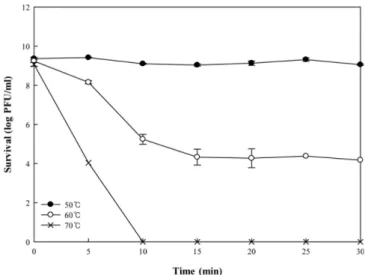
thermal stability, ΦLP12116 was exposed to high temperatures of 50, 60, and 70oC for 30 min (Fig. 2). ΦLP12116 was stable at 50oC, as shown by the fact that the reduction of phage was less than 0.31 log PFU/mL. Other reports on Lac. plantarum phages also have a viability of 95% at 50oC for 30 min (Marcó et al., 2012; Chen et al., 2016). However, the viability of ΦLP12116 was reduced from 9.1 log PFU/mL to 4.0 log PFU/mL within 5 min of heat treatment and completely inactivated out after 10 min at 70oC. Thus, ΦLP12116 would be stable below 50oC such as in the refrigerator.
Salt concentration is widely known as one of the factors affecting kimchi fermentation. It has been reported that kimchi is generally fermented at approximately 2-3% of salt concentration (Mhin and Kwon, 1984). To examine the effect of high salinity on phages, the phage was exposed to three different salinity conditions, and viability was determined. The plaque after exposure at 5% and 10% salinity was shown and the number was not significantly different compared to control exposure. ΦLP12116 showed 92% survival at 10% salinity (data not shown). Accordingly, salinity during kimchi environment might have no influence on phage inactivation.
Viability of ΦLP12116 after exposure to various pH and organic acids
Survival under acidic conditions was investigated at pH 3, 4, and 5 for 48 h. This experiment was performed at two different temperatures of 4oC and 30oC (Fig. 3). The pH stability test indicated that the survival rate of ΦLP12116 was 99.8% after 48 h exposure at pH 5 and 4oC. The survival rate of ΦLP12116 at pH 4 and 4oC was 98.1%, which indicated that the phage was stable at pH 4 and 5 at 4oC. However, the phage was completely inactivated after 24 h of exposure to pH 3 at 4oC. The above results indicated that the phage was stable under acidic conditions above pH 3 at 4oC. However, the stabilities at 30oC were lower than those at 4oC. Therefore, ΦLP12116 might be stable in acidic condition under low temperatures.
ΦLP12116 was exposed to various lactic acid concentrations of 0.1, 0.5, and 1.0%. ΦLP12116 showed a survival rate of 96.4% at 0.1, 0.5, and 1.0% lactic acid at 4oC for 48 h (Fig. 4). No Fig. 1. Morphology of bacteriophage ΦLP12116 by transmission
electron microscopy (50,000× magnification; size bars, 20 nm and 100 nm).
Fig. 2. Viability of bacteriophage ΦLP12116 in log PFU/mL after exposure to 50, 60, and 70oC for 30 min.
significant reduction for the viability of ΦLP12116 occurred at these concentrations. At 30oC, ΦLP12116 exhibited a survival rate of 94.8% with 0.1% lactic acid. ΦLP12116 also showed 76.8% and 28.1% survival in lactic acids of 0.5% and 1%, respectively. These results indicated that the phage was stable at a high lactic acid concentration at 4oC for up to 48 h.
Acetic acid is present in smaller quantities than lactic acid in kimchi. During Dongchimi fermentation, the acetic acid content reaches approximately 0.2% after 30 days, and the range of acetic acid is 0.1-0.2% (Cho et al., 2015). The phage was exposed to acetic acid at 0.1, 0.5, and 1.0% (w/w). At any concentration of acetic acid at 4oC for 48 h, ΦLP12116 exhibited no significant difference in survival rate compared to non-treatment control. However, after 48 h at 30oC, the survival rate of ΦLP12116 decreased to 94.8% at 0.1% acetic acid, 85.5% at 0.5% acetic acid, and 59.5% at 1.0% acetic acid (data not shown). The above results indicated that ΦLP12116 was more sensitive to lactic acid than acetic acid and was inactivated more easily on lactic acid. Phage ΦT25 is also resistant to a wide range of pH; however, no survivors can be detected after incubation at pH 2 for 30 min at 37oC (Sunthornthummas et al., 2017). The viability of ΦLP12116 might be maintained at high organic acid and low temperatures similar to the other reports.
Several mechanisms have been reported on the acid tolerance for LAB, which are the neutralization processes like (a) the arginine dihydrolase system and the malolactic fermentation, (b) the biofilm formation and membrane modification, (c) the proton pump like F1-F0-ATPase and amino acid decarboxylation, and (d) protection and repair of cellular macromolecules (Wang et al., 2018). However, no report is shown for the different tolerance toward lactic and acetic acid, which may come from the different proton dissociation constant of lactic acid (pKa=3.86) and acetic acid (pKa=4.75). Lactic acid is a stronger acid than acetic acid to destroy the cell neutrality more easily when the organic acids come into the cell cytosol.
Growth inhibition of Lac. plantarum by ΦLP12116 in kimchi extract
Various LAB, including W. koreensis, Leu. mesenteroides, Lac. sakei, and Lac. plantarum, are known to be involved in late kimchi fermentation (Lim et al., 1989). In particular, it has been reported that Lac. plantarum produces a large amount of acid, which is related to over-ripened kimchi fermentation (Lee and Lee, 2010). To control the growth of Lac. plantarum, ΦLP12116 and Lac. plantarum KCCM 12116 were co-cultivated. Lac. plantarum was inoculated into kimchi extract broth for 15 days at 4oC, and the population reduction of Lac. plantarum by phage was analyzed (Fig. 5). Lac. plantarum in the non-phage-treated broth grew steadily for 15 days. The number of Lac. plantarum increased slowly until day 5, but after that, bacteria grew rapidly and reached 5.9 log CFU/mL at day 15. However, the number of Lac. Fig. 4. Viability of bacteriophage ΦLP12116 in log PFU/mL after exposure to lactic acid of 0.1, 0.5, and 1.0% at 4oC (A) and 30oC (B) for 24 and 48 h.
Fig. 5. Growth inhibition of Lac. plantarum in kimchi broth by treating bacteriophage of ΦLP12116 at 4oC for 15 days. Symbols; cross, phage number in PFU/mL; closed circle, Lac. plantarum only in CFU/mL; open circle, Lac. plantarum with phage in CFU/mL.
plantarum decreased until day 4 in phage-treated broth. The number was 3.4 log CFU/mL at the start, but became 2.9 log CFU/mL at day 4. The bacteria began to grow again slowly and reached 3.7 log CFU/mL at day 15. During fermentation for 15 days, the number of phages showed no significant change from 8 log PFU particles/mL. Based on these data, it was clear that the growth of Lac. plantarum KCCM 12116 was inhibited by ΦLP12116 in kimchi broth, indicating growth inhibition by ΦLP12116 against Lac. plantarum. However, the number of Lac. plantarum was not reduced to zero level. As a response to phages, bacteria have developed many anti-phage mechanisms such as clustered regularly interspaced short palindromic repeats (CRISPRs) and CRISPR-associated genes (cas) systems, restriction-modification systems, superinfection exclusion (Sie) systems, and abortive infection systems (Labrie et al., 2010). Battle between bacteria and phages leads to the co-evolution between the two entities and then bacteria may be adapted and be resistant to phages.
There are many reports on reducing the over-acidification of kimchi to extend the edible period and maintain its flavor by control of Lac. plantarum. Physical and chemical trials, in addition to starter development and bacteriocin addition, have been conducted, but have not reached a satisfactory solution. This research suggests that growth for Lac. plantarum known as an acid-producing strain at the late fermentation in kimchi might be controlled by using the phage. However, the phage cocktail infecting Lac.platarum strains is needed because of the narrow host spectrum.
Acknowledgment
This research was supported by the National Research Foundation of Korea (grant 2020R1F1A107000111).
Conflict of Interest
The authors have no financial conflict of interest to declare.
References
Acker HW. Tailed bacteriophages: The Order Caudovirales. Adv. Virus Res. 51: 135-201 (1999)
Ainsworth S, Sadovskaya I, Vinogradov E, Courtin P, Guerardel Y, Mahony J, Grard T, Cambillau C, Chapot-Chartier MP, Sinderen DV. Differences in lactococcal cell wall polysaccharide structure are major determining factors in bacteriophage sensitivity. mBio 5: 1-11 (2014)
Ali Y, Koberg S, Heßner S, Sun X, Rabe B, Back A, Neve H, Heller KJ. Temperate Streptococcus thermophilus phages expressing superinfection exclusion proteins of the Ltp type. Front. Micro-biol. 5: 98-98 (2014)
Baptista C, Santos MA, São-José C. Phage SPP1 reversible adsorp-tion to Bacillus subtilis cell wall teichoic acids accelerates virus recognition of membrane receptor YueB. J. Bacteriol. 190: 4989-4996 (2008)
Caso JL, Reyes-Gavilan CGDS, Herrero M, Montilla A, Rodriguez A, Suarez J. Isolation and characterization of temperate and viru-lent bacteriophages of Lactobacillus plantarum. J. Dairy Sci. 78: 741-750 (1995)
Chang JY, Chang HC. Improvement in the quality and shelf life of kimchi by fermentation with the induced bacteriocin-producing
strain, Leuconostoc citreum GJ7 as a starter. J. Food Sci. 75: M103-M110 (2010)
Chen X, Xi Y, Zhang H, Wang M, Fan Y, Wu W. Characterization and adsorption of Lactobacillus virulent phage P1. J. Dairy Sci. 99: 6995-7001 (2016)
Cho JH, Lee SJ, Choi JJ, Chung CH. Chemical and sensory profiles of Dongchimi (Korean watery radish kimchi) liquids based on descriptive and chemical analyses. Food Sci. Biotechnol. 24: 497-506 (2015)
da Silva Duarte V, Giaretta S, Campanaro S, Treu L, Armani A, Tar-rah A, Oliveira de Paula S, Giacomini A, Corich V. A Cryptic non-inducible prophage confers phage-immunity on the Strepto-coccus thermophilus M17PTZA496. Viruses 11: 7 (2019)
Han JS, Kang J. Retardation of Kimchi fermentation by addition of glucono-δ-lacton. J. Korean Soc Food Sci. Nutr. 33: 553-559 (2004)
Jończyk E, Klak M, Międzybrodzki R, Górski A. The influence of external factors on bacteriophages-Review. Folia Microbiol. 56:191-200 (2011)
Jung JY, Lee SH, Kim JM, Park MS, Bae JW, Hahn Y, Madsen EL, Jeon,CO. Metagenomic analysis of kimchi, a traditional Korean fermented food. Appl. Environ. Microbiol. 77: 2264-2274 (2011) Jurczak-Kurek A, Gąsior T, Nejman-Faleńczyk B, Bloch S, Dydecka
A, Topka G, Necel A, Jakubowska-Deredas M, Narajczyk M, Richert M. Mieszkoswska A, Wrobel B, Wegrzn G, Wegryn A. Biodiversity of bacteriophages: Morphological and biological properties of a large group of phages isolated from urban sewage. Sci. Rep. 6: 34338-34354 (2016)
Kim HY, Bong YJ, Jeong JK, Lee S, Kim BY, Park KY. Heterofer-mentative lactic acid bacteria dominate in Korean commercial kimchi. Food Sci. Biotechnol. 25: 541-545 (2016)
Kleppen HP, Holo H, Jeon SR, Nes IF, Yoon SS. Novel Podoviridae family bacteriophage infecting Weissella cibaria isolated from kimchi. Appl. Environ. Microbiol. 78: 7299-7308 (2012)
Kong CS, Bak SS, Rhe SH, Park KY. Standardization of manufac-tured method and lactic acid bacteria growth and CO2 levels of Nabak kimchi at different fermentation temperatures. J. Korean Soc. Food Sci. Nutr. 34: 707-714 (2005)
Kong SJ. Weissella-Leuconostoc succession with bacteriophage during kimchi fermentation and bacteriophage characterization. MS thesis, Gachon University, Seongnam, Korea (2019)
Kong SJ, Park JH. Acid tolerance and morphological characteristics of five Weissella cibaria bacteriophages isolated from kimchi. Food Sci. Biotechnol. 29: 873-8781 (2020)
Kutter E. Phage host range and efficiency of plating. Vol. 1, pp 141-149. In: Bacteriophages Methods and Protocols. Clokie MRJ, Kropinski AM (eds). Humana Press, New York, USA. (2009) Labrie SJ, Samson JE, Moineau S. Bacteriophage resistance
mecha-nisms. Nat. Rev. Microbiol. 8: 317-327 (2010)
Lee K, Lee Y. Effect of Lactobacillus plantarum as a starter on the food quality and microbiota of kimchi. Food Sci. Biotecnol. 19: 641-646 (2010)
Lee KH, Byun MW. Quality changes of kimchi manufactured with sanitized materials by ozone and gamma irradiation during stor-age. J. Korean Soc. Food Sci. Nutr. 36: 216-221 (2007)
Lee KH, Lee JH. Isolation of Leuconostoc and Weissella species inhibiting the growth of Lactobacillus sakei from Kimchi. Korean J. Microbiol. Biotechnol. 39: 175-181 (2011)
Lim CR, Park HK, Han HU. Revaluation of isolation and identifica-tion of Gram-positive bacteria in kimchi. Kor. J. Microbiol. 27: 404-414 (1989)
Lu Z, Breidt F, Plengvidhya V, Fleming HP. Bacteriophage ecology in commercial sauerkraut fermentations. Appl. Environ. Micro-biol. 69: 3192-3202 (2003)
Lu Z, Perez-Diaz IM, Hayes JC, Breidt F. Bacteriophage ecology in a commercial cucumber fermentation. Appl. Environ. Microbiol. 78: 8571-8578 (2012)
Lunde M, Aastveit AH, Blatny JM, Nes IF. Effects of diverse envi-ronmental conditions on ΦLC3 prophage stability in Lactoccocus lactis. Appl. Environ. Microbiol. 71: 721-727 (2005)
Mahony J, Cambillau C, van Sinderen D. Host recognition by lactic acid bacterial phages. FEMS Microbiol. Rev. 41: S16-S26 (2017)
Manohar P, Tamhankar AJ, Lundborg CS, Ramesh N. Isolation, char-acterization and in vivo efficacy of Escherichia phage myPSH1131. PLoS One 13: e0206278 (2018)
Marcó MB, Garneau M, Tremblay JE, Quiberoni D, Moinneau A. Characterization of two virulent phages of Lactobacillus planta-rum. Appl. Environ. Microbiol. 78: 8719-8734 (2012)
Mheen TI, Kwon TW. Effect of temperature and salt concentration on kimchi fermentation. Korean. J. Food Sci. Technol. 16: 443-450 (1984)
Ortman AC, Suttle CA. Determination of virus abundance by epiflu-orescence microsopy. Vol. 1, pp87-95. In: Bacteriophages Meth-ods and Protocols. Clokie MRJ, Kropinski AM (eds). Humana Press, New York, USA. (2009)
Park EJ, Chun J, Cha CJ, Park WS, Jeon CO, Bae JW. Bacterial community analysis during fermentation of ten representative kinds of kimchi with barcoded pyrosequencing. Food Microbiol. 30: 197-204 (2012)
Park JG, Kim JH, Park JN, Kim YD, Kim WG, Lee JW, Hwang HJ, Byun MW. The effect of irradiation temperature on the quality improvement of Kimchi, Korean fermented vegetables, for its shelf stability. Radiat. Phys. Chem. 77: 497-502 (2008)
Park WJ. Succession of lactic acid bacteria and bacteriophage during Dongchimi fermentation and bacteriophage characterization. MS thesis, Gachon University, Seongnam, Korea (2017)
Pringsulaka O, Patarasinpaiboon N, Suwannasai N, Atthakor W, Rangsiruji A. Isolation and characterisation of a novel Podoviri-dae-phage infecting Weissella cibaria N 22 from Nham, a Thai fermented pork sausage. Food Microbiol. 28: 518-525 (2011) Pujato SA, Guglielmotti DM, Martinez-Garcia M, Quiberoni A,
Mojica FJM. Leuconostoc mesenteroides and Leuconostoc pseu-domesenteroides bacteriophages: Genomics and cross-species host ranges. Int. J. Food Microbiol. 257: 128-137 (2017)
Rattanachaikunsopon P, Phumkhachorn P. Bacteriophages ΦLPN014 infecting Lactobacillus plantarum N014, A potential starter cul-ture for NHAM fermentation. Ann. Exp. Bio. 2: 1-7 (2014) Renata GK, Leuschner EKA, Hammes WP. Characterization of a
vir-ulent Lactobacillus sake phage PWH2. Appl. Microbiol. Biotech-nol. 34: 255-260 (1993)
Shon KH, Lee HJ. Effect of high pressure treatment on the quality and storage of kimchi. Int. Food Sci. Technol. 33: 359-365 (1998)
Sunthornthummas S, Doi K, Rangsiruji A, Sarawaneeyaruk S, Pring-sulaka O. Isolation and characterization of Lactobacillus paraca-sei LPC and phage ΦT25 from fermented milk. Food Control 73: 1353-1361 (2017)
van Houte S, Buckling A, Westra ER. Evolutionary ecology of pro-karyotic immune mechanisms. Microbiol. Mol. Biol. R. 80: 745-763 (2016)
Wang C, Cui Y, Qu X. Mechanisms and improvement of acid resis-tance in lactic acid bacteria. Arch. Microbiol. 200: 195-201 (2018)
Wang L, Zhu Z, Qian H, Li Y, Chen Y, Ma P, Gu B. Comparative genome analysis of 15 clinical Shigella flexneri strains regarding virulence and antibiotic resistance. AIMS Microbiol. 5: 205-222 (2019)
Weitz JS, Poisot T, Meyer JR, Flores CO, Valverde S, Sullivan MB, Hochberg ME. Phage-bacteria infection networks. Trends Micro-biol. 21: 82-91 (2013)