Bloodstream Infections and Clinical Significance of Healthcare- associated Bacteremia: A Multicenter Surveillance Study
in Korean Hospitals
Recent changes in healthcare systems have changed the epidemiologic paradigms in many infectious fields including bloodstream infection (BSI). We compared clinical characteristics of community-acquired (CA), hospital-acquired (HA), and healthcare- associated (HCA) BSI. We performed a prospective nationwide multicenter surveillance study from 9 university hospitals in Korea. Total 1,605 blood isolates were collected from 2006 to 2007, and 1,144 isolates were considered true pathogens. HA-BSI accounted for 48.8%, CA-BSI for 33.2%, and HCA-BSI for 18.0%. HA-BSI and HCA- BSI were more likely to have severe comorbidities. Escherichia coli was the most common isolate in CA-BSI (47.1%) and HCA-BSI (27.2%). In contrast, Staphylococcus aureus (15.2%), coagulase-negative Staphylococcus (15.1%) were the common isolates in HA-BSI. The rate of appropriate empiric antimicrobial therapy was the highest in CA-BSI (89.0%) followed by HCA-BSI (76.4%), and HA-BSI (75.0%). The 30- day mortality rate was the highest in HA-BSI (23.0%) followed by HCA-BSI (18.4%), and CA-BSI (10.2%). High Pitt score and inappropriate empirical antibiotic therapy were the independent risk factors for mortality by multivariate analysis. In conclusion, the present data suggest that clinical features, outcome, and microbiologic features of causative pathogens vary by origin of BSI. Especially, HCA-BSI shows unique clinical characteristics, which should be considered a distinct category for more appropriate antibiotic treatment.
Key Words: Bloodstream infection; Bacteremia; Community-acquired; Hospital- acquired; Healthcare-associated
Jun Seong Son1, Jae-Hoon Song2,13, Kwan Soo Ko3,13, Joon Sup Yeom4, Hyun Kyun Ki5, Shin-Woo Kim6, Hyun-Ha Chang6, Seong Yeol Ryu7, Yeon-Sook Kim8, Sook-In Jung9, Sang Yop Shin10, Hee Bok Oh11, Yeong Seon Lee11, Doo Ryeon Chung2, Nam Yong Lee12, and Kyong Ran Peck2 Division of Infectious Diseases1, East-West Neo Medical Center, Kyunghee University School of Medicine, Seoul;
Division of Infectious Diseases2, Samsung Medical Center, Department of Molecular Cell Biology3, Sungkyunkwan University School of Medicine, Seoul; Division of Infectious Diseases4, Kangbuk Samung Hospital, Sungkyunkwan University School of Medicine, Seoul; Division of Infectious Diseases5, Konkuk University Hospital, Seoul; Division of Infectious Diseases6, Kyungpook National University Hospital, Daegu; Division of Infectious Diseases7, Keimyung University Dongsan Medical Center, Daegu; Division of Infectious Diseases8, Chungnam National University Hospital, Daejeon; Division of Infectious Diseases9, Chonnam National University Medical School, Gwangju; Division of Infectious Diseases10, Jeju National University Hospital, Cheju; Center for Infectious Diseases, Korea Centers for Disease Control and Prevention11, Seoul; Department of Laboratory Medicine12, Samsung Medical Center, Sungkyunkwan University School of Medicine, Seoul;
Asian-Pacific Research Foundation for Infectious Diseases (ARFID)13 in Samsung Medical Center, Seoul, Korea Received: 19 May 2009
Accepted: 6 January 2010 Address for Correspondence:
Jae-Hoon Song, M.D.
Division of Infectious Diseases, Samsung Medical Center, Sungkyunkwan University, 81 Irwondong-gil, Gangnam-gu, Seoul 135-710, Korea Tel: 82-2-3410-0320, Fax: 82-2-3410-0328
E-mail: [email protected]
This study was supported by a grant of the Korea Centers for Disease Control and Prevention (Serial number: 2007-E-00129-00, 2007- E00037-00).
DOI: 10.3346/jkms.2010.25.7.992 • J Korean Med Sci 2010; 25: 992-998 Infectious Diseases, Microbiology & Parasitology
INTRODUCTION
Bloodstream infection (BSI) is potentially life-threatening con- dition with a case fatality rate of 30-40% (1). If BSI is clinically suspected, clinicians should choose antibiotics based on the type of infection, underlying disease, patient age, infecting patho- gen, and site of acquisition of infection (2).
BSI can be classified as community-acquired (CA), hospital-
acquired (HA), or healthcare–associated (HCA) depending on the site of acquiring infection and risk factors. Of 3 epidemio- logic categories, HCA-BSI is a recently developed category that is traditionally classified as CA-BSI (3-5).
Clinically, the characteristics of HA-BSI can be separated from those of CA-BSI in many factors such as pathogen distribution, type of infection and their susceptibility patterns. Staphlylococ- cus and catheter-related infection were predominant in HA-BSI,
while Escherichia coli and urinary tract infection were generally predominant in CA-BSI (6-8). With regard to HCA-BSI, previous studies showed that the clinical characteristics were not similar to those of CA-BSI (3, 5, 9). And the susceptibility patterns of HCA-BSI were similar to those of HA-BSI (5, 9). Generally, the concept of HCA-BSI has been accepted to be an independent epidemiologic category of BSI. However, so far the detailed data of clinical characteristics of HCA-BSI were not sufficient. Also, the independent risk factors for mortality of each BSI have not been compared yet.
In this study, we compared clinical characteristics of 3 BSI groups including HCA-BSI, instead of investigating one patho- gen or one source. And we performed additional analysis to evaluate risk factors for mortality according to the epidemio- logic type of infection.
MATERIALS AND METHODS Study design and data collection
This study was a prospective nationwide surveillance of BSI in 9 university hospitals in various regions of Korea from October 2006 to September 2007. The participating institutions included Kyunghee East-West Neo Medical Center (800-bed, Seoul), Sam- sung Medical Center (1,270-bed, Seoul), Kangbuk Samung Hos- pital (620-bed, Seoul), Konkuk University Hospital (820-bed, Seoul), Kyungpook National University Hospital (850-bed, Dae- gu), Keimyung University Dongsan Medical Center (910-bed, Daegu), Chungnam National University Hospital (980-bed, Dae- jeon), Chonnam National University Hospital (850-bed, Gwangju), and Jeju National University Hospital (300-bed, Jeju). The fol- lowing data were collected from all cases of BSI; demographic information, underlying disease, comorbid conditions, micro- biological data, sources of infection, and other important clini- cal parameters at the time of infection. The main clinical out- come was measured by the 30-day mortality rate.
Study population
All blood isolates during the study period from the participating hospitals were reviewed for clinical significance. Cases that show- ed clinical features of Systemic Inflammatory Response Syn- drome (SIRS) were included in this study. SIRS was defined by the presence of at least two of the following signs and symptoms:
tachycardia (pulse rate >90 beats/min); tachypnea (respiratory rate >20/min); fever (body temperature >38°C) or hypothermia (body temperature <36°C); leukocytosis (white blood cell count
>12,000 cells/μL) or leukopenia (white blood cell count <4,000 cells/μL) (10). The site of infection causing BSI was estimated by infectious disease specialist at participating institutions. All underlying diseases and comorbid conditions for each patient were included in the analysis. And only the first bacteremic epi- sode for each patient was included.
Definitions
BSI cases were classified as CA, HA, or HCA depending on the site of acquiring infection and risk factors. HA-BSI was defined if blood culture was positive >48 hr from admission or who had been discharged from an acute care hospital within the past 10 days. HCA-BSI was defined if patients had a history of hospital- ization for 2 or more days in the previous 90 days; receipt of in- travenous (IV) medication or home wound care in the previous 30 days; receipt of hemodialysis; or residence in a nursing home or long-term care facility (3, 5, 9 ). CA-BSI was defined if patients had the first positive blood culture <48 hr after admission and who did not have any risk factors for HCA-BSI. Neutropenia was defined as an absolute neutrophil count below 500/μL. Cortico- steroid use was noted only if the patient had recently received the equivalent of 30 mg of prednisone daily for at least seven days, or 20 mg each day for 14 days. Receipt of immunosuppres- sant was defined as use of any immunosuppressive drug (e.g., cyclosporine, antineoplastic chemotherapy) in the previous 30 days. Prior antibiotic use was defined as use of antimicrobial agent for >3 days in the previous 30 days. High alcohol uptake was noted if the patient had ingested >100 g of alcohol every day.
Severe sepsis was defined as sepsis associated with the presence of at least one or more following signs of organ dysfunction; met- abolic acidosis, acute alteration in mental status, oliguria, and/
or adult respiratory distress syndrome (11). The Pitt score was calculated within 2 days prior to or on the day of first positive blood culture on the basis of 5 parameters (fever, hypotension, mechanical ventilation, cardiac arrest, and mental status) (12).
The initial empirical antibiotic therapy was considered ‘appro- priate’ if the initial antibiotics, which were administered within 24 hr after acquisition of blood culture samples, included at least one antibiotic that was active in vitro and when the dosage and route of administration confirmed with current medical stan- dards. Otherwise, initial antibiotic therapy was considered ‘in- appropriate’ (13). The ‘improvement’ of treatment outcome was defined as partial resolution of presenting signs and test results (e.g., fever, elevated leukocyte) of the infection (14).
Microbiological tests
All isolates identified from each hospital were transported to the central laboratory of the Asian-Pacific Research Foundation for Infectious Diseases (ARFID), Seoul, Republic of Korea. Anti- biotic susceptibility testing was performed in this laboratory using the modified broth microdilution method. Minimum in- hibitory concentration (MIC) breakpoints and quality control protocols were used according to the standards established by the Clinical and Laboratory Standards Institute (CLSI) (15).
Statistical analysis
Student’s t-test was used to compare continuous variables (age and PITT score), and chi-square or Fisher’s exact test was used
to compare categorical variables (underlying diseases, comor- bid conditions, microorganism distributions, sources of infec- tion, and treatment outcome). For pairwise comparisons, 3×2 table (3 epidemiologic type of BSI) was broken down into three 2×2 tables (CA vs. HCA, HA vs. HCA, CA vs. HA). To determine the independent risk factors for mortality, a multiple logistic re- gression analysis was used. P values <0.05 were considered sta- tistically significant. The SPSS for Windows software package (version 11.5) was used for this analysis.
Ethics
This study was performed with an exemption of the deliberation permitted by the Institutional Review Board at East-West Neo Medical Center (2009-075). All data collected from this study were kept confidential.
RESULTS
Patient characteristics
A total of 1,605 blood isolates were obtained from 9 hospitals between October 2006 and September 2007. Of these 1,605 iso- lates, 461 isolates (28.7%) were not considered true pathogens because there were no compatible clinical features of SIRS.
Among 1,144 isolates included in the study, HA-BSI accounted for 48.8% (558/1,144), CA-BSI for 33.2% (380/1,144), and HCA- BSI for 18.0% (206/1,144).
The mean age was the highest in patients with CA-BSI (57.0±
20.7), followed by HCA-BSI (53.7±20.7), and HA-BSI (47.3±23.7)
(Table 1). With regard to underlying diseases, solid tumor and hematologic malignancy was the most common disease in HCA- BSI group (54.9%) and HA-BSI group (24.6%), respectively. Com- pared with other groups, patients with CA-BSI were significant- ly less likely to have solid tumor (24.2%, 32.4%, 54.9% in CA-BSI, HA-BSA, and HCA-BSI, P<0.005, respectively) and hematologic malignancy (3.4%, 24.6%, 16.0% in CA-BSI, HA-BSA, and HCA- BSI, P<0.005, respectively).
Patients with HA-BSI or HCA-BSI were more likely to have severe comorbidities (neutropenia, receipt of steroids, receipt of immunosuppressant, or prior antibiotic use) compared with CA-BSI group (for neutropenia, 8.9%, 28.3%, 25.2% in CA-BSI, HA-BSI, and HCA-BSI, P<0.005; for receipt of steroid, 4.7%, 28.1%, 24.3%, P<0.005; for receipt of immunosuppressant, 9.2%, 33.2%, 31.6%, P<0.005; for prior antibiotic use, 7.1%, 54.7%, 14.6%, re- spectively, P<0.005).
Pathogen distribution
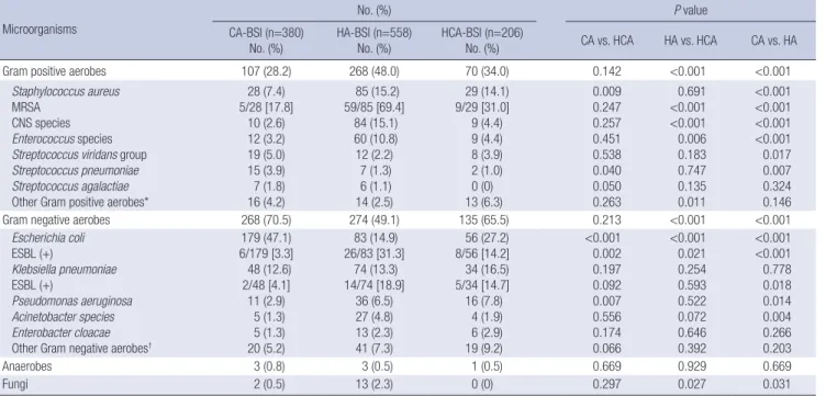
Of all BSI isolates, 59.2% were Gram-negative aerobes, and 38.9%
were Gram-positive aerobes. As a total, E. coli (27.8%) was the most common isolate followed by Klebsiella pneumoniae (13.6
%), S. aureus (12.4%), coagulase-negative Staphylococcus (9%), Enterococcus species (7.1%) (Table 2). The prevalence of E. coli was the highest in CA-BSI group, and the incidence of E. coli var- ied by type of BSI (47.1%, 14.9%, 27.2% in CA-BSI, HA-BSA, and HCA-BSI, respectively, P<0.005). Of CA-BSI isolates, the inci- dence of Gram-negative aerobes (70.5%) was more than two times higher than that of Gram-positive aerobes (28.2%). E. coli Table 1. Patient characteristics of bloodstream infection by epidemiologic type of infection
Characteristics
No. (%) P value
CA-BSI (n=380)
No. (%) HA-BSI (n=558)
No. (%) HCA-BSI (n=206)
No. (%) CA vs. HCA HA vs. HCA CA vs. HA
Demographics Age (Mean year±SD) Male sex
57.0±20.7 184 (48.4)
47.3±23.7 339 (60.8)
53.7±20.7 131 (63.6)
0.251
<0.001
0.001 0.474
<0.001
<0.001 Underlying disease
Solid tumor
Hematologic malignancy Diabetes mellitus Chronic liver disease Chronic renal disease Congestive heart failure Stroke
92 (24.2) 13 (3.4) 75 (19.2) 59 (15.5) 16 (4.2) 3 (0.8) 16 (4.2)
181 (32.4) 137 (24.6) 96 (17.2) 54 (9.7) 37 (6.6) 12 (2.2) 28 (5.0)
113 (54.9) 33 (16.0) 35 (17.0) 38 (18.4) 11 (5.3) 2 (1.0) 8 (3.9)
<0.001
<0.001 0.461 0.364 0.534 0.820 0.849
<0.001 0.012 0.944 0.001 0.514 0.281 0.511
0.006
<0.001 0.376 0.007 0.115 0.103 0.566 Comorbid condition
Neutropenia Receipt of steroids Receipt of immunosuppressant Prior antibiotic use
History of transplantation High alcohol uptake Smoking Urinary catheter Central venous catheter Intra-abdominal catheter
34 (8.9) 18 (4.7) 35 (9.2) 27 (7.1) 11 (2.9) 26 (6.8) 27 (7.1) 0 (0) 0 (0) 0 (0)
158 (28.3) 157 (28.1) 185 (33.2) 305 (54.7) 51 (9.1) 14 (2.5) 41 (7.3) 185 (33.1) 336 (60.2) 134 (24.0)
52 (25.2) 50 (24.3) 65 (31.6) 30 (14.6) 5 (2.4) 5 (2.4) 8 (3.9) 38 (18.4) 58 (28.2) 29 (14.1)
<0.001
<0.001
<0.001 0.004 0.740 0.023 0.116 NA NA NA
0.399 0.286 0.676 0.000 0.002 0.949 0.083
<0.001
<0.001 0.003
<0.001
<0.001
<0.001
<0.001
<0.001 0.001 0.888 NA NA NA CA, community-acquired; HA, hospital-acquired; HCA, healthcare-associated; BSI, bloodstream infection; NA, not available.
(47.1%) and K. pneumoniae (12.6%) represent more than half of the isolates in CA-BSI followed by S. aureus (7.4%), Streptococ- cus viridans group (5.0%). Of HA-BSI isolates, 49.1% were Gram- negative aerobes, and 48.0% were Gram-positive aerobes. The most common isolate of HA-BSI was S. aureus (15.2%) followed by coagulase-negative Staphylococcus (15.1%), and E. coli (14.9
%). The pattern of isolates in HCA-BSI was more similar to that in CA-BSI than that in HA-BSI. Of HCA-BSI isolates, Gram-neg- ative aerobes (65.5%) were more common than Gram-positive aerobes (34.0%) like CA-BSI. However, isolate distributions of CA-BSI and HCA-BSI group were not identical. While E. coli (47.1% vs. 27.2% in CA-BSI vs. HCA-BSI, P<0.001) and Strepto- coccus pneumoniae (3.9% vs. 1.0% in CA-BSI vs. HCA-BSI, P=
0.04) were recovered more frequently in CA-BSI, S. aureus (7.4%
vs. 14.1% in CA-BSI vs. HCA-BSI, P=0.009) and P. aeruginosa (2.9% vs. 7.8% in CA-BSI vs. HCA-BSI, P=0.007) were more prev-
alent in HCA-BSI.
With regard to resistant pathogens, extended spectrum beta- lactamase (ESBL)-producing E. coli was most frequently recov- ered in patients with HA-BSI (31.3%), followed by HCA-BSI (14.2
%) and CA-BSI (3.3%) (HA-BSI vs. CA-BSI, P<0.001; HA-BSI vs.
HCA-BSI, P=0.021; HCA-BSI vs. CA-BSI, P=0.002). ESBL-pro- ducing K. pneumoniae was common in patients with HA-BSI (18.9%), followed by HCA-BSI (14.7%) and CA-BSI (4.1%). Al- though, it did not show the statistical difference for the preva- lence of ESBL-producing K. pneumoniae (HA-BSI vs HCA-BSI, P=0.593; HCA-BSI vs. CA-BSI, P=0.092) except between HA-BSI and CA-BSI (P=0.018). Methicillin-resistant Staphylococcus au- reus (MRSA) was higher in HA-BSI (69.4%), compared with HCA- BSI (31.0%, P<0.001) and CA-BSI group (17.8%, P<0.001).
Table 2. Microorganism distribution of bloodstream infection by epidemiologic type of infection
Microorganisms
No. (%) P value
CA-BSI (n=380)
No. (%) HA-BSI (n=558)
No. (%) HCA-BSI (n=206)
No. (%) CA vs. HCA HA vs. HCA CA vs. HA
Gram positive aerobes 107 (28.2) 268 (48.0) 70 (34.0) 0.142 <0.001 <0.001
Staphylococcus aureus MRSA
CNS species Enterococcus species Streptococcus viridans group Streptococcus pneumoniae Streptococcus agalactiae Other Gram positive aerobes*
28 (7.4) 5/28 [17.8]
10 (2.6) 12 (3.2) 19 (5.0) 15 (3.9) 7 (1.8) 16 (4.2)
85 (15.2) 59/85 [69.4]
84 (15.1) 60 (10.8) 12 (2.2) 7 (1.3) 6 (1.1) 14 (2.5)
29 (14.1) 9/29 [31.0]
9 (4.4) 9 (4.4) 8 (3.9) 2 (1.0) 0 (0) 13 (6.3)
0.009 0.247 0.257 0.451 0.538 0.040 0.050 0.263
0.691
<0.001
<0.001 0.006 0.183 0.747 0.135 0.011
<0.001
<0.001
<0.001
<0.001 0.017 0.007 0.324 0.146
Gram negative aerobes 268 (70.5) 274 (49.1) 135 (65.5) 0.213 <0.001 <0.001
Escherichia coli ESBL (+)
Klebsiella pneumoniae ESBL (+)
Pseudomonas aeruginosa Acinetobacter species Enterobacter cloacae Other Gram negative aerobes†
179 (47.1) 6/179 [3.3]
48 (12.6) 2/48 [4.1]
11 (2.9) 5 (1.3) 5 (1.3) 20 (5.2)
83 (14.9) 26/83 [31.3]
74 (13.3) 14/74 [18.9]
36 (6.5) 27 (4.8) 13 (2.3) 41 (7.3)
56 (27.2) 8/56 [14.2]
34 (16.5) 5/34 [14.7]
16 (7.8) 4 (1.9) 6 (2.9) 19 (9.2)
<0.001 0.002 0.197 0.092 0.007 0.556 0.174 0.066
<0.001 0.021 0.254 0.593 0.522 0.072 0.646 0.392
<0.001
<0.001 0.778 0.018 0.014 0.004 0.266 0.203
Anaerobes 3 (0.8) 3 (0.5) 1 (0.5) 0.669 0.929 0.669
Fungi 2 (0.5) 13 (2.3) 0 (0) 0.297 0.027 0.031
*Other Gram positive aerobes, Streptococcus pyogenes, and other group Streptococcus; †Other Gram negative aerobes,Klebsiella oxytoca, Serratia marcescens, Burkholderia cepacia, Proteus mirabilis, Citrobacter freundii, Morganella morganii, Enterobacter aerogenes, Haemophilus influenazae, and other.
CA, community-acquired; HA, hospital-acquired; HCA, healthcare-associated; BSI, bloodstream infection; ESBL, extended spectrum β-lactamase; MRSA, methicillin-resistant Staphylococcus aureus; CNS, coagulase-negative Staphylococcus.
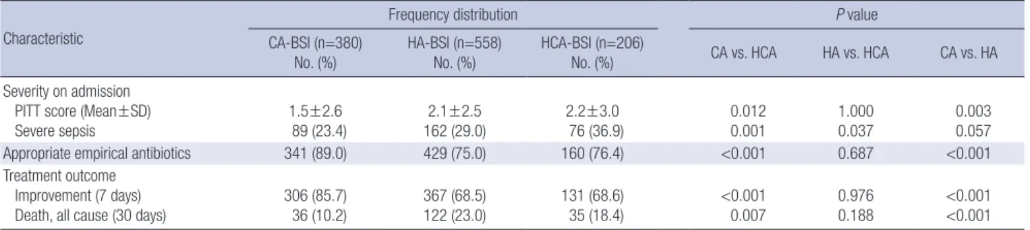
Table 3. Primary sources of bloodstream infection by epidemiologic type of infection
Source of infection
Frequency distribution P value
CA-BSI (n=380) No. (%)
HA-BSI (n=558) No. (%)
HCA-BSI (n=206)
No. (%) CA vs. HCA HA vs. HCA CA vs. HA
Primary bacteremia 22 (5.7) 297 (53.2) 82 (39.8) <0.001 0.001 <0.001
Intra-abdominal infection 116 (30.5) 98 (17.6) 56 (27.2) 0.396 0.003 <0.001
Urinary tract infection 119 (31.3) 32 (5.7) 30 (14.2) <0.001 <0.001 <0.001
Pneumonia 42 (11.1) 79 (14.2) 29 (14.1) 0.284 0.978 0.164
Soft tissue infection 13 (3.4) 15 (2.7) 6 (2.9) 0.740 0.866 0.517
Other* 68 (17.9) 37 (6.6) 3 (1.5) NA NA NA
*Other, Bone and joint infection, endocarditis, central nervous system infection, and other.
CA, community-acquired; HA, hospital-acquired; HCA, healthcare-associated; BSI, bloodstream infection; NA, not available.
Primary infection
The primary infections causing BSI significantly varied by type of BSI. The most common primary infection of CA-BSI were uri- nary tract infection (UTI) (31.3%) and intra-abdominal infec- tion (30.5%) (Table 3). In contrast, primary bacteremia (53.2%) was the most common in HA-BSI group followed by intra-ab- dominal infection (17.6%), and pneumonia (14.2%). The most common primary infection of HCA-BSI was primary bactere- mia (39.8%) like HA-BSI, and followed by intra-abdominal in- fection (27.2%) and UTI (14.2%).
Clinical outcomes
Severity index on admission measured by Pitt bacteremia score in patients with HCA-BSI (2.2±3.0) was significantly higher than that in CA-BSI group (1.5±2.6) (P=0.012) (Table 4). And, the in- cidence of severe sepsis in HCA-BSI group (36.9%) was also high- er than that in CA-BSI (23.4%, P=0.001).
The rate of appropriate empirical antibiotic therapy was the highest in CA-BSI group (89.0%), followed by HCA-BSI (76.4%),
and HA-BSI group (75.0%). Initial clinical improvement rate was more frequently observed in CA-BSI group (85.7%) than in HCA- BSI (68.64%, P<0.001) and HA-BSI group (68.5%, P<0.001).
The overall 30-day mortality rate in all patients with BSI was 18% (193/1,144). Mortality rate was significantly lower in CA- BSI group (10.2%) than in HCA-BSI (18.4%, P=0.007), and HA- BSI group (23.0%, P<0.001). Although, no significant difference was seen in mortality rate between HA-BSI and HCA-BSI group (P=0.188).
Risk factors for mortality
By univariate analysis, high Pitt score (>4), severe sepsis, inap- propriate empirical antibiotic therapy were the common risk factors for all patients with BSI (Table 5). For demographics and comorbidities, old age (≥65 yr), underlying solid tumor, stroke, high alcohol uptake, neutropenia were the significant risk fac- tors for mortality in CA-BSI group. In HA-BSI group, old age (≥65 yr), underlying congestive heart failure, receipt of immunosup- pressant, prior antibiotic use, history of transplantation were the Table 4. Clinical outcome of bloodstream infection by epidemiologic type of infection
Characteristic
Frequency distribution P value
CA-BSI (n=380)
No. (%) HA-BSI (n=558)
No. (%) HCA-BSI (n=206)
No. (%) CA vs. HCA HA vs. HCA CA vs. HA
Severity on admission PITT score (Mean±SD) Severe sepsis
1.5±2.6 89 (23.4)
2.1±2.5 162 (29.0)
2.2±3.0 76 (36.9)
0.012 0.001
1.000 0.037
0.003 0.057
Appropriate empirical antibiotics 341 (89.0) 429 (75.0) 160 (76.4) <0.001 0.687 <0.001
Treatment outcome Improvement (7 days) Death, all cause (30 days)
306 (85.7) 36 (10.2)
367 (68.5) 122 (23.0)
131 (68.6) 35 (18.4)
<0.001 0.007
0.976 0.188
<0.001
<0.001 CA, community-acquired; HA, hospital-acquired; HCA, healthcare-associated; BSI, bloodstream infection.
Table 5. Risk factors associated with 30 day mortality in bloodstream infection by epidemiologic type of infection
Risk factors CA-BS I HA-BSI HCA-BSI
Odds ratio (95% CI) P value Odds ratio (95% CI) P value Odds ratio (95% CI) P value
Age, ≥65 yr 2.44 (1.20-4.96) 0.011 1.62 (1.04-2.51) 0.030 1.87 (0.88-3.96) 0.095
Male sex 0.7 (0.35-1.40) 0.313 0.85 (0.56-1.3) 0.469 0.65 (0.29-1.45) 0.292
Solid tumor 2.47 (1.21-5.05) 0.011 1.23 (0.80-1.88) 0.337 1.89 (0.88-4.06) 0.099
Congestive heart failure 0.89 (0.86-0.93) 0.558 3.44 (0.98-12.1) 0.041 0.17 (0.12-0.23) 0.003
Stroke 3.83 (1.13-12.93) 0.020 1.18 (0.48-2.86) 0.713 0.62 (0.07-5.22) 0.659
High alcohol uptake 1.22 (1.22-9.0) 0.013 0.76 (0.73-0.8) 0.081 0.81 (0.75-0.87) 0.337
Neutropenia 2.59 (1.03-6.47) 0.035 0.98 (0.63-1.54) 0.960 2.05 (0.95-4.42) 0.063
Receipt of immunosuppressant 1.19 (0.39-3.61) 0.751 0.56 (0.35-0.89) 0.015 1.27 (0.59-2.74) 0.529
Prior antibiotic use 0.75 (0.17-3.32) 0.706 2.03 (1.32-3.1) 0.001 0.67 (0.21-2.06) 0.485
History of transplantation 0.89 (0.86-0.92) 0.256 0.33 (0.13-0.86) 0.018 1.11 (0.12-10.25) 0.926
PITT score >4 54.07 (20.59-141.96) <0.001 5.96 (3.56-9.99) <0.001 17.21 (6.83-43.36) <0.001
Severe sepsis 10.59 (4.92-22.77) <0.001 7.44 (4.77-11.61) <0.001 5.5 (2.48-12.18) <0.001
Inappropriate empirical antibiotics 6.77 (2.88-15.91) <0.001 2.4 (1.53-3.77) <0.001 6.66 (2.91-15.22) <0.001 Pathogen
Escherichia coli
Fungi 0.37 (0.17-0.79)
0.89 (0.86-0.93) 0.009
0.633 0.61 (0.32-1.16)
3.44 (0.98-12.1) 0.133
0.041 0.97 (0.43-2.2)
NA NA 0.957 NA Source of infection
Pneumonia
Urinary tract infection 3.82 (1.67-8.7)
0.32 (1.12-0.84) 0.001
0.017 4.16 (2.51-6.88)
0.68 (0.25-1.83) <0.001
0.447 5.25 (2.16-12.77)
0.51 (0.14-1.8) <0.001 0.290 CA, community-acquired; HA, hospital-acquired; HCA, healthcare-associated; BSI, bloodstream infection; NA, not available.
significant risk factors. In HCA-BSI group, underlying congestive heart failure was the significant risk factor. For infecting patho- gens, E. coli was the negative predictive factor for 30-day mortal- ity in CA-BSI group. Fungal infection was the significant risk fac- tor for mortality in HA-BSI group. For primary infection, pneu- monia was the significant risk factor for all patients with BSI.
By multivariate analysis, high Pitt score (CA-BSI, OR 28.3, P<
0.001; HA-BSI, OR 2.2, P=0.003; HCA-BSI, OR 7.8, P=0.001) and ineffective empirical antibiotic therapy (CA-BSI, OR 5.1, P=0.01;
HA-BSI, OR 2.2, P=0.003; HCA-BSI, OR 6.0, P=0.001) were the common independent risk factors in all patients with BSI (Table 6). Severe sepsis (OR 5.5, 95% CI 3.2-9.5, P<0.001), pneumonia (OR 3.0, 95% CI 1.6-5.6, P<0.001), and prior antibiotic use (OR 1.7, 95% CI 1.0-2.9, P=0.03) were the independent risk factors in patients with HA-BSI.
DISCUSSION
This study was a nationwide surveillance of BSI in Korean hos- pitals. In this study, we compared clinical and microbiological characteristics of 3 types of BSI.
With regard to prevalent pathogens by the epidemiologic type, Gram-negative bacilli (especially E. coli and K. pneumoniae) in CA-BSI were due to higher incidence of UTI and intra-abdomi- nal infection, while Gram-positive cocci (especially staphylo- cocci) in HA-BSI were due to primary bacteremia which were frequently associated with cather-related infection. In HCA-BSI, Gram-negative bacilli were also the prevalent pathogen like CA- BSI, because the incidences of UTI and intra-abdominal infec- tion were higher than that of primary bacteremia. However, the proportion of Gram-negative bacilli in HCA-BSI was differed from that in CA-BSI.
Cheong et al. (16) previously reported that the prevalence of ESBL-producing E.coli in HCA-BSI (6.7%) was higher than that in CA-BSI (3.2%), although it did not show the statistical differ-
ence between 2 groups. In addition, Kang et al. (17) reported that more than half (63.2%) of community-onset BSIs caused by ESBL-producing E. coli were HCA-BSI. Our study also showed the similar result that the prevalence of ESBL-producing E. coli in HCA-BSI and HA-BSI group was higher than that in CA-BSI.
With regard to S. aureus bacteremia, previous reports showed that the prevalence of MRSA was the highest in in HA-BSI (33.8- 61%) followed by HCA-BSI (29.4-52%) and CA-BSI (11-21.2%) (3, 10, 18). And, our study also showed the similar result.
In general, early and appropriate antibiotic therapy signifi- cantly affects the overall mortality in BSI (19, 20). Previous stud- ies reported that 10-40% of BSI cases received inappropriate empirical therapy, which was more frequent in cases of HA-BSI and HCA-BSI (9, 21, 22). Our data was also consistent with this finding that initial antibiotic therapy was more frequently inap- propriate in HA-BSI and HCA-BSI cases than in CA-BSI. Given the different pathogens and antimicrobial resistance in HCA- BSI compared with CA-BSI, empirical antibiotic choice should be different in HCA-BSI from those in CA-BSI cases.
Previously, high severity index on admission and inappropri- ate empirical antibiotic therapy were known as significant risk factors in BSI (16, 19, 23). Pazos et al. (23) reported that septic shock, and inappropriate antibiotic therapy were found to be risk factors for mortality in overall BSI. And, Cheong et al. (16) also reported that high Charlson’s index, high Pitt score, and acute renal failure were found to be risk factors for mortality in E. coli bacteremia. In our study, high Pitt score and inappropri- ate empirical antibiotic therapy were shown to be the common independent risk factors in every BSI group including HCA-BSI on multivariate analysis.
There is a limitation in this study. Our data were obtained from a few university hospitals that might lead to the bias of pathogen distribution, antimicrobial resistance or primary infection.
In conclusion, the present data suggest that clinical features, clinical outcome, and microbiologic features of causative patho- gens vary by epidemiologic type of BSI. Especially, HCA-BSI shows a distinct entity of BSI with its unique epidemiology, mi- crobiology, and treatment outcomes, which shares more simi- larity with HA-BSI. The only independent risk factors for mor- tality associated with HCA-BSI could not be found. However, considering the inappropriate empirical antibiotic therapy as a major independent risk factor for mortality, clinicians should be aware of this category for more appropriate antibiotics based on possible resistant pathogens.
REFERENCES
1. Leibovici L, Samra Z, Konigsberger H, Drucker M, Ashkenazi S, Pitlik SD. Long-term survival following bacteremia or fungemia. JAMA 1995;
274: 807–12.
2. Pedersen G, Schønheyder HC, Sørensen HT. Source of infection and Table 6. Independent risk factors for mortality in bloodstream infection by epidemiologic
type of infection
Risk factors Adjusted odds ratio
(95% CI) P value
Patients with CA-BSI PITT score >4 Severe sepsis
Inappropriate empirical antibiotics
28.35 (7.11-113.01) 3.8 (1.09-13.19)
5.15 (1.33-19.9)
<0.001 0.035 0.017 Patients with HA-BSI
PITT score >4 Severe sepsis Pneumonia Prior antibiotic use
Inappropriate empirical antibiotics
2.24 (1.17-4.29) 5.56 (3.24-9.52) 3.05 (1.65-5.63) 1.75 (1.03-2.98) 2.24 (1.3-3.85)
0.003
<0.001
<0.001 0.038 0.003 Patients with HCA-BSI
PITT score >4
Inappropriate empirical antibiotics
7.87 (2.33-26.59) 6.04 (2.16-16.87)
0.001 0.001 CA, community-acquired; HA, hospital-acquired; HCA, healthcare-associated; BSI, bloodstream infection.
other factors associated with case fatality in community-acquired bacte- remia-a Danish population-based cohort study from 1992 to 1997. Clin Microbiol Infect 2003; 9: 793-802.
3. Friedman ND, Kaye KS, Stout JE, Stout JE, McGarry SA, Trivette SL, Briggs JP, Lamm W, Clark C, MacFarquhar J, Walton AL, Reller LB, Sex- ton DJ. Healthcare-associated bloodstream infections in adults: a reason to change the accepted definition of community-acquired infections. Ann Intern Med 2002; 137: 791–7.
4. Siegman-Igra Y, Fourer B, Orni-Wasserlauf R, Golan Y, Noy A, Schwartz D, Giladi M. Reappraisal of community-acquired bacteremia: a propos- al of a new classification for the spectrum of acquisition of bacteremia.
Clin Infect Dis 2002; 34: 1431-9.
5. Shorr AF, Tabak YP, Killian AD, Gupta V, Liu LZ, Kollef MH. Healthcare- associated bloodstream infection: a distinct entity? Insights from a large U.S. database. Crit Care Med 2006; 34: 2588-95.
6. Weinstein MP, Towns ML, Quartey SM, Mirrett S, Reimer LG, Parmigiani G, Reller LB. The clinical significance of positive blood cultures in the 1990s:
a prospective comprehensive evaluation of the microbiology, epidemiol- ogy, and outcome of bacteremia and fungemia in adults. Clin Infect Dis 1997; 24: 584-602.
7. Lark RL, Chenoweth C, Saint S, Zemencuk JK, Lipsky BA, Plorde JJ. Four year prospective evaluation of nosocomial bacteremia: epidemiology, microbiology, and patient outcome. Diagn Microbiol Infect Dis 2000; 38:
131-40.
8. Lark RL, Saint S, Chenoweth C, Zemencuk JK, Lipsky BA, Plorde JJ. Four- year prospective evaluation of community-acquired bacteremia: epide- miology, microbiology, and patient outcome. Diagn Microbiol Infect Dis 2001; 41: 15-22.
9. McDonald JR, Friedman ND, Stout JE, Sexton DJ, Kaye KS. Risk factors for ineffective therapy in patients with bloodstream infection. Arch Intern Med 2005; 165: 308-13.
10. Rangel-Frausto MS, Pittet D, Costigan M, Hwang T, Davis CS, Wenzel RP. The natural history of the systemic inflammatory response syndrome (SIRS): a prospective study. JAMA 1995; 273: 117-23.
11. American College of Chest Physicians/Society of Critical Care Medicine Consensus Conference: definitions for sepsis and organ failure and guidelines for the use of innovative therapies in sepsis. Crit Care Med 1992; 20: 864-74.
12. Chow JW, Yu VL. Combination antibiotic versus monotherapy for gram- negative bacteremia: a commentary. Int J Antimicrob Agents 1999; 11:
7–12.
13. Kang CI, Kim SH, Park WB, Lee KD, Kim HB, Kim EC, Oh MD, Choe KW.
Bloodstream infections caused by antibiotic-resistant gram-negative ba-
cilli: risk factors for mortality and impact of inappropriate initial anti- microbial therapy on outcome. Antimicrob Agents Chemother 2005; 49:
760–6.
14. Kasiakou SK, Michalopoulos A, Soteriades ES, Samonis G, Sermaides GJ, Falagas ME. Combination therapy with intravenous colistin for man- agement of infections due to multidrug-resistant Gram-negative bacteria in patients without cystic fibrosis. Antimicrob Agents Chemother 2005;
49: 3136-46.
15. Clinical and Laboratory Standards Institute. Performance Standards for Antimicrobial Susceptibility Testing: Fifteenth Informational Supplement.
Wayne, PA: CLSI, 2005. Document No. M100-S15.
16. Cheong HS, Kang CI, Kwon KT, Heo ST, Wi YM, Kim ES, Lee JS, Ko KS, Chung DR, Lee NY, Song JH, Peck KR. Clinical significance of healthcare- associated infections in community-onset Escherichia coli bacteraemia.
J Antimicrob Chemother 2007; 60: 1355-60.
17. Kang CI, Cheong HS, Chung DR, Peck KR, Song JH, Oh MD, Choe KW.
Clinical features and outcome of community-onset bloodstream infec- tions caused by extended-spectrum beta-lactamase-producing Escherich- ia coli. Eur J Clin Microbiol Infect Dis 2008; 27: 85-8.
18. Lesens O, Hansmann Y, Brannigan E, Hopkins S, Meyer P, O’Connel B, Prévost G, Bergin C, Christmann D. Healthcare-associated Staphylococ- cus aureus bacteremia and the risk for methicillin resistance: is the Cen- ter for Disease Control and Prevention definition for community acquired bacteremia still appropriate? Infect Control Hosp Epidemiol 2005; 26:
204-9.
19. Ramphal R. Importance of adequate initial antimicrobial therapy. Che- motherapy 2005; 51: 171-6.
20. Du B, Long Y, Liu H, Chen D, Liu D, Xu Y, Xie X. Extended spectrum beta- lactamase-producing Escherichia coli and Klebsiella pneumoniae blood- stream infection: risk factors and clinical outcome. Intensive Care Med 2002; 28: 1718–23.
21. Elhanan G, Sarhat M, Raz R. Empiric antibiotic treatment and the misuse of culture results and antibiotic sensitivities in patients with community- acquired bacteraemia due to urinary tract infection. J Infect 1997; 35: 283-8.
22. Byl B, Clevenbergh P, Jacobs F, Jacobs F, Struelens MJ, Zech F, Kentos A, Thys JP. Impact of infectious diseases specialists and microbiological data on the appropriateness of antimicrobial therapy for bacteremia. Clin In- fect Dis 1999; 29: 60-6.
23. Pazos Añón R, Fernández Rodríguez R, Paz Vidal I, Tinajas A, Cantón I, Abel V, González R, Martínez R, Gayoso P, Fernández Alvarez O. Prog- nostic factors of bacteremia: prospective study. An Med Interna 2001; 18:
415-20.