INTRODUCTION
Nasal polyposis (NP) is a chronic inflammatory disease of the upper airway featuring inflammatory cell infiltration, tis- sue remodeling that includes extracellular matrix (ECM) accu- mulation and fibrosis (1). The inflammatory cellular infiltrate in nasal polyps has been shown to consist of eosinophils, lym- phocytes, plasma cells, and mast cells to an extent which is similar to that observed in the bronchial mucosa of asthmat- ics (2-4), suggesting that inflammatory mechanisms of the two diseases may be similar (5). According to experimental models, it seems reasonable to speculate that the formation and growth of NPs require ECM accumulation (6).
The matrix metalloproteinases (MMPs) appear to be respon- sible for edema and cell transmigration, and ECM remodeling in asthmatic airways (7). Among the matrix metalloprotein- ases (MMPs), MMP-9, especially active MMP-9 expression, was clearly enhanced in nasal polyps compared with control mucosa (8). The activation of MMPs is inhibited by tissue inhibitors of metalloproteinases (TIMPs) that form a 1:1 complex with MMPs (9). Loss of coordination in the expres- sion of MMPs and TIMPs is believed to generate tissue degra-
dation in inflammatory diseases. Eosinophil is one of the major sources of MMPs; MMP-9 was overexpressed by eosinophils accumulating in airway walls of asthmatics (10). Several in vitro studies have demonstrated that MMP-2 and MMP-9 were produced and activated by mast cells, and the possible involvement of mast cells in connective tissue degradation and fibrosis was suggested (11, 12).
Transforming growth factor- (TGF- ) is a chemoattrac- tant for fibroblasts, stimulating fibroblast proliferation, and enhancing collagen laydown by fibroblasts. Of the TGF- isoforms, TGF- 1 synthesized by infiltrating eosinophils may contribute to stromal fibrosis and basement membrane thick- ening which are characteristic of nasal polyp (13). Another in vitro study showed that TGF- 1 induces MMP-9 proenzyme and MMP-2 (14). There have been few studies in relation to infiltrating cell activations and TGF- 1 on the specific effects of MMP-2, MMP-9, and TIMP-1 in nasal polyps, which sh- ares certain similarities with asthma. The aim of the present study was to investigate the association between MMPs and TIMP-1, and inflammatory cell activation markers in nasal polyp tissue in relation to TGF- 1.
Young-Mok Lee*, Sun-Sin Kim,
Hyun-Ah Kim, Yu-Jin Suh, Soo-Keol Lee, Dong-Ho Nahm, Hae-Sim Park
Department of Respiratory Medicine and Allergy, Soonchunhyang University Hospital*, Seoul;
Department of Allergy and Clinical Immunology, Ajou University School of Medicine, Suwon, Korea
Address for correspondence Hae-Sim Park, M.D.
Department of Allergy and Rheumatology, Ajou University School of Medicine, San-5, Wonchondong, Paldalgu, Suwon 442-749, Korea Tel : +82.31-219-5196, Fax : +82.31-219-5154 E-mail: [email protected]
97
Eosinophil Inflammation of Nasal Polyp Tissue: Relationships with Matrix Metalloproteinases, Tissue Inhibitor of Metalloproteinase-1, and Transforming Growth Factor- 1
Eosinophil and mast cell infiltrations are consistent findings in nasal polyp tissue. Pre- vious studies have shown that matrix metalloproteinases (MMPs) may be involved in eosinophil infiltration in airway mucosa of asthmatic patients, and that transforming growth factor-beta1 (TGF- 1) induces extracellular matrix deposition in nasal polyp tissue. The aim of this study was to evaluate the role of MMPs and tissue-inhibitor of metalloproteinase-1 (TIMP-1) in association with TGF- 1, eosinophils and mast cell activation in nasal polyp tissue. Nasal polyp tissues from 20 patients who under- went polypectomies were collected and prepared into tissue homogenate. Eosinophil cationic protein (ECP) and tryptase levels were measured by CAP system (Pharma- cia, Sweden). MMP-2, MMP-9, TIMP-1 and TGF- 1 levels were measured by en- zyme-liked immunosorbent assay. MMP-2 was the predominant form of MMPs, fol- lowed by MMP-9 and TIMP-1. There were significant correlations between ECP, and MMP-9, MMP-2, TGF- 1 and tryptase, but not with TIMP-1. Significant correlations were noted between tryptase, and MMP-2, MMP-9, and TGF- 1, but not with TIMP- 1. Close correlations were noted between TGF- 1, and MMP-9 and MMP-2, but not with TIMP-1. MMP-2, MMP-9, and TGF- 1 may contribute to eosinophil and mast cell migrations into nasal polyp tissue.
Key Words : Nasal Polyps; Matrix Metalloproteinases; Tissue-Inhibitor of Metalloproteinase-1; Transforming Growth Factor beta; Eosinophils
Received : 2 August 2002 Accepted : 4 November 2002
MATERIALS AND METHODS Subjects
Nasal polyps were obtained from 20 patients with bronchial asthma, between the ages of 27 and 56 yr (40.0±3.4 yr [mean
±SEM]), who had complained of asthmatic symptoms dur- ing visits to the Allergy Clinic of Ajou University Hospital, Suwon, Korea (Table 1). Nasal polyps were identified by ante- rior rhinoscopy in all patients. Asthma was diagnosed accord- ing to the American Thoracic Society guidelines for the diag- nosis of asthma (15). Skin prick test with common aeroaller- gens (Bencard, U.K.) was performed and atopy was defined as showing positive response (≥3
+
by Allergen/Histamine ratio) to two or more inhalant allergens. None of the study subjects had used topical steroids for at least 4 weeks prior to obtaining the polyp. The polyps, soon after surgical removal, were washed with normal saline to remove stagnant mucus.The middle portion of the polyp was cut off and immediately sent to a pathology laboratory to prepare the paraffin-embed- ded tissue. Half of them were made into tissue homogenate.
All the subjects gave their informed consent as regulated by the Ajou University Hospital, Suwon, Korea.
Preparation of nasal polyp tissue
The polyp tissue was frozen at -70℃ immediately after the operation. When prepared for the experiments, the samples were thawed, and ground in a homogenizer (POLYTRON, Switzerland) with phosphate-buffered saline (PBS, pH7.5) including 1% Triton X-100. They were centrifuged and the separated supernatant was kept at -70℃to measure ECP, tryp- tase, MMP-2, MMP-9, TIMP-1, and TGF- 1 levels.
Enzyme-Linked Immunosorbent Assays for MMP-2, MMP-9, TIMP-1, and TGF- 1
Commercially available kits were used (MMP-2, MMP-9, and TIMP-1:RPN 2617, RPN 2614, and RPN 2611, respec- tively; Amersham Pharmacia Bioteck, U.K., TGF- 1:Quan- tikine; R&D Systems, Minneapolis, U.S.A.). The MMP-2 and MMP-9 ELISA detects free proMMP-2, proMMP-9, and pro MMP-9-TIMP-1 complexes. It does not detect active MMP- 2 or MMP-9. The TIMP-1 kit detects the total TIMP-1, free
TIMP-1, and MMP-9-TIMP-1 complexes. For all assays, opti- cal density was measured with a spectrophotometer set at 450 nm. Quantification was performed by interpolation from a standard curve. The detection limit was 0.37 ng/mL for MMP- 2, 0.6 ng/mL for MMP-9, 1.25 ng/mL for TIMP-1, and 7 pg/
mL for TGF- 1.
Levels of protein were also measured by nephelometry (16) with a Beckman Array system (Beckman Instruments Inc., Brea, Calif). The data of MMPs, TIMP-1, and TGF- 1 con- centrations in tissue homogenate of nasal polyp were present- ed as the ratio of protein measured, and were expressed as ng/
mg protein concentration.
ECP and tryptase assay
ECP and tryptase in tissue homogenate of nasal polyp were measured by fluoroimmunoassay using the Pharmacia CAP system (Pharmacia and Upjohn, Uppsala, Sweden). The low- est detection limits for ECP and tryptase measurements were 2 ng/mL and 4 ng/mL, repectively. The data of ECP and try- ptase concentrations were presented as the ratio of protein mea- sured, and were expressed as ng/mg protein concentration.
Statistical analysis
The Mann-Whitney U test and ANOVA were applied us- ing the SPSS version 7.0 (Chicago, U.S.A.) to evaluate the sta- tistical differences among the data. The Pearson correlation analysis was applied to evaluate the statistical significance be- tween the two values. A p value of 0.05 or less was regarded as significant.
RESULTS Clinical features of the study subjects
The characteristics of the study subjects are shown in Table 1. No significant differences were noted in MMP-2, MMP-9, TIMP-1, TGF- 1 levels, and in molar ratio of MMP-9/TIMP- 1 according to atopy status, duration of nasal symptom, and recurrence (p>0.05, respectively).
Correlations of MMP-2, MMP-9, and TIMP-1 levels, and inflammatory cell markers
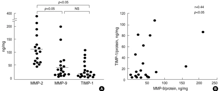
The MMP-2 level (109.3±17.9 ng/mg protein [mean± SEM]) was the highest in polyp tissue homogenate, followed by the MMP-9 (40.8±11.1) and TIMP-1 (26.9±7.2) levels (p<0.05). However, there was no significant difference between MMP-9 and TIMP-1 levels (p>0.05; Fig. 1A). The MMP-9 level in polyp tissue homogenate was significantly correlated with the TIMP-1 level (r=0.44, p<0.05; Fig. 1B). The ECP level in polyp tissue homogenate was significantly correlated
*Values are mean±S.E.
Number 20
Sex (male:female) 14:6
Age (yr)* 40.0±3.4
Atopy (presence/absence) 6/14
Duration of nasal symptom (yr)* 2.8±0.4 Recurrence (presence/absence) 8/12 Table 1.Characteristics of study subjects
with the MMP-2 (r=0.61, p<0.05) and MMP-9 (r=0.65, p<
0.05) levels, but not with the TIMP-1 level (r=0.42, p>0.05) as shown in Fig. 2. Significant correlations were noted between
the tryptase level in polyp tissue homogenate, and MMP-2 (r=0.47, p<0.05), and MMP-9 (r=0.62, p<0.05), but not with TIMP-1 (r=0.04, p>0.05) level as shown in Fig. 3.
ng/mg
MMP-2 MMP-9 TIMP-1
p<0.05 p<0.05
NS 400
200
150
100
50
0
MMP-9/protein, ng/mg
Fig. 1.Comparison of MMP-2, MMP-9, and TIMP-1 levels in nasal polyp tissue homogenate (A) and the correlation between MMP-9 and TIMP-1 level (B). Horizontal bars represent mean values.
TIMP-1/protein, ng/mg
50 100 150 200 250
p<0.05 r=0.44 120
100
80
60
40
20
0
A B
Fig. 2.Correlations between ECP, and MMP-2 (A), MMP-9 (B) and TIMP-1 (C) levels in nasal polyp tissue.
MMP-2/protein, ng/mg
400 350 300 250 200 150 100 50 0
ECP/protein, ng/mg r=0.61
p<0.05
0 20 40 60 80
MMP-9/protein, ng/mg
250
200
150
100
50
0
ECP/protein, ng/mg r=0.65
p<0.05
0 20 40 60 80
TIMP-1/protein, ng/mg
120
90
60
30
0
ECP/protein, ng/mg r=0.42
p>0.05
0 20 40 60 80
A B C
Fig. 3.Correlations between tryptase, and MMP-2 (A), MMP-9 (B) and TIMP-1 (C) levels in nasal polyp tissue.
MMP-2/protein, ng/mg
400 350 300 250 200 150 100 50 0
Tryptase/protein, ng/mg r=0.47 p<0.05
0 1,000 2,000 3,000 4,000
MMP-9/protein, ng/mg
250
200
150
100
50
0
Tryptase/protein, ng/mg r=0.62 p<0.05
0 1,000 2,000 3,000 4,000
TIMP-1/protein, ng/mg
120
90
60
30
0
Tryptase/protein, ng/mg r=0.04 p>0.05
0 1,000 2,000 3,000 4,000
A B C
Correlations of TGF- 1, MMPs, and TIMP-1 levels, and inflammatory cell markers
As shown in Fig. 4, close correlations were also noted bet- ween the TGF- 1 level in polyp tissue homogenate, and the MMP-2 (r=0.82, p<0.05) and MMP-9 (r=0.92, p<0.05) levels, but not with the TIMP-1 level (r=0.31, p>0.05). Fig. 5 shows the correlations between the ECP, tryptase, and TGF- 1 levels.
The ECP level in polyp tissue homogenate was significantly cor- related with the tryptase (r=0.58, p<0.05; Fig. 5A) and TGF- 1 levels (r=0.62, p<0.05; Fig. 5C). Tryptase was significant- ly correlated with the TGF- 1 level (r=0.73, p<0.05; Fig. 5B).
DISCUSSION
Inflammatory processes within the mucosa of the upper res- piratory tract are believed to play an important role in the devel- opment of nasal polyp. Nasal mucosa infiltration by numerous eosinophils and mast cells is the most characteristic feature of nasal polyposis (17). Higher ECP and eosinophil counts were noted in patients with polyposis, suggesting that an excessive
response by activated eosinophils may induce a more profound inflammatory process of the nasal polyp (18). Mast cells act as a key effector in allergic reactions with an ability to release both tryptase and cytokines in response to allergen (19). Nasal polyp has tryptase-containing mast cells in both epithelial and stromal layers, suggesting that activated mast cells which de- granulate and release tryptase could participate in nasal polyp generation (17). Another previous study demonstrated that activated mast cells induced the release of eosinophil cationic protein (20). In this study, tryptase was detected and signif- icantly correlated with ECP in nasal polyp tissue, which was responsible for chronic inflammatory response of nasal polyps.
A previous study demonstrated the presence of elevated lev- els of pro- and active form of MMP-9 in nasal polyps by im- munohistochemistry and zymography, while MMP-2 expres- sion remained almost equivalent to both nasal polyp tissue and control mucosa (8). In this study, however, MMP-2 was the predominant form of MMPs in nasal polyp tissue, followed by MMP-9. MMP-9 was reported in an in vitro system to play a crucial role in the transmigration of eosinophils (21), and to be overexpressed by eosinophils accumulating in airway walls of asthmatics (10). After eosinophils migrated to nasal mucosa
Fig. 5.Correlations between ECP and tryptase (A), TGF- 1 and tryptase (B), and ECP and TGF- 1 (C) levels in nasal polyp tissue.
ECP/protein, ng/mg
80
60
40
20
0
Tryptase/protein, ng/mg r=0.58 p<0.05
0 1,000 2,000 3,000 4,000
Tryptase/protein, ng/mg
5,000
4,000
3,000
2,000
1,000
0
TGF- 1/protein, ng/mg r=0.73 p<0.05
0 2 4 6 8 10 12
ECP/protein, ng/mg
80
60
40
20
0
TGF- 1/protein, ng/mg r=0.62 p<0.05
0 2 4 6 8 10 12
A B C
Fig. 4.Correlations between TGF- 1, and MMP-2 (A), MMP-9 (B) and TIMP-1 (C) level in nasal polyp tissue.
MMP-2/protein, ng/mg
400
300
200
100
0
TGF- 1/protein, ng/mg r=0.82 p<0.05
0 2 4 6 8 10 12
MMP-9/protein, ng/mg
200
150
100
50
0
TGF- 1/protein, ng/mg r=0.92 p<0.05
0 2 4 6 8 10 12
TIMP-1/protein, ng/mg
120
90
60
30
0
TGF- 1/protein, ng/mg r=0.31 p>0.05
0 2 4 6 8 10 12
A B C
by their own MMP-2 and -9, they would trigger the synthe- sis of MMP-2 and -9, which may be involved in the develop- ment and maintenance of airway inflammation in asthmat- ics. Tryptase, which is the specific and major enzyme of human mast cell granules (22), degrades fibronectin and cleaves pro MMP-2 to the activated form (23). Recently, an in vitro study demonstrated that human cultured mast cells could express MMP-9 (11). As shown in the previous study (11, 23), mast cells localized in nasal mucosa expressed MMP-2 and MMP- 9 in pathological condition and these findings support the view that the mast cells may influence ECM degradation and remo- deling in allergic inflammation. In this study, MMP-2 and MMP-9 were significantly correlated with ECP as well as tryp- tase, which suggests the possibility that MMP-2 and MMP-9 may be involved in migration of eosinophils and mast cells into nasal polyp tissue. This data also suggests that mast cells and eosinophils might be, either directly or indirectly, respon- sible for the production of MMPs. MMP-2 may not only be synthesized abundantly in inflamed nasal mucosa, whether they are pro- or active form, but it may also play a role in nasal polyp formation and growth.
Recently, MMP-9 and TIMP-1 expressions were reported in the epithelial cells of nasal mucosa and infiltrating eosinophils as well as bronchial tissue in asthmatics (7, 24, 25). The above data demonstrated that the extent of MMP-9 expression was greater than that of TIMP-1. Moreover, MMP-9 expression significantly correlated with eosinophil infiltration in nasal mucosa. In healthy subjects, MMP-9 was co-secreted with TIMP-1 in 1:1 stoichiometry, and previous reports showed the imbalance between MMP-9 and TIMP-1 in airway inflam- mation (25, 26). In this study, the MMP-9 level was higher than the TIMP-1 level and significantly correlated with the TIMP-1 level. However, the TIMP-1 level or the molar ratio of MMP-9/TIMP-1 was not different according to clinical para- meters, and it was not correlated with cellular activation markers. This result suggests that TIMP-1 is co-secreted with MMP-9 in nasal polyp tissue, and the MMP-9/TIMP-1 imbal- ance may play a role in regulating inflammatory response in nasal polyp tissue.
Recent studies demonstrated that TGF- 1 was strongly expressed in inflammatory nasal mucosa and allergic rhinitis tissues but not in normal nasal mucosa, and also found that eosinophils represented a major source of TGF- 1 in nasal polyp tissue (13, 27). Recently, Sehgal and Thompson (14) found that TGF- 1 stimulates the MMP-2 and MMP-9 secretory activity in human prostate cell lines. Another in vitro study showed that MMP-9, as well as MMP-2, proteolytically cleave latent TGF- , providing a novel and potentially important mecha-nism for TGF- activation (28). Furthermore, in pati- ents with chronic airway inflammation, TGF- 1 correlated with the number of macrophage and mast cells in bronchio- lar epithelium (29). In nasal polyposis, especially mature nasal polyp, mast cells are more abundant in the submucosa, and they degranulate in human mature nasal polyps (30). Our data
also showed that the TGF- 1 level in nasal polyp tissue was significantly correlated with MMP-2, MMP-9, ECP, and tryp- tase. According to the above results, we suggest the hypothesis that TGF- 1, in conjunction with MMP-2 and MMP-9, might be involved in ECM degradation and fibrosis in nasal polyp tissue where eosinophils and mast cells are the most prevalent cells.
In conclusion, this study has identified the presence of MMP- 2, MMP-9, and TGF- 1 in nasal polyp tissue in relation to eosinophil and mast cell activation. Both MMP-2 and MMP- 9 may play a role in migration of eosinophils and mast cells into nasal polyp tissue. Further studies will be needed to elu- cidate the role of TGF- 1 in nasal polyp inflammation.
REFERENCES
1. Stierna PLE. Nasal polyps: relationship to infection and inflammation.
In:Settipane GA, Lund VJ, Berstein JM, Tos M, eds. Nasal Polyps:
Epidemiology, Pathogenesis and Treatment. OceanSide Publications;
Providence, RI; 1997: 119-26.
2. Hamilos DL. Nasal polyps as immunoreactive tissue. Allergy Asthma Proc 1996; 17: 293-6.
3. Stoop AE, van der Heijden HA, Biewenga J, van der Baan S. Lympho- cytes and non-lymphoid cells in human nasal polyps. J Allergy Clin Immunol 1991; 87: 470-5.
4. Kawabori S, Denburg JA, Schwarz LB, Irani AA, Wong D, Jordana G, Evans S, Dolovich J. Histochemical and immunohistochemical charac- teristic of mast cells in nasal polyps. Am J Respir Cell Mol Biol 1992;
6: 37-43.
5. Djukanovic R, Wilson JW, Britten KM, Wilson SJ, Walls AF, Roche WR, Howarth PH, Holgate ST. Quantitation of mast cells and eosi- nophils in the bronchial mucosa of symptomatic atopic asthmatics and healthy control subjects using immunohistochemistry. Am Rev Respir Dis 1990; 142: 863-71.
6. Norlander T, Westrin KM, Fukami M, Stierna P, Carlsoo B. Experi- mentally induced polyps in the sinus mucosa: a structural analysis of the initial stages. Laryngoscope 1996; 106: 196-203.
7. Hoshino M, Takahashi M, Takai Y, Sim J. Inhaled corticosteroids decrease subepithelial collagen deposition by modulation of the bal- ance between matrix metalloproteinase-9 and tissue inhibitor of met- alloproteinase-1 expression in asthma. J Allergy Clin Immunol 1999;
104: 356-63.
8. Lechapt-Zalcman E, Coste A, d’Ortho MP, Frisdal E, Harf A, Lafuma C, Escudier E. Increased expression of matrix metalloproteinase-9 in nasal polyps. J Pathol 2001; 193: 233-41.
9. Nagase H. Activation mechanisms of matrix metalloproteinase. Biol Chem 1997; 378: 151-60.
10. Ohno I, Ohtani H, Nitta Y, Suzuki J, Hoshi H, Honma M, Isoyama S, Tanno Y, Tamura G, Yamauchi K, Nagura H, Shirato K. Eosinophils as a source of matrix metalloproteinase-9 in asthmatic airway inflam- mation. Am J Respir Cell Mol Biol 1997; 16: 212-9.
11. Kanbe N, Tanaka A, Kanbe M, Itakura A, Kurosawa M, Matsuda H.
Human mast cells produce matrix metalloproteinase 9. Eur J Immunol
1999; 29: 2645-9.
12. Fang KC, Wolters PJ, Steinhoff M, Bidgol A, Blount JL, Caughey GH.
Mast cell expression of gelatinase A and B is regulated by kit ligand and TGF- 1. J Immunol 1999; 162: 5528-35.
13. Elovic A, Wong DT, Weller PF, Matossian K, Galli SJ. Expression of transforming growth factor- and 1 messenger RNA and product by eosinophils in nasal polyps. J Allergy Clin Immunol 1994; 93: 864-9.
14. Sehgal I, Thompson TC. Novel regulation of type IV collagenase (ma- trix metallo proteinase-9 and -2) activities by transforming growth factor 1 in human prostate cancer cell lines. Mol Biol Cell 1999; 10:
407-16.
15. American Thoracic Society. Standards for the diagnosis and care of patients with chronic obstructive pulmonary disease (COPD) and asth- ma. Am Rev Respir Dis 1987; 136: 225-44.
16. Rhichie RF, Alper CA, Graves J, Pearson N, Larson C. Automated quantitation of proteins in serum and other biologic fluids. Am J Clin Pathol 1973; 59: 151-9.
17. Park HS, Nahm DH, Park K, Suh KS, Yim HE. Immunohistochemi- cal characterization of cellular infiltrate in nasal polyp from aspirin- sensitive asthmatic patients. Ann Allergy Asthma Immunol 1998; 81:
219-24.
18. Keith PK, Conway M, Evans S, Wong DA, Jordana G, Pengelly D, Dolovich J. Nasal polyps: effects of seasonal allergen exposure. J Aller- gy Clin Immunol 1994; 93: 567-74.
19. Redington AE, Howarth PH. Mast cells, cytokines and asthma. Can Respir Dis 1994; 1: 1-9.
20. Okayama Y, Kobayashi H, Ashman LK, Holgate ST, Church MK, Mori M. Activation of eosinophil with cytokines produced by lung mast cells. Int Arch Allergy Immunol 1997; 114(Suppl 1): 75-7.
21. Okada S, Kita H, George TJ, Gleich GJ, Leiferman KM. Migration of eosinophils through basement membrane components in vitro: role
of matrix metalloproteinase-9. Am J Respir Cell Mol Biol 1997; 17:
519-28.
22. Broide DH, Lotz M, Cuomo AJ, Coburn DA, Federman EC, Wasser- man SI. Cytokines in symptomatic asthma airways. J Allergy Clin Im- munol 1992; 89: 958-67.
23. Schwartz LB, Bradford TR, Irani AM, Dblois G, Craig SS. The major enzymes of human mast cell secretory granules. Am Rev Respir Dis 1987; 135: 1186-9.
24. Lee HM, Choi JH, Choi CS, Hwang SJ, Lee SH. Expression of MMP- 9 and TIMP-1 in the nasal mucosa of allergic rhinitis. Korean J Oto- laryngol 2000; 43: 604-9.
25. Mautino G, Capony F, Bousquet J, Vignola AM. Balance in asthma between matrix metalloproteinases and their inhibitors. J Allergy Clin Immunol 1999; 104: 530-3.
26. Lemjabbar H, Gosset P, Lamblin C, Tillie I, Hartmann D, Wallaert B, Tonnel AB, Lafuma C. Contribution of 92 kDa gelatinase/type IV col- lagenase in bronchial inflammation during status asthmaticus. Am J Respir Crit Care Med 1999; 159: 1298-307.
27. Mautino G, Henriquet C, Jaffuel D, Bousquet J, Capony F. Tissue in- hibitor of metalloproteinase-1 levels in bronchoal veolar fluid from asth- matics subjects. Am J Respir Crit Care Med 1999; 160: 324-30.
28. Yu Q, Stamenkovic I. Cell surface-localized matrix metalloproteinase- 9 proteolytically activates TGF- and promotes tumor invasion and angiogenesis. Genes Dev 1999; 14: 163-76.
29. de Boer WI, van Schadewijk A, Sont JK, Sharma HS, Stolk J, Hiem- stra PS, van Krieken JH. Transforming growth factor 1 and recruit- ment of macrophage and mast cells in airways in chronic obstructive pulmonary disease. Am J Respir Crit Care Med 1998; 158: 1951-7.
30. Drake-Lee A, Price J. Mast cell ultrastructure in the inferior turbinate and stroma of nasal polyps. J Laryngol Otol 1997; 111: 340-5.