INTRODUCTION
Cells, whether normal or malignant, have the ability to
‘sense’ low oxygen condition, probably via a heme flavo-oxido- reductase protein or even through hypoxia-stimulated release of reactive oxygen species from mitochondria (1), which acti- vates a signaling pathway for the expression of the hypoxia- regulated genes. A widespread oxygen sensing system exists in mammalian cells. This regulates the expression of a diverse group of genes including erythropoietin, glucose transporters, glycolytic pathway enzymes, vascular endothelial growth fac- tor (VEGF), transferrin, heme oxygenase, and inducible nitric oxide synthetase, many of which are known to be up-regu- lated in cancer (2). In tumor cell lines, hypoxia-regulated gene expression has been shown to involve the stabilization, nucle- ar accumulation, and DNA binding of the transcription fac- tors hypoxia inducible factors (HIFs). These proteins are het- erodimers consisting of two different, hypoxia-inducible alpha subunits (HIF-1 and HIF-2 ) and a common, constitutive beta subunits (HIF-1 ) identical to aryl hydrocarbon nuclear receptor translocator (ARNT) (3, 4). The critical determinant of HIF activity is the level of HIF-1 or HIF-2 protein, since ARNT or HIF-1 is constitutively present (5). On expo-
sure to hypoxia, both HIF-1 and HIF-2 proteins accumu- late rapidly in the nucleus and bind to short DNA sequences called hypoxia-response elements near or in oxygen-sensitive genes, stimulating gene expression. Although the exact mech- anism of oxygen sensing remains to be elucidated, the cell probably senses its oxygen concentration through reactive oxygen species, so that stabilization of HIF-1 or HIF-2 protein is said to be redox induced (6). With reoxygenation, they disappear and their cellular levels are determined main- ly by the rate of ubiquitin-proteasomal degradation (7).
Regions of hypoxia are known to exist within many tumors, and the extent of tumor hypoxia correlates with prognosis in a number of tumor types (8, 9). Hypoxia in the tumor micro- environment is sufficient to activate HIF-dependent gene ex- pression (10). A major role of HIF proteins in determining gene expression, tumor angiogenesis, and growth has been demonstrated in xenograft experiments with a cell line defi- cient in ARNT (11). Description of the distribution within human tissue of HIF-1 has primarily been of mRNA. But the nuclear accumulation of HIF-1 protein can be also detect- ed immunohistochemically and has been shown to occur in human malignancies and their metastases (12). Recently Gia- tromanolaki et al. (13) reported that the immunohistochemi-
Chang Hun Lee, Min Ki Lee*, Chi Duk Kang�, Young Dae Kim�, Do Youn Park, Jee Yeon Kim, Mee Young Sol, Kang Suek Suh
Departments of Pathology, Internal Medicine*, Biochemistry�and Thoracic Surgery�, College of Medicine, Pusan National University, Busan, Korea
Address for correspondence Chang Hun Lee, M.D.
Department of Pathology, College of Medicine, Pusan National University, 1-10 Ami-dong, Seo-gu, Busan 602-739, Korea
Tel : +82.51-240-7718, Fax : +82.51-256-1780 E-mail : [email protected]
196
Differential Expression of Hypoxia Inducible Factor-1 and Tumor Cell Proliferation Between Squamous Cell Carcinomas and
Adenocarcinomas Among Operable Non-Small Cell Lung Carcinomas
This study aimed to evaluate whether the elevated level of hypoxia-inducible factor- 1 (HIF-1 ) correlated with histologic types, angiogenesis, tumor cell proliferation, and clinical parameters in common non-small cell lung carcinomas (NSCLCs). We performed immunohistochemical stains using paraffin-embedded tissue blocks from 84 cases of operable NSCLC [No. of squamous cell carcinoma (SCC), 45; No. of adenocarcinoma (AC), 39]. HIF-1 expression was related with histologic types (66.7% in SCCs vs 20.5% in ACs, p<0.001), but not with lymph node status, tumor stage, vascular endothelial growth factor expression, microvessel density (MVD), and proliferating cell nuclear antigen (PCNA) index (p>0.05, respectively). As for the histologic types, MVD and PCNA index were significantly higher in SCCs than in ACs (p=0.009 and p=0.016, respectively). Among HIF-1 positive carcinomas, MVD was significantly higher in HIF-1 positive SCCs than in HIF-1 positive ACs (p=
0.023). The overall survival curves were not associated with HIF-1 expression or any other histologic parameters (p>0.05). These findings suggest that HIF-1 expres- sion in NSCLCs may play a differential role according to histologic types, but its prog- nostic significance is indeterminate.
Key Words : Carcinoma, Non-Small Cell Lung; Hypoxia-inducible Factor-1 ; Endothelial Growth Fac- tors; Antigens, CD34; Proliferating Cell Nuclear Antigen; Immunohistochemistry
Received : 15 November 2002 Accepted : 17 January 2003
cal expression of HIF-1 and HIF-2 proteins in non-small cell lung carcinomas (NSCLCs) was related to the up-regula- tion of various angiogenic factors including VEGF and with poor prognosis. They described that HIF-1 and HIF-2 ex- pression was observed in both cytoplasmic and nuclear por- tions, and regarded both expression patterns as positivity for HIF-1 and HIF-2 proteins (13). However, the impact of HIF-1 protein on tumor growth kinetics of NSCLC is not still defined.
In the present study we studied the differential immuno- histochemical expression of HIF-1 protein according to com- mon histologic types of NSCLCs when only nuclear HIF-1 staining was adopted as positivity. In addition, HIF-1 expres- sion was also analysed in comparison with microvessel density (MVD), VEGF expression, proliferating cell nuclear antigen (PCNA) index, and clinical prognostic parameters in NSCLCs.
MATERIALS AND METHODS
Eighty-four NSCLC samples used in this study were obtai- ned from patients who underwent lobectomy or pneumonec- tomy for operable lung cancers. They all were diagnosed at the Department of Pathology, Pusan National University Hospital between 1998 and 2000. The tumors were composed of 45 cases of squamous cell carcinoma (SCC) and 39 of ade- nocarcinoma (AC).
The latter was composed of acinar or papillary ACs of peri- pheral type. Seven cases of them showed focal bronchioloalve- olar carcinoma (BAC) pattern. As for ACs with BAC pattern, separate subtyping was not performed because they were gen- erally known to show similar biologic behavior with usual ACs.
Resected lung tissues were fixed immediately in 10% buf- fered formalin (pH 7.0) and embedded in paraffin, and 4 m- thick serial sections were prepared. For histopathological diag- nosis, one of these sections was stained with hematoxylin-eosin.
The others were used for immunohistochemistry. The patho- logical diagnosis was based on light microscopic examination, according to the WHO classification (14).
Immunohistochemical staining
Sections from paraffin-embedded blocks were transferred to poly-L-lysine coated glass slides and air-dried overnight at 37℃. They were dewaxed in xylene (three changes), rehydrat- ed in a graded series of decreasing ethanol concentrations, and then rinsed in Tris-buffered saline (50 mM Tris/HCl, pH 7.4, containing 100 mM sodium chloride). For the blockage of endogenous peroxidase activity, sections were immersed for 20 min in methanol containing 0.3% hydrogen peroxide.
Table 1 presents the summary of immunohistochemical panel used in this study. For the detection of HIF-1 , tissue sections were immersed in 10 mmol/L citrate buffer (pH 6.0), and sub- jected to microwave irradiation for 5 min×3. After antigen
retrieval, a cooling-off period of 30 min was followed, and HIF-1 antibody was incubated with the tissue sections one hr at 20℃in moisture chamber. Thereafter, the catalysed signal amplification system (DAKO Co., Carpinteria, CA, U.S.A.) was used according to the manufacturer’s instructions.
All other antibodies to CD34, VEGF and PCNA were used without antigen retrieval treatment. They were incubated with the tissue sections overnight at 4℃, and then immunohis- tochemical procedures were performed by Histostain Plus kit (Zymed Laboratories Inc., South San Francisco, CA, U.S.A.) using the standard streptavidin-biotin complex method. The reaction products were visualized by exposing sections to 3,3- diaminobenzidine for HIF-1 , and to 3-amino-9-ethylcarbazole for the others. Nuclei were lightly counterstained for about 20 sec with Meyer’s hematoxylin. Sections were then mount- ed in diluted malinol after the application of Universal Mount (DAKO, Carpinteria, CA, U.S.A.). Tissue samples incubat- ed with non-immune serum served as negative controls. Breast cancer tissue sections with strong nuclear HIF-1 expression were used as positive controls.
Immunohistochemical interpretation
All immunohistochemical evaluation was performed by two independent observers. Interobserver variability was minimal (p<0.05 by 2test). HIF-1 staining was regarded as positiv- ity if the tumor cells of more than 1% within tumor tissue showed completely darkly stained nuclei. Cytoplasmic stain- ing, observed occasionally, was ignored because active HIF- 1 is located only in the nucleus (15). Angiogenic activity was assessed by estimating CD34-positive microvessels in the sur- rounding stroma of invasive tumor nests as described in the literature (16). In short, in four adjacent fields of vision in the most vascularized area, microvessels were counted at ×200 magnification using an Olympus microscope (BX 50, Olym- pus Optical Co., Japan), and then MVD was expressed as the mean value of microvessels/mm2 for each case. For PCNA evaluation, we examined at least 500 tumor cells, to determine whether the nuclei of the cells were positive for the PCNA stain- ing at high power (×400) after screening for areas of highest intense staining at low power (×100). We did not divide the
MAb, monoclonal antibody; poly, polyclonal antibody; MW, microwaving with 10 mM/L citrate buffer, pH 6.0 (3×5 min.); ND, not done; *, Novus Biological Inc., Littleton, CO, USA; �, NeoMarkers, Union City, CA, USA; �, Santa Cruz Biotechnology, Inc., Santa Cruz, CA, USA; �, Sigma Chemical Co., St. Louis, Mo, USA.
Antigen Antigen Source
retrieval Dilution Antibody clone
HIF-1 Mouse Mab (H1a67) 1:1,000 MW Novus*
CD34 Mouse Mab (QBEnd/10) 1: 50 ND NeoMarkers� VEGF Goat poly (A20) 1: 50 ND Santa Cruz� PCNA Mouse Mab (PC10) 1: 1,500 ND Sigma� Table 1. Immunohistochemical panel used in this study
PCNA-expression level. Instead we used the percentage of PCNA-positive cells (PCNA index) for all analyses. For eval- uation of VEGF expression a score corresponding to the sum of both (a) staining intensity (0, negative; 1, weak; 2, inter- mediate; 3, strong) and (b) percentage of positive cells (0, 0%
positive cells; 1, <25% positive cells; 2, 26-50% positive cells;
3, >50% positive cells) was determined. Their sum reached a maximum score of 6. A score greater than 2 was regarded as positive (17).
Statistical analysis
To evaluate whether the elevated level of HIF-1 correlated with histologic types, VEGF expression, lymph node status, and clinical stages, we performed 2test for trend. Associa- tions between HIF-1 expression and PCNA index and MVD were analysed with Student t-test. Within the cases of HIF- 1 positive NSCLC, we also evaluated the relationship between histologic types and VEGF expression by 2test and between histologic types and PCNA index and MVD by Mann Whit- ney U test. Finally with relation to histologic parameters in- cluding HIF-1 expression, histologic type, VEGF expression, MVD, and PCNA index, each survival curve was analyzed using the Kaplan-Meier methodology. Then the log-rank test was used to determine statistical differences between life tables.
All analyses were performed with the SPSS statistical package on a personal computer, Release 10.0.1 (SPSS Inc., Chicago, IL). p values less than 0.05 were regarded as statistically sig- nificant. All statistical tests were two-sided.
RESULTS
Relationship between HIF-1 expression and clinico- pathologic parameters in SCCs and ACs of lung
Immunoreactivity of HIF-1 was found in 38 of 84 cases of pulmonary SCCs and ACs (45.2% in positivity). For histolog- ic type, SCCs showed HIF-1 positivity in 30 out of 45 cases (66.7%), whereas ACs showed the positivity in 8 out of 39 cases (20.5%). HIF-1 expression was significantly different between the two histologic types (p<0.001; Table 2). Among
SCC, squamous cell carcinoma; AC, adenocarcinoma; LNM, lymph node metastasis; MVD, microvessel density. * and �, expressed as mean±SD.
Parameters Total
(No. of cases)
HIF-1
Negative Positive p value
Histologic SCC 45 15 30 <0.001
types AC 39 31 8
LNM Yes 34 20 14 0.753
No 47 26 21
Stage I 39 20 19 0.570
II 14 8 6
III 28 18 10
VEGF Negative 27 18 9 0.131
Positive 57 28 29
MVD (/mm2)* 84 17.8±10.1 20.5± 9.7 0.215
PCNA (%)� 84 46.2±22.0 54.9±22.3 0.077
Table 2. Relationship between HIF-1 expression and clinico- pathologic parameters in squamous cell carcinomas and ade- nocarcinomas of the lung
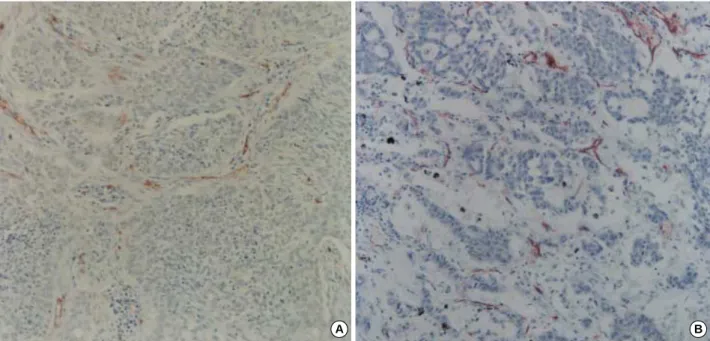
Fig. 1.Immunostaining for HIF-1 protein. (A) Squamous cell carcinoma shows dark brown nuclear staining, predominantly around necrot- ic area of the tumor (×40). (B) Moderately differentiated adenocarcinoma reveals positive nuclear reaction in a randomly scattered fash- ion (×100). N, necrotic area.
A B
N
N
39 SCC cases showing HIF-1 positivity, 22 cases exhibited the positive reaction predominantly around necrotic zones of tumor nests (Fig. 1A), and the others revealed the positivity in both around necrosis and non-necrotic area. In comparison with SCCs, five of the positive eight cases of ACs revealed the pos- itive reaction only in non-necrotic areas (Fig. 1B), and three of them showed the combined positive reaction in both non- necrotic areas and around necrotic zones of tumor nests (Table 3). In ACs the positive reaction displayed the tenden-
cy to occur in histologically poorly differentiated tumor por- tions. In both histologic types, HIF-1 positive tumor cells was focally scattered in small clusters. In contrast to SCCs, ACs more frequently showed cytoplasmic immunoreactivity which was regarded as negative reaction. Some non-neoplastic respiratory epithelial tissues neighboring to carcinomas also displayed focal nuclear positive reaction.
With the whole cases of this study, HIF-1 expression was not correlated with lymph node status, tumor stage, VEGF ex- pression, MVD, or PCNA index (p>0.05, respectively; Table 2).
VEGF expression, MVD, and PCNA index according to SCCs and ACs of lung
Immunoreactivity for VEGF showed no association be- tween the two histologic types (68.9% in SCCs vs 66.7% in ACs, p=0.828; Table 4). In the positive cases the reaction was noted in the cytoplasm of tumor cells over the large areas of tumors (Fig. 2). In addition to tumor cells, stromal cells also displayed positive reaction. CD34 immunoreactivity for MVD was heterogeneous, with no difference between the central
Fig. 2.Immunostaining for VEGF. (A) Squamous cell carcinoma shows reddish brown cytoplasmic staining in a diffuse fashion (×200). (B) Adenocarcinoma reveals the positive cytoplasmic reaction in a rather focal pattern (×200).
A B
SCC, squamous cell carcinoma; AC, adenocarcinoma. *, positive nucle- ar staining presenting in both around necrosis and non-necrotic area.
Histologic types
Distribution of HIF-1 staining Only around
necrosis
Only non-necrotic
area Combined*
SCC 22 0 8
AC 0 5 3
Table 3. Distribution of HIF-1 staining in squamous cell carci- nomas and adenocarcinomas of the lung
SCC, squamous cell carcinoma; AC, adenocarcinoma; MVD, microves- sel density.
Histologic types
VEGF (No. of cases) Negative Positive
PCNA (%) MVD (/mm2)
SCC 14 31 21.7±9.4 55.6±21.9
AC 13 26 16.0±9.8 43.9±21.6
p value 0.828 0.009 0.016
Table 4. VEGF expression, MVD, and PCNA index in squamous cell carcinomas and adenocarcinomas of the lung
SCC, squamous cell carcinoma; AC, adenocarcinoma; MVD, microves- sel density.
Histologic types VEGF (Positivity, %) MVD (/mm2) PCNA (%)
SCC 24/30 (80.0) 21.5±8.5 54.2±20.7
AC 5/8 (62.5) 12.7±7.4 42.2±24.4
p value 0.363 0.023 0.227 Table 5. VEGF expression, microvessel density, and PCNA index according to HIF-1 positive squamous cell carcinomas and adenocarcinomas of the lung
and marginal tumor areas. MVD revealed a significant dif- ference beween the two histologic types (21.7±9.4 microves- sels/mm2in SCCs vs 16.0±9.8 microvessels/mm2in ACs, p=0.009; Fig. 3; Table 4). The PCNA index also showed a significant difference between the two histologic types (55.6
±21.9% in SCCs vs 43.9±21.6% in ACs, p=0.016; Fig.
4; Table 4).
VEGF expression, MVD, and PCNA index according to HIF-1 positive SCCs and ACs
VEGF expression was present in 24 of 30 cases (80.0%) of HIF-1 positive SCCs, and in 5 of 8 cases (62.5%) of HIF-1 positive ACs. Compared with HIF-1 positive AC, HIF-1 positive SCCs showed the tendency of higher VEGF expres- sion, but there was no association between histologic types
Fig. 3.Immunostaining for CD34. (A) Squamous cell carcinoma shows increased CD34-positive microvessels in the interfaces between carcinoma nests and stoma (×100). (B) Adenocarcinoma also reveals increased CD34-positive microvessels around the tumor (×100).
A B
Fig. 4.Immunostaining for PCNA. (A) Squamous cell carcinoma shows diffuse strong positivity in the nuclei of the tumor cells (×100). (B) Adenocarcinoma reveals the positive nuclear reaction in a rather focal pattern (×100).
A B
(p=0.363; Table 5). MVD was significantly higher in HIF-1 positive SCCs than in HIF-1 positive ACs (p=0.023; Table 5). The PCNA index turned out to exhibit no difference be- tween the two histologic types showing HIF-1 positivity (p=0.227; Table 5).
Survival analysis
During the course of this study (maximal follow-up, 132 months; minimal follow-up, 1 month; median follow-up, 23 months), 9 cases were lost to follow-up. In this analysis all pa- tients died of the disease, and data on postoperative treatment were not available. As for MVD and PCNA index, we used the median values (17.8 microvessels/mm2and 43.0%, respec- tively) as cut-off points in the univariate analysis of survival.
Firstly, with regard to HIF-1 expression, 14 of the positive 34 patients were censored and 20 died (mean and median survival times, 47.3 and 29.0 months, respectively). Nine of the negative 41 patients were censored and 32 died (mean and median survival times, 36.9 and 24.0 months, respectively).
The overall survival was not associated with HIF-1 expres- sion (p=0.442 by log-rank test). It also had no relation with histologic types, VEGF expression, MVD, or PCNA index (p=0.672, p=0.277, p=0.890, and p=0.573 by log-rank test, respectively).
DISCUSSION
HIF-1 protein is known to activate the transcription of genes encoding transferrin, VEGF, endothelin-1, and inducible nitric oxide synthetase, which are implicated in vasodilation, neovascularization, and tumor metastasis, and plays an essen- tial role in oxygen homeostasis (2, 18, 19). In the present study, we investigated the differential expression of HIF-1 protein among common histologic types of NSCLC. In the cases of SCC, HIF-1 positive cells were predominantly located around tumor necrosis. The predominant perinecrotic expression of HIF-1 protein indicates that the hypoxic tumor microen- vironment may directly contribute for induction of HIF-1 activity in these cancers. Nevertheless, some positively stain- ing cells were also present in non-necrotic tumor nests. In the lower parts of columnar or squamous metaplastic epithe- lium neighboring to the tumor, the focal expression of HIF- 1 protein was also observed, which would be consistent with the presence of low pH and hypoxia. In the cases of AC, how- ever, it was interesting that the increased level of HIF-1 pro- tein was noted more frequently in poorly differentiated areas than in necrotic areas. This finding may reflect the existence of alternative regulatory modes of HIF-1 expression. Altered patterns of gene expression in cancer could arise both from genetic alterations in the tumor cells and from stimulation by an abnormal microenvironment within the tumor. As a matter of fact, a growing line of evidence indicates that both
oncogene activation and tumor suppressor gene inactivation are also associated with increased HIF-1 expression (20-22).
In other words the persistent HIF-1 expression in poorly differentiated ACs may show the emergence of an aggressive phenotype with high oxygen consumption as a result of the transformation itself and not of the hypoxic environment. HIF- 1 expression has been shown to be enhanced by v-src (20) and in response to several growth factors, including insulin- like growth factors (IGFs) 1 and 2, basic fibroblast growth factor, and epidermal growth factor (23). Activation of the phosphatidylinositol 3-kinase/AKT/FRAP pathway, which mediates signals from a broad range of growth factors, has likewise been demonstrated to increase HIF-1 expression (24). Though the precise mechanism of these interfaces with the hypoxia-sensitive pathway is still not clear, those findings suggest a more general influence of growth-promoting stim- uli on HIF-1 activity. Thus, our results indicate that there may exist a different molecular event in tumor progression between SCCs and ACs.
Although the HIF-1 protein is commonly up-regulated in a variety of cancers, the positive staining is known not to be universal. At present there is no certain explanation for this discrepancy. However, the prolonged fixation on pellets of hypoxic cells is known to substantially compromise antigen detection, so that failure to stain some tumors might be arti- ficial. In the survey of tissue culture cells by immunoblot anal- ysis, Wiesener et al. (4) found that under maximal hypoxic stimulation, all cells had detectable levels of at least one HIF subunit, albeit the levels were quite variable. On the basis of the above finding, it is possible that relatively low levels of induced expression were still below the detection threshold in this immunohistochemical analysis, or that some tumors were relatively well oxygenated so that the HIF-1 protein was not induced in the sections examined. Some tumors that were negative for the HIF-1 protein in our cases also may express the HIF-1 protein at levels below the limits of detection by the current immunohistochemical methodolo- gy. Otherwise other transcription factors that may have simi- lar biological properties to HIF-1 , such as HIF-2 or HIF- 3 , may also mediate hypoxic adaptation in tumors (25, 26).
Zhong et al. (12) found that the HIF-1 expression was noted in premalignant lesions such as colonic adenoma, breast ductal carcinoma in situ, and prostatic intraepithelial neopla- sia, whereas every benign tumor was negative for the HIF-1 expression. They suggested that HIF-1 expression can occur very early in carcinogenesis. In our study, premalignant lesions were not included, but exceptionally a few of non-neoplastic epithelial cells adjacent to cancer showed the immunoreactiv- ity for the HIF-1 protein. Zhong et al. (12) also found a sig- nificant association of HIF-1 expression with Ki-67 prolif- eration index. But we could not confirm a significant associ- ation of HIF-1 expression with the PCNA index in our cases of SCC and AC, although the use of a different proliferation marker might be related with this discrepancy. We also found
that the PCNA index in overall SCCs was significantly high- er than that in overall ACs, but within the category of HIF- 1 positive carcinomas the index did not show a significant difference between the two histologic types. Maybe this incon- sistency stems from the hypothesis that pulmonary SCCs form a heterogeneous group of tumors with different biological properties and clinical behaviors (27). Feldser et al. (23) report- ed that the treatment of cultured prostatic carcinoma cells with insulin, IGF-1, or IGF-2 induced the expression of HIF- 1 protein, which was in turn required for expression of IGF- 2 mRNA, suggesting the involvement of HIF-1 protein in an autocrine growth factor loop. Thus, the HIF-1 expression may be associated with growth factors, which endowed tumors with a higher PCNA index.
Since HIF-1 stabilization up-regulates the expression of angiogenic and glycolytic pathways to restore oxygen homeo- stasis, the HIF-1 protein may have an important role for the survival and growth of cancer. We examined the expres- sion of the most representative angiogenic factor VEGF and MVD in SCCs and ACs. The MVD of SCCs was significant- ly higher than that of ACs, whereas the VEGF expression of SCCs showed no significant difference from that of ACs. Even within the category of HIF-1 positive carcinomas, this ten- dency was also observed between SCCs and ACs. Thus, the MVD that we assessed by CD34 immunoreactivity was relat- ed to a specific histologic type of HIF-1 positive carcino- mas. These findings somewhat reflect the impact of HIF-1 protein on the angiogenic process of common NSCLCs, espe- cially SCCs. Otherwise the tendency of increasing MVD in SCCs could be associated with tumor necrosis, which was more frequently found in these types of cancer. On the con- trary, Tsoli et al. (28), who used CD31 as the endothelial cell marker, reported no relationship between histologic types and MVD. This discrepancy is most likely associated with differences in the evaluation of the results, the use of differ- ent endothelial cell marker, and tumor heterogeneity. As concerned with VEGF, the lack of direct correlation between HIF-1 and VEGF expression might suggest that although hypoxia triggers the expression of VEGF through HIF-1 stabilization, the process of angiogenesis in pulmonary SCCs and ACs is also subject to other modulators such as platelet- derived endothelial cell growth factor, bcl-2, c-erbB-2, and MUC1 glycoprotein (29-31). But our result was in contrast with those of Giatromanolaki et al. (13), who reported the strong association between HIF-1 expression and VEGF expression. This discordance may be related with the differ- ent interpretation methods in immunohistochemical stain- ing. They regarded both nuclear and cytoplasmic staining as HIF-1 positivity. As far as we know, although the HIF- 1 protein is synthesized in the cytoplasm and also degrad- ed in the cytoplasm, it would be assumed that only nuclear HIF-1 is the active form (15). However, we consider that the relationship between HIF-1 and angiogenic factors needs to be clarified, with the development of more strict criteria
for immunohistochemical interpretation.
Finally, we observed no association between the overall survival of the patients and HIF-1 expression (p=0.442).
This result was rather different from that of Giatromanolaki et al. (13) who reported that HIF-1 positive group was marginally related (p=0.08) to poor outcome. In addition, we also found no association between the overall survival and histologic parameters including histologic types, VEGF ex- pression, MVD, and PCNA index. As concerned with MVD and VEGF expression, some workers suggested that the two parameters were not correlated with the survival (28, 32).
However, other researchers reported that both MVD and VEGF expression were linked to poor survival (29, 33, 34).
These discrepant observations may be explained by the dif- ferent methodologies applied, the different approaches to the estimated results, and the cut-off levels used (28).
In conclusion, we suggest that the HIF-1 expression, which can occur with hypoxic tumor environment in SCCs or with histological dedifferentiation in ACs, may play a differential role according to the histologic types of common NSCLCs.
Thus, it seems that the process of HIF-1 expression is con- trolled by different mechanisms between the two histologic types.
RERERENCES
1. Chandel NS, McClintock DS, Feliciano CE, Wood TM, Melendez JA, Rodriguez AM, Schumacker PT. Reactive oxygen species gener- ated at mitochondrial complex II stabilizes HIF-1 during hypoxia:
a mechanism of O2sensing. J Biol Chem 2000; 275: 25130-8.
2. Ratcliffe PJ, Ebert BL, Firth JD, Gleadle JM, Maxwell PH, Nagao M, O’Rourke JF, Pugh CW, Wood SM. Oxygen regulated gene expression: erythropoietin as a model system. Kidney Int 1997; 51:
514-26.
3. Wenger PH. Mammalian oxygen sensing, signaling and gene regu- lation. J Exp Biol 2000; 203: 1253-63.
4. Wiesener MS, Turley H, Allen WE, Willam C, Eckardt KU, Talks KL, Wood SM, Gatter KC, Harris AL, Pugh CW, Ratcliffe PJ, Maxwell PH. Induction of endothelial PAS protein-1 by hypoxia: characteri- zation and comparison with hypoxia-inducible factor-1 . Blood 1998;
92: 2260-8.
5. Wang GL, Jiang BH, Rue EA, Semenza GL. Hypoxia-inducible fac- tor-1 is a basic-helix-loop-helix-PAS heterodimer regulated by cellu- lar O2tension. Proc Natl Acad Sci USA 1995; 92: 5510-4.
6. Salceda S, Caro J. Hypoxia-inducible factor 1alpha (HIF-1alpha) pro- tein is rapidly degraded by the ubiquitin-proteasome system under normoxic conditions. Its stabilization by hypoxia depends on redox- induced changes. J Biol Chem 1997; 272: 22642-7.
7. Huang LE, Gu J, Schau M, Bunn HF. Regulation of hypoxia-inducible factor 1 alpha is mediated by an O2dependent degradation domain via the ubiquitin-proteasome pathway. Proc Natl Acad Sci USA 1998;
95: 7987-92.
8. Hockel M, Schlenger K, Hockel S, Aral B, Schaffer U, Vaupel P. Tumor
hypoxia in pelvic recurrences of cervical cancer. Int J Cancer 1998;
79: 365-9.
9. Stadler P, Becker A, Feldmann HJ, Hansgen G, Dunst J, Wurschmidt F, Molls M. Influence of the hypoxic subvolume on the survival of patients with head and neck cancer. Int J Radiat Oncol Biol Phys 1999;
44: 749-54.
10. Dachs GU, Patterson AV, Firth JD, Ratcliffe PJ, Townsend KM, Strat- ford IJ, Harris AL. Targeting gene expression to hypoxic tumor cells.
Nat Med 1997; 3: 515-20.
11. Maxwell PH, Dachs GU, Gleadle JM, Nicholls LG, Harris AL, Strat- ford IJ, Hankinson O, Pugh CW, Ratcliffe PJ. Hypoxia-inducible factor-1 modulates gene expression in solid tumors and influences both angiogenesis and tumor growth. Proc Natl Acad Sci USA 1997;
94: 8104-9.
12. Zhong H, De Marzo AM, Laughner E, Lim M, Hilton DA, Zagzag D, Buechler P, Isaacs WB, Semenza GL, Simons JW. Overexpres- sion of hypoxia-inducible factor 1 in common human cancers and their metastases. Cancer Res 1999; 59: 5830-5.
13. Giatromanolaki A, Koukourakis MI, Sivridis E, Turley H, Talks K, Pezzella F, Gatter KC, Harris AL. Relation of hypoxia inducible fac- tor 1 and 2 in operable non-small cell lung cancer to angiogenic/
molecular profile of tumours and survival. Br J Cancer 2001; 85: 881- 90.
14. Travis WD, Colby TV, Corrin B, Shimosato Y, Brambilla E, Collabora- tors from 14 countries. World Health Organization Pathology Panel:
World Health Organization. Histological Typing of Lung and Pleu- ral Tumors. International Histological Classification of Tumors. Third ed. Springer Verlag, Berlin, 1999.
15. Bos R, Zhong H, Hanrahan CF, Mommers EC, Semenza GL, Pine- do HM, Abeloff MD, Simons JW, van Diest PJ, van der Wall E. Lev- els of hypoxia-inducible factor-1 during breast carcinogenesis. J Natl Cancer Inst 2001; 93: 309-14.
16. Weidner N, Semple JP, Welch WR, Folkman J. Tumor angiogenesis and metastasis-correlation in invasive breast carcinoma. N Eng J Med 1991; 324: 1-8.
17. Mattern J, Koomagi R, Volm M. Association of vascular endothelial growth factor expression with intratumoral microvessel density and tumour cell proliferation in human epidermoid lung carcinoma. Br J Cancer 1996; 73: 931-4.
18. Semenza GL. Regulation of mammalian O2homeostasis by hypoxia- inducible factor 1. J Biol Chem 1995; 270: 1230-7.
19. Chun YS, Kim MS, Park JW. Oxygen-dependent and -independent regulation of HIF-1alpha. J Korean Med Sci 2002; 17: 581-8.
20. Jiang BH, Agani F, Passaniti A, Semenza GL. V-SRC induces expres- sion of hypoxia-inducible facor 1 (HIF-1) and transcription of genes encoding vascular endothelial growth factor and enolase 1: involve- mentof HIF-1 in tumor progression. Cancer Res 1997; 57: 5328-35.
21. Baas IO, Mulder JW, Offerhaus GJ, Vogelstein B, Hamilton SR. An evaluation of six antibodies for immunohistochemistry of mutant p53 gene products in archival colorectal neoplasms. J Pathol 1994;
172: 5-12.
22. Maxwell PH, Wiesener MS, Chang GW, Clifford SC, Vaux EC, Cock- man ME, Wykoff CC, Pugh CW, Maher ER, Ratcliffe PJ. The tumor
suppressor protein VHL targets hypoxia-inducible factors for oxygen- dependent proteolysis. Nature (Lond) 1999; 399: 271-5.
23. Feldser D, Agani F, Iyer NV, Pak B, Ferreira G, Semenza GL. Recip- rocal positive regulation of hypoxia-inducible factor 1 and insulin- like growth factor 2. Cancer Res 1999; 59: 3915-8.
24. Zhong H, Chiles K, Feldser D, Laughner E, Hanrahan C, Georgescu MM, Simons JW, Semenza GL. Modulation of hypoxia-inducible factor 1 expression by the epidermal growth factor/phosphatidyl inositol 3-kinase/PTEN/ AKT/FRAP pathway in human prostate cancer cells: implications for tumor angiogenesis and therapeutics.
Cancer Res 2000; 60: 1541-5.
25. Hogenesch JB, Chan WK, Jackiw VH, Brown RC, Gu YZ, Pray-Grant M, Perdew GH, Bradfield CA. Characterization of a subset of the basic-helix-loop-helix-PAS superfamily that interacts with components of the dioxin signaling pathway. J Biol Chem 1997; 272: 8581-93.
26. Gu YZ, Moran SM, Hogenesch JB, Wartman L, Bradfield CA. Molec- ular characterization and chromosomal localization of a third -class hypoxia inducible factor subunit, HIF3 . Gene Expr 1998; 7: 205-3.
27. Mattern J, Koomagi R, Volm M. Biologial characterization of sub- groups of squamous cell lung carcinomas. Clin Cancer Res 1999; 5:
1459-63.
28. Tsoli E, Zacharatos P, Dasiou-Plakida D, Peros J, Evangelou K, Zavras AI, Yannoukakos D, Konstantopoulou I, Asimacopoulos PJ, Kittas C, Gorgoulis VG. Growth index is independent of microvessel den- sity in non-small-cell lung carcinomas. Hum Pathol 2002; 33: 812-8.
29. O’Byrne KJ, Koukourakis MI, Giatromanolaki A, Cox G, Turley H, Steward WP, Gatter K, Harris AL. Vascular endothelial growth factor, platelet-derived endothelial cell growth factor and angiogen- esis in non-small-cell lung cancer. Br J Cancer 2000; 82: 1427-32.
30. Koukourakis MI, Giatromanolaki A, O’Byrne KJ, Cox J, Krammer B, Gatter KC, Harris AL. bcl-2 and c-erbB-2 proteins are involved in the regulation of VEGF and of thymidine phosphorylase angiogenic activ- ity in non-small cell lung cancer. Clin Exp Metastasis 2000; 17: 545- 54.
31. Giatromanolaki A, Koukourakis MI, Sivridis E, O’Byrne K, Cox G, Thorpe PE, Gatter KC, Harris AL. Co-expression of MUC1 glycopro- tein with multiple angiogenic factors in non-small cell lung cancer suggests co-activation of angiogenic and migratory pathways. Clin Cancer Res 2000; 6: 1917-21.
32. Decaussin M, Sartelet H, Robert C, Moro D, Claraz C, Brambilla C, Brambilla E. Expression of vascular endothelial growth factor (VEGF) and its two receptors (VEGF-R1-Flt1 and VEGF-R2-Flk1/KDR) in non-small cell lung carcinomas (NSCLCs): correlation with angio- genesis and survival. J Pathol 1999; 188: 369-77.
33. Giatromanolaki A, Koukourakis MI, Kakolyris S, Turley H, O’Byrne K, Scott PA, Pezzella F, Georgoulias V, Harris AL, Gatter KC. Vas- cular endothelial growth factor, wild-type p53, and angiogenesis in early operable non-small cell lung cancer. Clin Cancer Res 1998; 4:
3017-24.
34. Fontanini G, Vignati S, Boldrini L, Chine S, Silvestri V, Lucchi M, Mussi A, Angeletti CA, Bevilacqua G. Vascular endothelial growth factor is associated with neovascularization and influences progression of non-small cell lung carcinoma. Clin Cancer Res 1997; 3: 861-5.
. .
. .