Giant Intramyocardial Aneurysm in a Patient with Intercoronary
Communication between the Left Circumflex Artery and Right Coronary Artery:
A Case Report
우관상동맥과 좌회선지간 교통이 있는 환자에서 나타난 거대 심근내 동맥류: 증례 보고
Yu Hyun Lee, MD , Noh Hyuck Park, MD* , Ji Yeon Park, MD , Seon-Jeong Kim, MD
Department of Radiology, Myongji Hospital, Hanyang University College of Medicine, Goyang, Korea
Coronary artery aneurysm is a rare disease. It occurs in the epicardial space, mostly along the course of major coronary arteries. Here, we report a case of a giant incidental aneurysm em- bedded in the basal posterior wall of the left ventricle. A 43-year-old woman was referred to our institution for the evaluation of cardiac palpitations that had been present from the previous 2 months. She reported no medical history (such as Kawasaki’s disease or hypertension) or pre- vious operative history. Echocardiogram and subsequent cardiac CT revealed a giant aneurysm in the left ventricle, with a direct fistulous connection to a dilated and tortuous left circumflex artery, which showed direct communication with the straight right coronary artery.
Received April 15, 2019 Revised June 6, 2019 Accepted June 19, 2019
*Corresponding author Noh Hyuck Park, MD Department of Radiology, Myongji Hospital, Hanyang University College of Medicine, 55 Hwasu-ro, 14beon-gil, Deogyang-gu, Goyang 10475, Korea.
Tel 82-31-810-7167 Fax 82-31-810-6537 E-mail [email protected] This is an Open Access article distributed under the terms of the Creative Commons Attribu- tion Non-Commercial License (https://creativecommons.org/
licenses/by-nc/4.0) which permits unrestricted non-commercial use, distribution, and reproduc- tion in any medium, provided the original work is properly cited.
ORCID iDs Yu Hyun Lee https://
orcid.org/0000-0002-7635-6762 Noh Hyuck Park
https://
orcid.org/0000-0003-4716-3491 Ji Yeon Park
https://
orcid.org/0000-0002-4933-0292 Seon-Jeong Kim
https://
orcid.org/0000-0001-9064-7155
ies. We encountered a patient with a giant aneurysm embedded in the basal posterior wall of the left ventricle (LV), with a direct fistulous connection to a dilated, tortuous left circumflex artery (LCX). Additionally, the LCX directly communicated with the right coronary artery (RCA) at the crux cordis. To our knowledge, this phenomenon has not been reported; herein, we describe this rare and incidental finding by focusing on its presentation in echocardio- gram (ECG)-gated multidetector computed tomography (MDCT).
CASE REPORT
A 43-year-old woman was referred to our institution for the evaluation of palpitation that had been present for 2 months. Notably, the palpitation occurred in the resting condition and ex- hibited a duration of 20 minutes. The patient had no remarkable medical history such as hy- pertension, diabetes, or trauma; however, she had received hormone replacement therapy for premature menopause throughout the prior 18 months. Her blood pressure was 122/72 mm Hg and her pulse rate was 103 beats/min. Chest radiography showed normal heart configura- tion and size.
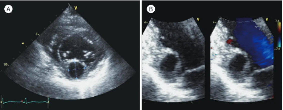
ECG showed normal sinus rhythm and 24 hours Holter monitoring showed ventricular pre- mature contraction. On ECG (Vivid 7 ultrasound, GE Healthcare, Chicago, IL, USA), a 2.3 × 2.0-cm-sized encapsulated, homogeneous, and anechoic round mass was identified at the bas- al posterior wall of the LV (Fig. 1A, B). Subsequently, further evaluation comprised cardiac CT, which was performed by using a 64-slice MDCT (Lightspeed VCT, GE Healthcare); this ex- amination revealed a 2.1 × 2.4-cm-sized intramyocardial coronary aneurysm (IMCA) with thin cap at the basal posterior wall of the LV (Fig. 1C). Additionally, a diffuse, dilated, and tortuous LCX was observed to communicate with the IMCA (Fig. 1D, E); there was direct intercoronary communication (ICC) between LCX and RCA at crux cordis (Fig. 1F). The RCA was straight without evidence of abnormal dilation; MDCT did not demonstrate any myocardial thicken-
A B
Fig. 1. A giant intramyocardial aneurysm with intercoronary communication in a 43-year-old woman, pre- senting with palpitation for 2 months.
A. Short axis view of echocardiogram shows a round, anechoic cystic mass (2.3 × 2.0-cm in size) at the bas- al posterior wall of the LV.
B. Long axis view of the echocardiogram with color Doppler shows that the intracardial cystic mass does not communicate with the color-filled LV.
LV = left ventricle
ing or stenosis in the coronary arteries. The coronary sinus was normal without any abnor- C
E
D
F
Fig. 1. A giant intramyocardial aneurysm with intercoronary communication in a 43-year-old woman, pre- senting with palpitation for 2 months.
C. An axial image of cardiac CT shows a round, intracardial cystic mass (arrow) without evidence of commu- nication with the LV at the basal posterior wall of the LV.
D, E. 3D reconstruction (D) and maximum intensity projection image (E) show a diffuse, dilated, and tortu- ous LCX (arrow) communicating with the intramyocardial coronary aneurysm (dashed arrow).
F. 3D reconstruction image shows a direct communication (dashed arrow) between the dilated and tortu- ous LCX (arrow) and RCA (arrow) at the crux cordis.
D = dimensional, LCX = left circumflex artery, LV = left ventricle, RCA = right coronary artery
tions; with these causes, most CAAs occur along major coronary arteries in the epicardial space (1-4). IMCA as exhibited in our patient is very rare with an unknown incidence; extensive re- view of the literature revealed a case report that introduced IMCA arising from the septal branch of the left anterior descending artery (LAD), secondary to primary percutaneous transluminal coronary angioplasty (5). The authors of that report suggested that intramyocardial localization of the aneurysm resulted in different angiographic, pathophysiological, and clinical features of the disease, compared with epicardial coronary aneurysm (5). Most CAAs are asymptomatic;
in those that are symptomatic, thrombus is frequently found within the aneurysm (1). Rupture of the CAA can be a life-threatening condition (2). Therefore, early diagnosis of CAA is critical.
Another remarkable finding of our case is the ICC between LCX and RCA at the crux cor- dis; this comprises a very rare subset of coronary artery anomalies, involving unidirectional or bidirectional blood flow between two or more coronary arteries (6, 7). The true prevalence in the general population is not known; however, coronary angiographic findings have shown in- cidences of 0.002% in 126595 patients and 0.02% in 9726 patients (8, 9).
Two types of ICC have been reported thus far: 1) between LAD and posterior descending arteries in the distal interventricular groove, and 2) between the LCX and RCA in the posteri- or atrioventricular groove (6), as in our patient. The practical significance of ICCs and their conse- quences remain unknown. Some authors speculate that these connections may play a protec- tive role for the myocardium upon the development of significant coronary artery obstruction in one of the connecting vessels. Importantly, myocardial ischemia can result from the coro- nary steal phenomenon by unidirectional flow (6). Collateral vessels and ICC are quite differ- ent: collaterals develop in obstructive coronary artery disease, are typically less than 1 mm in diameter, and appear tortuous and twisted with a corkscrew shape, whereas intercoronary anas- tomosis in the absence of obstructive lesions tends to be straight or gently curved (7). Histo- logically, collaterals that develop in the presence of obstructive coronary artery disease are composed of endothelium supported by poorly organized collagen, muscle, and elastic fi- bers; in contrast, ICCs are similar to an epicardial vessel with a well-defined muscular layer.
(10). Persistence of fetal coronary circulation has been suggested as the underlying mechanism for the development of ICC. A true intercommunication in the coronary system is benign, and may serve as a collateral source if a coronary artery obstruction develops (6).
We suspect either of two possibilities for intramyocardial CAA with this finding. First, spon- taneous closure of a preexisting fistula between the LAD artery and LV may have resulted in aneurysmal dilation at the distal intramyocardial fistulous segment; second, the straight RCA, with narrower caliber than the LCX, has higher pressure relative to the LCX, such that persis- tent unidirectional or bidirectional flow with dominant flow from RCA to LCX may cause di- lation and tortuosity of LCX, resulting in a giant intramyocardial CAA.
In conclusion, we have reported a rare instance of an IMCA in a patient with ICC between a dilated tortuous LCX and straight RCA.
Author Contributions
Conceptualization, P.N.H., P.J.Y., K.S.; supervision, P.N.H.; writing—original draft, L.Y.H.; and writ- ing—review & editing, P.N.H.
Conflicts of Interest
The authors have no potential conflicts of interest to disclose.
REFERENCES
1. Abou Sherif S, Ozden Tok O, Tas¸köylü Ö, Goktekin O, Kilic ID. Coronary artery aneurysms: a review of the epidemiology, pathophysiology, diagnosis, and treatment. Front Cardiovasc Med 2017;4:24
2. Johnson PT, Fishman EK. CT angiography of coronary artery aneurysms: detection, definition, causes, and treatment. AJR Am J Roentgenol 2010;195:928-934
3. Jeudy J, White CS, Kligerman SJ, Killam JL, Burke AP, Sechrist JW, et al. Spectrum of coronary artery aneu- rysms: from the radiologic pathology archives. Radiographics 2018;38:11-36
4. Edwards NFA, Wijesekera VA, Anderson BA, Habibian M, Burstow DJ, Walters DL, et al. A rare case of a giant coronary sinus with focal aneurysm secondary to multiple fistulous connections arising from a dilated, tortuous left circumflex coronary artery. CASE (Phila) 2018;2:99-102
5. Gungor B, Gurkan U, Alper AT, Bolca O. Intramyocardial coronary aneurysm: a distinct clinical entity. Int J Cardiol 2011;153:e39-40
6. Oliveira MD, Cavalcanti RR, Kajita AH, Miranda T, Kajita LJ, Horta PE, et al. Direct communication between the left circumflex and the right coronary arteries: a very rare coronary anomaly circulation. Cardiovasc Di- agn Ther 2016;6:87-91
7. Kwon SH, Kim EJ, Woo JS, Kim SJ, Youn HC, Oh JH. Communication between the right and circumflex cor- onary arteries discovered incidentally by multidetector computed tomography. J Korean Soc Radiol 2016;
75:214-217
8. Yamanaka O, Hobbs RE. Coronary artery anomalies in 126,595 patients undergoing coronary arteriogra- phy. Cathet Cardiovasc Diagn 1990;21:28-40
9. Gur M, Yilmaz R, Demirbag R. Unidirectional communication between the circumflex and right coronary arteries: a very rare coronary anomaly and cause of ischemia. Int J Cardiovasc Imaging 2006;22:339-342 10. Kim SH, Kim DH, Choi WG, Woo SI, Choi IS, Kwan J, et al. Intercoronary communication between the cir-
cumflex and right coronary arteries coexisted with coronary vasospasm. Korean Circ J 2013;43:488-490
우관상동맥과 좌회선지간 교통이 있는 환자에서 나타난 거대 심근내 동맥류: 증례 보고
이유현 · 박노혁* · 박지연 · 김선정
관상동맥류는 드문 질환으로 대개 주관상동맥의 주행을 따라 심외막공간에서 발생한다. 본 논문에서 우리는 우연히 발견된 좌심실의 하기저벽에 생긴 거대동맥류 사례를 보고하고자 한다. 가와사키병이나 고혈압을 비롯한 과거력이 없는 43세 여자 환자가 두 달간 지속된 심계 항진을 주소로 내원하였다. 심초음파와 심장 컴퓨터단층촬영에서 좌심실에 거대동맥류가 있 었고 이것은 좌회선지와 직접 동맥루를 이루었으며, 좌회선지는 우관상동맥과 교통하였다.
한양대학교 의과대학 명지병원 영상의학과