Successful Endovascular Treatment of an Infected Aortic Aneurysm Induced by Klebsiella pneumoniae
Klebsiella pneumonia로 인해 발생한 감염성 동맥류의 성공적인 혈관 내 치료
Hong Gwon Byun, MD1 , Yook Kim, MD1* , Jung Hwan Lee, MD1 , Jisun Lee, MD1 , Kil Sun Park, MD2
1Department of Radiology, Chungbuk National University Hospital, Cheongju, Korea
2Department of Radiology, Chungbuk National University College of Medicine, Cheongju, Korea
Aortic aneurysms infected by Klebsiella pneumoniae are rarely seen. We describe a 50-year-old man with infected aortic aneurysm that was successfully treated with endovascular aneurysm repair (EVAR). Diagnosis was confirmed using blood culture and computed tomography (CT).
Intravenous antibiotics were immediately administered, with improvements in clinical findings and negative blood cultures before the procedure. Twenty-four months after the procedure, the patient was stable and serial CT revealed regression of the infected aortic aneurysm. There- fore, after controlling bacteremia and fever with targeted antibiotic therapy, EVAR can be con- sidered as an alternative for patients who have serious comorbidities and are ineligible for con- ventional surgery.
Index terms Aneurysm, Infected; Endovascular Procedure; Klebsiella Infections; Antibiotics
INTRODUCTION
Infected aortic aneurysm is defined as the infectious destruction of the arterial wall with the formation of a saccular outpouching lesion that is connected to the arterial lu- men. It is a rare, life-threatening condition, with an incidence of 0.7–3% among all aor- tic aneurysms. Untreated or delayed treatment of infected aortic aneurysms often leads to fulminant sepsis, spontaneous arterial rupture, and death (1, 2).
Traditional treatment option of the infected aortic aneurysm is surgical resection and debridement of the infected aorta and surrounding tissue, and in situ or extra-anatomi- cal bypass, followed by long-term antibiotic therapy. However, surgical management is
Received June 30, 2019 Revised September 6, 2019 Accepted September 14, 2019
*Corresponding author Yook Kim, MD
Department of Radiology, Chungbuk National University Hospital,
776 1sunhwan-ro, Seowon-gu, Cheongju 28644, Korea.
Tel 82-43-269-6365 Fax 82-43-269-6479 E-mail sixtwin@hanmail.net This is an Open Access article distributed under the terms of the Creative Commons Attribu- tion Non-Commercial License (https://creativecommons.org/
licenses/by-nc/4.0) which permits unrestricted non-commercial use, distribution, and reproduc- tion in any medium, provided the original work is properly cited.
ORCID iDs Hong Gwon Byun https://
orcid.org/0000-0002-3227-1900 Yook Kim
https://
orcid.org/0000-0003-2162-419X Jung Hwan Lee
https://
orcid.org/0000-0002-8815-4092 Jisun Lee
https://
orcid.org/0000-0002-6264-7171 Kil Sun Park
https://
orcid.org/0000-0002-2639-3522
associated with high risk and mortality rates (22–36%) (2, 3).
In recent decades, endovascular aortic repair (EVAR) for thoracic or abdominal aortic an- eurysms has become widespread, with satisfactory results (4). Recent studies have reported that the endovascular treatments for infected aortic aneurysm are simpler than conventional treatments and may be effective for long-term prognosis (5). They may be good alternative treatments for infected aortic aneurysm; nevertheless, placement of an endovascular graft in an infected field remains a matter of controversy (2, 4).
Therefore, we present a case of a successful endovascular treatment, combined with tar- geted antibiotic therapy for infected aortic aneurysm induced by Klebsiella pneumoniae (K. pneumoniae), without complications on long-term follow-up.
CASE REPORT
A 50-year-old man with pancreatic cancer and hepatic metastasis presented to the emer- gency room with high fever (39.2℃). Laboratory examinations revealed white blood cell (WBC) count and C-reactive protein (CRP) level elevated at 19.24 × 10³/µL (normal range 4.0–
10.0 × 10³/µL) and 21.52 mg/dL (normal concentration < 0.3 mg/dL), respectively. Abdominal contrast-enhanced computed tomography (CT) revealed a 3.9-cm diameter saccular aneu- rysm arising from the left lateral aspect of the infrarenal aorta with surrounding thrombus and periaortic inflammation (Fig. 1A). Based on imaging and the presence of infection, we made a diagnosis of infected aortic aneurysm. Initially, the patient was treated with empiri- cal intravenous antibiotics (piperacillin/tazobactam) for 7 days until specific bacterial growth on blood culture.
EVAR was chosen as the treatment of choice because the patient had a high risk of periop- erative death and limited life expectancy.
Before endovascular treatment, K. pneumoniae grew on the initial blood culture. The anti- biotics were changed (ceftriaxone and ciprofloxacin) according to the sensitivity identified from the preoperative blood culture. Four days later, his body temperature was returned to normal levels, and the CRP level was markedly decreased. The WBC count normalized after 1 week. Finally, no bacterial growth was detected on numerous follow-up blood cultures dur- ing specific antibiotic treatments for 12 days (Fig. 1B).
Follow-up CT was performed 2 weeks later to evaluate the infected aortic aneurysm prior to EVAR. This study revealed that the size of the enhancing portion of the infected aortic an- eurysm increased from 2.7 cm to 3.9 cm, and shape changed into a more irregular and in- creased outpouching enhancing lesion at left lateral aspect of infected aortic aneurysm, sug- gesting impending rupture (Fig. 1C). Immediate endovascular treatment was performed using a 22 mm diameter and 8 cm length stent graft (Hercules: S&G Biotech Inc. Seongnam, Korea) (Fig. 1D). The patient had an uneventful operative and postoperative course. An im- mediate follow-up CT revealed complete isolation of the infected aortic aneurysm with no signs of complications such as endoleak (Fig. 1E). The patient was treated with postoperative antibiotics for 2 days without any signs of infection. Twelve-month follow-up CT showed con- siderable decrease in aortic inflammation with near total isolation and shrinkage of the an- eurysm. To date, 24 months after endovascular graft implantation, the patient was complete-
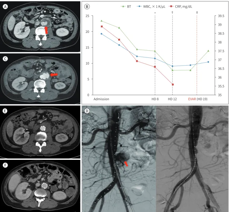
Fig. 1. A 50-year-old man with Klebsiella pnemonia induced aortic aneurysm. Primary diagnosis is based on abdominal CT.
A. Initial abdominal enhanced axial CT image shows a 3.9-cm diameter saccular aneurysm (arrow) arising from the left lateral aspect of the in- frarenal aorta with surrounding thrombus and periaortic inflammation, suggesting an infected aortic aneurysm.
B. Trending chart of the infection markers according to a timeline. CRP, WBC count, and BT are highly elevated on initial examination. Infec- tion markers markedly decrease after using bacterial-specific antibiotics. Endovascular treatment is administered after normalization of in- fection markers and absence of bacterial growth on blood culture.
C. The pre-procedure follow-up CT angiogram 2 weeks later demonstrates an increase in the size of the saccular aneurysm and changes in the shape, suggesting impending rupture (arrow).
D. Abdominal aorta angiography demonstrates contrast pulling to the left side of the infrarenal abdominal aorta (arrow). After implantation of stent graft including the infected aortic anerusymal site, the anurysm is completely isolated on control angiography.
E. Immediate follow-up CT scan reveals complete isolation of the infected aortic aneurysm with no signs of complications such as endoleak.
F. The follow up CT scan after 24 months of stent graft insertion demonstrates no significant complications associated with the procedure.
*Time when the antibiotic treatment was changed (changed on HD 8).
†Time when the fever was subsided (normalization of BT on HD 12).
‡No evidence of bacterial growth on follow-up blood culture and performance of the EVAR on HD 19.
BT = body temperature, CRP = C-reactive protein, CT = computed tomography, EVAR = endovascular aortic repair, HD = hospital days, WBC = white blood cell
D C
E
F
A B BT WBC, × 1 K/µL CRP, mg/dL
25
20
15
10
5
0
39.5 39 38.5 38 37.5 37 36.5 36 35.5 Admission HD 8 HD 12 EVAR (HD 19) 35
* † ‡
ly asymptomatic with no signs of procedure-related complications such as infection, aneurysmal growth, endoleak, or graft fracture (Fig. 1F).
DISCUSSION
Several bacterial species are involved in inflammatory processes, the most common being Staphylococcus aureus (40%), Salmonella (15%), Streptococci (8%), Escherichia coli (7%), and in some cases, Klebsiella, Haemophilus influenza, and Mycobacterium tuberculosis. As a re- sult of bacteremia, atheromatous lesions inside the aneurysm may become infected, leading to necrosis of the aortic wall and subsequent aneurismal rupture. Typical symptoms of infec- tion include fever, elevated WBC count and CRP level, as well as severe back and abdominal pain (1, 6). As demonstrated in our case, there were two consecutive preoperative CT scans taken within the period of 3 weeks, and there was an increase in the sac size, suggesting im- pending rupture within a short period of time. This is a characteristic of a rapidly enlarging infected aortic aneurysm. Given that rupture is highly associated with poor prognosis after endovascular treatment, EVAR should be performed immediately without delay. Short-term repeated follow-up CT and evaluation of infection parameters (e.g., CRP) are necessary be- fore performing EVAR (3).
Resection of the infected aorta and in situ graft replacement, together with long-term in- travenous antibiotic therapy is considered the standard of care (7, 8). Nevertheless, tradition- al open surgery entails complex techniques and procedures. Therefore, this surgical inter- vention is associated with high rates of morbidity and mortality. By contrast, endovascular treatment of infected aortic aneurysm has significant advantages over open surgery in that it avoids a large incision, full heparinization, extracorporeal circulation, aortic cross-clamping, interference with respiratory function, and the need for substantial blood transfusion (3). A systemic review of outcomes after EVAR for infected aortic aneurysm showed that endovas- cular treatment of infected aortic aneurysm is feasible and it is a durable treatment option for most patients (2, 7). Nevertheless, the release of the endoprosthesis in the infected area is controversial and violates general surgical principles; these objections do not pertain to EVAR. If the infection persists after EVAR, it is likely to produce subsequent irremediable di- saster. We suggest that EVAR can be performed in selected patients with infected aortic an- eurysm. First, this type of repair should be considered in patients at high risk for major com- plications after surgery (e.g., due to age or multiple comorbidities). Second, it could also be performed in a patient with a ruptured aneurysm as a temporary measure to quickly achieve hemodynamic stability and as a bridge to subsequent definite surgical treatment (2, 7, 8, 9).
Based on our experience handling the present case, we can propose several explanations for the successful use of EVAR in infected aortic aneurysms. First, immediate introduction of broad spectrum antibiotics is required as soon as an infected aortic aneurysm is suspected.
This is usually followed by appropriate antibiotics determined by culture and sensitivity test- ing, which might eradicate many bacteria. Therefore, many authors suggest that EVAR is fea- sible when antibiotic suppression has achieved negative blood cultures prior to surgery (2, 3, 5, 7, 8). If active infections are evident, there is a high risk of recurrence of infection after the procedure. Therefore, patients who have extensive infection are preferably excluded from
endovascular repair. In the present case, the patient had high fever and increased levels of parameters (e.g., CRP) at the time of operation, suggesting that the patient had an active in- fection. Therefore, the endovascular option could be performed when the infection was treated, confirmed by adjustment of fever, CRP, and absence of microbes on blood and tissue cultures after treatment with antibiotics targeted against K. pneumoniae. Second, prolonged postoperative antibiotic therapy is also advocated as a key component for success; however, there is no consensus regarding the optimal duration of antibiotic therapy (9). Most com- monly, parenteral antibiotics are given for 2 to 8 weeks; however, whether lifelong oral anti- biotics are necessary is a matter of debate (2, 3, 8). During the 24-month follow-up in our case, the patient did not report any clinical manifestations, a finding supported by CT. Anti- biotics were maintained for a short period of 2 days after treatment according to the sugges- tion of our communicable disease specialist to avoid adverse effects; we have not seen any sign of graft infection.
Based on our experience with this patient, we propose that EVAR might be an alternative therapy in selected patients who are not candidates for surgical replacement because of un- derlying disease when bacteremia and fever are controlled after treatment with targeted an- tibiotics for 3 weeks during hospitalization. Compared to conventional surgical treatment, EVAR with appropriate antibiotic therapy is a simpler, less traumatic and more efficient pro- cedure.
Author Contributions
Conceptualization, B.H.G., K.Y.; data curation, B.H.G., K.Y., L.J.H.; investigation, all authors; project administration, K.Y.; supervision, K.Y.; visualization, B.H.G., K.Y., L.J.H.; writing—original draft, B.H.G., K.Y.; and writing—review & editing, all authors.
Conflicts of Interest
The authors have no potential conflicts of interest to disclose.
REFERENCES
1. Lee WK, Mossop PJ, Little AF, Fitt GJ, Vrazas JI, Hoang JK, et al. Infected (mycotic) aneurysms: spectrum of imaging appearances and management. Radiographics 2008;28:1853-1868
2. Huang YK, Ko PJ, Chen CL, Tsai FC, Wu CH, Lin PJ, et al. Therapeutic opinion on endovascular repair for my- cotic aortic aneurysm. Ann Vasc Surg 2014;28:579-589
3. Kan CD, Lee HL, Yang YJ. Outcome after endovascular stent graft treatment for mycotic aortic aneurysm: a systematic review. J Vasc Surg 2007;46:906-912
4. Lederle FA, Freischlag JA, Kyriakides TC, Matsumura JS, Padberg FT Jr, Kohler TR, et al. Long-term compar- ison of endovascular and open repair of abdominal aortic aneurysm. N Engl J Med 2012;367:1988-1997 5. Sörelius K, Mani K, Björck M, Sedivy P, Wahlgren CM, Taylor P, et al. Endovascular treatment of mycotic aor-
tic aneurysms: a European multicenter study. Circulation 2014;130:2136-2142
6. Chen YJ, Chen SY, Wang JT, Hsueh PR. Mycotic aneurysm caused by gas-forming serotype K5 Klebsiella pneumoniae. Int J Infect Dis 2009;13:e47-e48
7. Silverberg D, Halak M, Yakubovitch D, Reinitz ER, Garniek A, Rimon U, et al. Endovascular management of mycotic aortic aneurysms. Vasc Endovascular Surg 2010;44:693-696
8. Sedivy P, Spacek M, El Samman K, Belohlavek O, Mach T, Jindrak V, et al. Endovascular treatment of infect- ed aortic aneurysms. Eur J Vasc Endovasc Surg 2012;44:385-394
9. Beltran-Ordonez IS, Yian TS, Hou A, Hai EW. Mycotic aneurysm of infrarenal aorta: a case report and review of literature. J Biosci Med (Irvine) 2015;3:88
Klebsiella pneumonia로 인해 발생한 감염성 동맥류의 성공적인 혈관 내 치료
변홍권1 · 김 육1* · 이정환1 · 이지선1 · 박길선2
Klebsiella pneumoniae에 의해 발생하는 감염성 대동맥류는 드문 것으로 알려져 있다. 저 자들은 감염성 대동맥류가 발생한 50세 남성에 대해 성공적으로 이루어진 혈관 내 치료를 보 고하고자 한다. 진단은 혈액 배양 검사와 전산화단층촬영을 통해 이루어졌다. 시술 전 투여 된 항생제로 인해 임상증상과 혈액 배양이 개선되었다. 시술 후 24개월 동안 환자는 안정된 상태였으며 감염성 대동맥류의 감소가 일련의 전산화단층촬영을 통해 확인되었다. 따라서 기저 질환으로 인해 외과적 치료를 할 수 없는 환자에서 혈관 내 치료는 선택적 항생제 사용 후 균혈증과 열이 조절되는 경우 감염성 대동맥류에 대한 치료 방법이 될 수 있다.
1충북대학교병원 영상의학과, 2충북대학교 의과대학 영상의학교실