Effect of Airflow Limitation on Acute Exacerbations in Patients with Destroyed Lungs by Tuberculosis
History of treatment for tuberculosis (TB) is a risk factor for obstructive lung disease.
However, it has been unclear whether the clinical characteristics of patients with destroyed lung by TB differ according to the presence or absence of airflow limitation. The objective of the study was to evaluate differences in acute exacerbations and forced expiratory volume in 1 second (FEV1) decline in patients with destroyed lung by TB according to the presence or absence of airflow limitation. We performed a retrospective cohort study and enrolled patients with destroyed lung by TB. The presence of airflow limitation was defined as FEV1/forced vital capacity (FVC) < 0.7. One hundred and fifty-nine patients were enrolled, and 128 (80.5%) had airflow limitation. The proportion of patients who experienced acute exacerbation was higher in patients with airflow limitation compared to those without (89.1 vs. 67.7%, respectively; P = 0.009). The rate of acute exacerbation was higher in patients with airflow limitation (IRR, 1.19; 95% CI, 1.11-1.27). Low body mass index (X vs. X + 1; HR, 0.944; 95% CI, 0.895-0.996) in addition to airflow limitation (HR, 1.634; 95% CI, 1.012-2.638), was an independent risk factor for acute exacerbation.
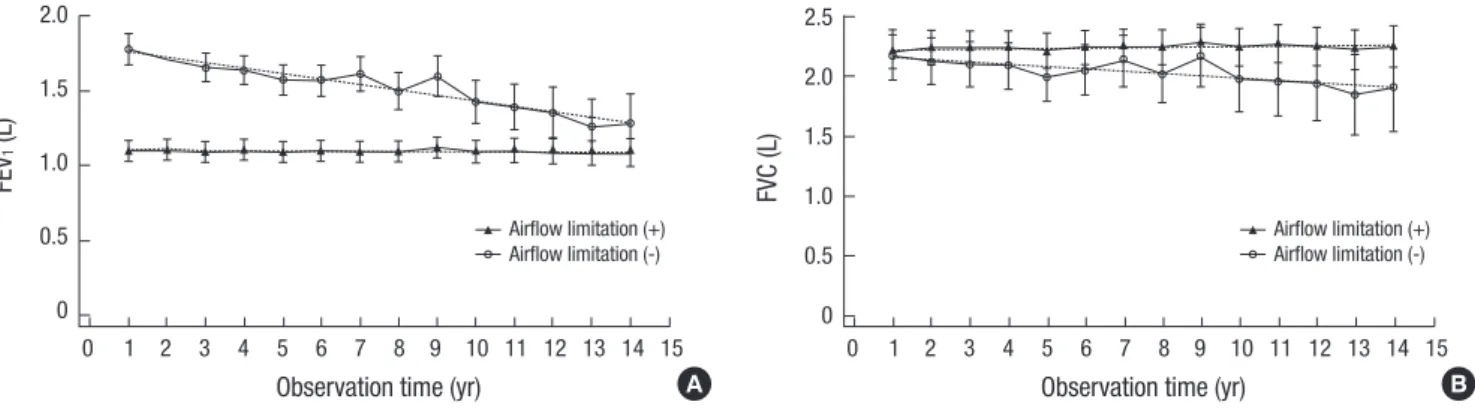
The annual decline of FEV1 was 2 mL in patients with airflow limitation and 36 mL in those without (P < 0.001). In conclusion, the presence of airflow limitation is an independent risk factor for acute exacerbation in patients with the destroyed lung by TB.
Keywords: Acute Exacerbation; Airflow Limitation; Pulmonary Function; Tuberculosis Soo Jung Kim, Jinwoo Lee, Young Sik Park,
Chang-Hoon Lee, Sang-Min Lee, Jae-Joon Yim, Young Whan Kim, Sung Koo Han, and Chul-Gyu Yoo Division of Pulmonary and Critical Care Medicine, Department of Internal Medicine, Seoul National University Hospital; Seoul National University College of Medicine, Seoul, Korea
Received: 30 October 2014 Accepted: 3 February 2015 Address for Correspondence:
Chul-Gyu Yoo, MD
Division of Pulmonary and Critical Care Medicine, Department of Internal Medicine, Seoul National University College of Medicine, 101 Daehak-ro, Jongno-gu, Seoul 110-744, Korea Tel: +82.2-2072-3760, Fax: +82.2-762-9662
E-mail: [email protected]
http://dx.doi.org/10.3346/jkms.2015.30.6.737 • J Korean Med Sci 2015; 30: 737-742
INTRODUCTION
Tuberculosis (TB) is the second most common cause of death due to infectious disease globally (1). It can cause pulmonary sequelae after microbiological cure; these are characterized by bronchial and parenchymal destruction, including broncho- vascular distortion, bronchiectasis, emphysematous changes, and fibrotic bands (2). These changes, which are collectively called destroyed lung by TB, result in respiratory functional dis- ability (3).
The pulmonary physiologic sequelae in these patients are obstructive, restrictive, or mixed changes. The prevalence of each abnormality varies among reports. Plit et al. (3) reported residual airflow limitation or a restrictive pattern in 28% and 24% of patients, respectively. By contrast, Chae et al. (4) report- ed obstructive ventilatory defects, including mixed defects, in 86.4% of patients with destroyed lung by TB.
Although the prevalence of airflow limitation in patients with destroyed lung by TB differs markedly (3-5), destroyed lung by TB is a risk factor for obstructive lung disease (5, 6). The possi- ble etiology of airflow limitation in these patients is either smok- ing itself (7) or peripheral airway collapse and subsequent air trapping due to parenchymal destruction (8).
Considering the similar physiological abnormalities, the clin-
ical manifestations of patients with destroyed lung by TB with airflow limitation could be similar to those of patients with chro- nic obstructive pulmonary disease (COPD). A few studies have compared the clinical characteristics of those two groups of pa- tients. Lee and Chang (9) reported that a positive bronchodila- tor response was observed more frequently in patients with COPD than in patients with chronic airflow obstruction sec- ondary to TB. Another study reported that patients with destroy- ed lung by TB showed more diminished pulmonary function and needed more tracheostomies than did patients with COPD in intensive care unit setting (10).
In a substantial number of patients with destroyed lung by TB, pulmonary physiological defects were observed to be pure- ly obstructive changes. Some previous studies have shown both similarities and differences in the clinical manifestations of pa- tients with destroyed lung by TB and airflow limitation and those with COPD. However, whether the clinical course and long-term outcomes of patients with destroyed lung by TB dif- fer according to the presence or absence of airflow limitation remains unclear. The objective of the present study was to eval- uate the impact of airflow limitation on the rate of acute exacer- bation and forced expiratory volume in 1-second (FEV1) decline in patients with destroyed lung by TB.
MATERIALS AND METHODS Subjects
We enrolled patients with pulmonary TB who were followed up at Seoul National University Hospital from January, 2005 to De- cember, 2011. Destroyed lung by TB was defined as follows:
previous history of antituberculosis treatment, negative acid- fast bacilli smear and mycobacterial culture for tuberculosis on the sputum sample, and parenchymal destruction in greater than 25% of a hemithorax on chest radiograph. Patients who were followed up for more than 3 yr and who had more than two spirometries during the followed-up period were enrolled.
The following patients were excluded from the study: those with active TB, nontuberculous mycobacteria infection, or other lung disease—such as pneumoconiosis, idiopathic fibrosis, and lung cancer. Patients with a history of lung resectional surgery were also excluded.
Study design
We performed a retrospective cohort study. Demographic, ra- diologic, and clinical parameters were compared between pa- tients with and without airflow limitation.
The presence of airflow limitation was defined as FEV1/ forced vital capacity (FVC) less than 70%. Extent of destruction was graded as three categories. If the extent of destruction on chest radiograph was less than one-third of the hemithorax, it was classified as grade 1. If the extent of destruction was between one- and two-thirds, it was classified as grade 2. If the extent of destruction was more than two-thirds, it was classified as grade 3.
Acute exacerbation was defined as an increase in or new on- set of more than one respiratory symptom (cough, sputum, or dyspnea) requiring a prescription of antibiotics, systemic ste- roid, or both or hospitalization. Frequent exacerbators were de- fined as patients who had two or more acute exacerbations per year. All exacerbations were separated by ≥ 1 month.
Statistical analysis
Differences in parameters between two groups were evaluated using the chi-square test or Student’s t-test. The rate of acute ex- acerbation was assessed using a Poisson regression model. In- dependent risk factors for exacerbation (with their 95% confi- dences interval [CIs]) were estimated by Cox regression analy- sis, including BMI as a continuous variable. Lung function de- cline was assessed using a mixed linear regression model.
Ethics statement
The present study was reviewed and approved by the institution- al review board for human research and was performed in accor- dance with Good Clinical Practice guidelines (H-1204-084-406).
RESULTS
Demographic data
In total, 386 patients were screened, and 159 were enrolled (Fig.
1). The baseline characteristics, lung function, and extent of de- struction are shown in Tables 1 and 2.
Among the 159 patients, 128 (80.5%) had airflow limitation at Table 1. Demographic characteristics of patients with destroyed lung by tuberculosis
Variables No. of patients (n = 159)
Female 74 (46.5)
Age (yr) 56.55 ± 10.81
Ever-smoker 51/118 (43.2)
Pack-years 10.97 ± 18.49
(n = 106)
BMI (kg/m2) 21.46 ± 3.92
Comorbidities Diabetes mellitus Hypertension Chronic renal failure Coronary artery disease Heart failure
Arrhythmia
13 (8.2) 45 (28.3)
2 (1.3) 4 (2.5) 9 (5.7) 13 (8.2)
Data are shown as No. (%) or mean ± SD. SD, standard deviation; BMI, body mass index.
Table 2. Lung function and extent of parenchymal destructions
Variables No. of patients (n = 159)
Lung function FVC (L) FVC (% predicted) FEV1 (L) FEV1 (% predicted) FEV1/FVC ratio (%) DLCO (% predicted)
2.20 ± 0.81 62.9 ± 17.5 1.20 ± 0.53 48.4 ± 19.8 56.5 ± 17.3 72.4 ± 19.0
(n = 30) Extent of destruction
Grade 1 Grade 2 Grade 3
68 (42.8) 47 (29.5) 44 (27.7)
Data are shown as No. (%) or mean ± SD. SD, standard deviation; FVC, forced vital capacity; FEV1, forced expiratory volume in 1 second; DLCO, Diffusing capacity for carbon monoxide.
386 patients were screened
Total 159 patients
227 patients were excluded
88 patients: parenchymal destruction less than 25% of a hemithorax on chest radiography 2 patients: active TB
33 patients: NTM infection
35 patients: previous lung resectional surgery 8 patients: other lung diseases
(pneumoconiosis, IPF, lung cancer) 61 patients: less than 3 yr follow-up
Fig. 1. Inclusion of patients. NTM, nontuberculous mycobacteria; IPF idiopathic fibrosis.
the time of diagnosis of destroyed lung by TB. In patients with airflow limitation, 60 patients were classified pure obstructive pattern without total lung capacity (TLC). Of the remaining 68 patients, TLC was estimated in 12 patients. Four patients had obstructive pattern and 8 patents had mixed pattern. In patients without airflow limitation, 26 patients showed restrictive pat- tern on spirometry and 5 patients had normal spirometry. The baseline characteristics of patients with or without airflow limi- tation were not significantly different, except for a higher body mass index (BMI) in patients with air flow limitation (21.83 ± 3.69 vs. 19.94 ± 4.50; P = 0.016) (Table 3).
Baseline lung function was evaluated. The % predicted value, but not absolute value, of FVC was significantly higher, and both the % predicted and absolute values of FEV1 was significantly lower in patients with airflow limitation compared to those with- out. When subgroup analysis was performed between 64 pa- tients with pure obstructive pattern and 8 patients with mixed pattern, FEV1 as well as FVC were higher in patients with pure obstructive pattern whether expressed as absolute (1.26 ± 0.48 vs. 0.90 ± 0.17 L, P < 0.001; 2.68 ± 0.82 vs. 1.88 ± 0.32 L, P < 0.001, respectively) or predicted values (51.73 ± 18.38 vs. 32.75% ± 8.63%, P < 0.001; 75.77 ± 15.11 vs. 51.13% ± 8.71%, P < 0.001, respectively). There was no significant difference in the extent of destruction between the two groups (Table 4).
Acute exacerbation
The proportion of patients who experienced acute exacerbation at least once during the period was higher in patients with air- flow limitation compared to those without (89.1 vs. 67.7%; P = 0.009) (Table 5). The duration of follow up was not significantly different between the two groups (Table 4). When only 55 never smokers with airflow limitation were compared with those with- out, the proportion of patients who experienced acute exacer- bation was higher in patients with airflow limitation than in those without (89.1 vs. 67.7%; P = 0.015). The proportion of patients who experienced acute exacerbation was not associated with the ex- tent of lung destruction (89.7% vs. 72.3% vs. 90.9%; P = 0.868).
After adjusting for age, gender, BMI, smoking history, and ex- tent of destruction, the rate of acute exacerbation (number of events/person-year) was higher in patients with airflow limita- tion than in those without (incidence rate ratio [IRR], 1.46; 95%
CI, 1.37-1.55). When FEV1 was adjusted for, the incidence rate of acute exacerbation remained higher in patients with airflow limitation (IRR, 1.19; 95% CI, 1.11-1.27; Table 5). Among patients with acute exacerbation, 8/114 (7.0%) of those with airflow lim- itation were frequent exacerbators. By contrast, no frequent ex- acerbator existed among patients without airflow limitation;
however, this was not statistically significant.
Predictive factors for acute exacerbation were evaluated by Cox regression analysis. After adjusting for age, gender, and ex-
Table 3. Demographic characteristics in patients with or without airflow limitation Variables Airflow limitation (+)
(n = 128)
Airflow limitation (-) (n = 31) P value
Female 61 (47.7) 13 (41.9) 0.567
Age (yr) 56.52 ± 9.64 56.55 ± 11.11 0.986
Ever-smoker 44/99 (44.4) 7/19 (36.8) 0.540
Pack-years 11.12 ± 19.03
(n = 87)
19.00 ± 10.26 (n = 19)
0.856
BMI (kg/m2) 21.83 ± 3.69 19.94 ± 4.50 0.016
Duration of follow up, months 121 ± 115 102 ± 33 0.384 Comorbidities
Diabetes mellitus Hypertension Chronic renal failure Coronary artery disease Heart failure
Arrhythmia
11 (6.5) 38 (29.7)
2 (1.6) 4 (3.1) 8 (6.3) 12 (9.4)
2 (6.5) 7 (22.6)
0 (0) 0 (0) 1 (3.2) 1 (3.2)
1.000 0.431 1.000 1.000 1.000 0.466 Data are shown as No. (%) or mean ± SD. SD, standard deviation; BMI, body mass index.
Table 4. Lung function and extent of parenchymal destruction in patients with or without airflow limitation
Variables Airflow limitation (+)
(n = 128) Airflow limitation (-)
(n = 31) P value Lung function
FVC (L) FVC (% predicted) FEV1 (L) FEV1 (% predicted) FEV1/FVC ratio (%) DLCO (% predicted)
2.26 ± 0.81 64.8 ± 17.3 1.10 ± 0.44 45.0 ± 16.7 50.2 ± 12.3 73.5 ± 19.0 (n = 26)
1.95 ± 0.76 55.1 ± 18.4 1.61 ± 0.68 62.7 ± 24.7 82.5 ± 9.1 65.0 ± 19.8
(n = 4)
0.060 0.007
< 0.001
< 0.001
< 0.001 0.441 Extent of destruction
Grade 1 Grade 2 Grade 3
60 (46.9) 35 (27.3) 33 (25.8)
8 (25.8) 12 (38.7) 11 (35.5)
0.104
Data are shown as No. (%) or mean ± SD. SD, standard deviation; FVC, forced vital capacity; FEV1, forced expiratory volume in 1 second; DLCO, Diffusing capacity for carbon monoxide.
Table 5. Frequency of acute exacerbation in patients with or without airflow limitation
Exacerbation Airflow limitation (+) (n = 128) Airflow limitation (-) (n = 31) P value
Incidence 114 (89.1) 21 (67.7) 0.009
Incidence rate, No. of events/person-year 0.50 (0-8.0) 0.38 (0-1.29) < 0.001*
Frequent exacerbator 8/114 (7.0) 0/21 (0) 0.357
Treatment with mechanical ventilation 12/114 (10.5) 4/21 (19.0) 0.276
Data are shown as No. (%) or median [range]. *Poisson regression, IRR, 1.19; 95% CI, 1.11-1.27, adjusted by age, gender, BMI, smoking history, extent of destruction, and ini- tial FEV1. IRR, incidence rate ratio; CI, confidence interval; BMI, body mass index; FEV1, forced expiratory volume in 1 second.
tent of destruction, the hazard ratio (HR) for acute exacerbation in patients with airflow limitation was 1.634. The HR of BMI (X vs. X + 1) was 0.994 (Table 6).
Annual decline of lung function
The decline of FEV1 was lower in the group with airflow limita- tion than in the group without (-2 vs. -36 mL/yr; P < 0.001). No significant difference in FVC decline was noted between the two groups (+0.7 vs. -19 mL/yr; P = 0.210) (Fig. 2). After adjust- ing for initial FEV1, the difference in FVC remained non-signifi- cant (+0.6 vs. -19 mL/yr; P = 0.198).
DISCUSSION
Our data suggest that airflow limitation is a risk factor for acute exacerbation in destroyed lung by TB. One of the most striking physiological sequelae of destroyed lung by TB is airflow limita- tion. However, the prevalence of airflow limitation in these pa- tients has been reported to vary from 28 to 86.4% (3, 4). In the present study, the prevalence of airflow limitation in enrolled patients was 80.5%. With the addition of previously excluded patients, the prevalence of airflow limitation was 76.9%. The variable prevalence of airflow limitation could be a result of dif- ferent definitions of destroyed lung by TB. Although we enrolled patients with parenchymal destruction in greater than 25% of one hemithorax, other studies did not define the extent of de- struction (3) or determined a different reference value of de-
struction (4).
Although airflow limitation was reported to be present in as high as 86.4% of patients with destroyed lung by TB, its patho- genesis is not fully understood. The best-known cause of airflow limitation is smoking (7); therefore, this could also be a factor in airflow limitation in destroyed lung by TB. However, more than half of patients with airflow limitation in the present study were never smokers, and there was no difference in the proportion of ever-smokers between two groups. Thus, the development of airflow limitation in patients with destroyed lung by TB could not be explained solely by smoking. This finding is supported by reports that airflow limitation developed after pulmonary TB independent of smoking status (11-13). A cross-sectional study documented that airway obstruction was associated with TB in never smokers (12). Furthermore, the PREPOCOL study estab- lished a strong association between a history of TB and airflow obstruction that was higher than that with smoking (13). At the molecular level, destroyed lung by TB and COPD possess a com- mon pathway of parenchymal destruction by matrix metallo- proteinases (14).
In the current study, the incidence of acute exacerbation was higher in patients with airflow limitation than in those without, suggesting that airflow limitation is a risk factor for acute exac- erbation in destroyed lung by TB. This result was supported by Cox regression analysis. As the frequency of acute exacerbation is known to correlate with FEV1 in COPD patients (15-17), we evaluated the incidence of acute exacerbation after adjusting for FEV1. The incidence of acute exacerbation remained greater in patients with airflow limitation. By contrast, the extent of pa- renchymal destruction was not associated with the incidence of acute exacerbation. These findings do not preclude the asso- ciation of lung destruction and acute exacerbations. As a whole, 85% (135/159) of patients experienced acute exacerbation. More- over, patients with grade 3 of parenchymal destruction were more likely to experience acute exacerbations than those with grade 2 destruction (90.9% vs. 72.3%). These results suggest that parenchymal destruction can affect acute exacerbations, althou- Table 6. Predicting factors for acute exacerbation
Variables HR (95% CI) P value
Airflow limitation 1.634 (1.012-2.638) 0.044
Age 1.003 (0.987-1.020) 0.705
Gender (male vs. female) 1.482 (0.968-2.270) 0.070 BMI, kg/m2 (X vs. X+1) 0.944 (0.895-0.996) 0.035 Extent of destruction
Grade 1 vs. Grade 2
Grade 1 vs. Grade 3 0.615 (0.397-0.953) 0.758 (0.474-1.212)
0.089 0.030 0.247 HR, hazard ratio; CI, confidence interval; BMI, body mass index.
Fig. 2. Changes of pulmonary finction tests. (A) Annual decline of FEV1. The annual decline of FEV1 was lower in patients with airflow limitation than in those without (-2 vs. -36 mL/yr, respectively; P < 0.001). (B) Annual decline of FVC. There was no significance difference in the rate of FVC decline between the two groups (+0.7 vs. -19 mL/yr; P = 0.201).
FEV1 (L)
Observation time (yr)
0 1 2 3 4 5 6 7 8 9 10 11 12 13 14 15 2.0
1.5
1.0
0.5
0
Airflow limitation (+) Airflow limitation (-)
FVC (L)
Observation time (yr)
0 1 2 3 4 5 6 7 8 9 10 11 12 13 14 15 2.5
2.0 1.5 1.0 0.5 0
Airflow limitation (+) Airflow limitation (-)
A B
gh it may not be directly proportional to the degree of destruc- tion. Therefore, it seems that the impact of airflow limitation is more important than lung destruction regarding the issue of acute exacerbations.
In the current study, another risk factor for acute exacerba- tion in destroyed lung by TB was BMI. This is similar to a report that risk factors for acute exacerbation in COPD included low BMI. Previous study reported that low BMI was significantly as- sociated with failure of noninvasive ventilation and the need to intubate in acute exacerbation of pulmonary TB sequelae (18).
Additionally, in COPD, the BODE index (which combines the following four variables into a composite score: [B] BMI; [O] air- flow obstruction; [D] dyspnea; and [E] exercise capacity) was a predictor of mortality regarding the risk of death from both any cause and from respiratory causes (19). Additionally, a BMI < 21 kg/m2 was frequent in patients hospitalized due to acute exac- erbation of COPD (20).
Airflow limitation in COPD is characterized by a progressive nature. Some previous studies have reported that greater decline in absolute FEV1 occurred in the early stage of COPD. Tantucci and Modina (21) reviewed recent clinical trials to assess the de- cline of lung function in COPD patients with each stage accord- ing to the severity of airflow obstruction. They reported that COPD patients in the early stages had more lung function to lose than those in the most-advanced stage. The mean rates of FEV1 decline in Global Initiative for Obstructive Lung Disease (GOLD) stages II and III 47-79 mL/yr and 56-59 mL/yr, respec- tively, and < 35 mL/yr in GOLD stage IV. In the present study, the predicted value of FEV1 in patients with airflow limitation was 45.0% ± 16.7%, which is comparable to COPD of GOLD stage III. The rate of decline of FEV1 in patients with airflow lim- itation (-2 mL/yr) is likely to be lower than that reported in COPD patients of GOLD stage III; this could be explained by smoking status. In the ECLIPSE study, 36% of enrolled patients were cur- rent smokers, and the rate of FEV1 decline was greater in cur- rent smokers than in former smokers (22). In the present study, current smokers reported only 8% (9/118) of patients. The lower number of current smokers could be due to lower rate of FEV1
decline compared with that in COPD patients. The different rate of FEV1 decline may reflect differences in the nature of airflow limitation between destroyed lung by TB and COPD.
The decline of FEV1 was lower in destroyed lung by TB with airflow limitation than that without limitation in the present study. The decline of FEV1 is proportional to FEV1 in COPD. Name- ly, the larger the FEV1, the faster the rate of FEV1 decline. Because FEV1 was significantly lower in patients with airflow limitation, airflow limitation could be the reason for the lower rate of FEV1
decline. However, when FEV1 was adjusted for, the rate of FEV1
decline remained lower in patients with airflow limitation, im- plying that the underlying cause remains unclear. Two pharma- cological studies, TORCH (23) and UPLIFT (24) trials demon-
strated that medical treatment including combinations of in- haled corticosteroids and long-acting beta-agonists or anticho- linergic bronchodilator improved lung function in COPD pa- tients. Although the effect of medical treatment is not known in patients with destroyed lung by TB, it is very likely that patients with airflow limitation in destroyed lung by TB might have re- ceived more aggressive treatment with bronchodilators or anti- inflammatory drugs than those without. If these drugs can re- duce the rate of FEV1 decline in patients with destroyed lung by TB as in COPD patients, the lower rate of FEV1 decline in pati- ents with airflow limitation could be explained by the difference in pharmacotherapy.
Little is known of the clinical differences in destroyed lung by TB according to airflow limitation. To our knowledge, this is the first study to compare the clinical features, particularly acute exacerbation and FEV1 decline, of patients with destroyed lung by TB with and without airflow limitation. Our study possessed some limitations. First, it was of a retrospective design. We could not evaluate patients with mixed changes separately because the initial total lung capacity data were not available. Second, the coexistence of COPD cannot be excluded because we in- cluded patients with a smoking history. However, excluding ever smokers with airflow limitation, acute exacerbation also occurred more in patients with airflow limitation. Third, phar- macotherapy was not evaluated. In COPD patients, a large body of clinical evidence has shown that long-acting bronchodilators are effective in preventing exacerbations (25, 26) and decreas- ing the decline of FEV1 (27, 28). Therefore, the efficacy of phar- macotherapy in destroyed lung by TB with airflow limitation should be evaluated. A well-designed prospective study with- out these limitations should be conducted to confirm the re- sults of the present study.
In conclusion, the presence of airflow limitation is an inde- pendent risk factor for acute exacerbation in patients with the destroyed lung by TB. Lung function decline is more severe in the group without airflow limitation in the destroyed lung by TB.
DISCLOSURE
The authors have no conflicts of interest to disclose.
AUTHOR CONTRIBUTION
Study conception: Kim SJ, Yoo CG. Design, data collection and analysis: Kim SJ, Yoo CG, Lee J, Park YS, Lee CH, Lee SM, Yim JJ, Kim YW, Han SK. Drafting: Kim SJ, Yoo CG. Revision and appro- val of manuscript: all authors.
ORCID
Soo Jung Kim http://orcid.org/0000-0001-9141-7749
REFERENCES
1. Corbett EL, Watt CJ, Walker N, Maher D, Williams BG, Raviglione MC, Dye C. The growing burden of tuberculosis: global trends and interac- tions with the HIV epidemic. Arch Intern Med 2003; 163: 1009-21.
2. Pasipanodya JG, Miller TL, Vecino M, Munguia G, Garmon R, Bae S, Drewyer G, Weis SE. Pulmonary impairment after tuberculosis. Chest 2007; 131: 1817-24.
3. Plit ML, Anderson R, Van Rensburg CE, Page-Shipp L, Blott JA, Fresen JL, Feldman C. Influence of antimicrobial chemotherapy on spirometric parameters and pro-inflammatory indices in severe pulmonary tuber- culosis. Eur Respir J 1998; 12: 351-6.
4. Chae JN, Jung CY, Shim SW, Rho BH, Jeon YJ. CT Radiologic findings in patients with tuberculous destroyed lung and correlation with lung func- tion. Tuberc Respir Dis 2011; 71: 202-9.
5. Hnizdo E, Singh T, Churchyard G. Chronic pulmonary function impair- ment caused by initial and recurrent pulmonary tuberculosis following treatment. Thorax 2000; 55: 32-8.
6. Lee SW, Kim YS, Kim DS, Oh YM, Lee SD. The risk of obstructive lung disease by previous pulmonary tuberculosis in a country with interme- diate burden of tuberculosis. J Korean Med Sci 2011; 26: 268-73.
7. Chakrabarti B, Calverley PM, Davies PD. Tuberculosis and its incidence, special nature, and relationship with chronic obstructive pulmonary disease. Int J Chron Obstruct Pulmon Dis 2007; 2: 263-72.
8. Jordan TS, Spencer EM, Davies P. Tuberculosis, bronchiectasis and chron- ic airflow obstruction. Respirology 2010; 15: 623-8.
9. Lee JH, Chang JH. Lung function in patients with chronic airflow ob- struction due to tuberculous destroyed lung. Respir Med 2003; 97: 1237- 42.
10. Seo YK, Lee CH, Lee HK, Lee YM, Park HK, Choi SB, Kim HG, Jang HJ, Yum HK, Lee SH. Differences between patients with TB-destroyed lung and patients with COPD admitted to the ICU. Tuberc Respir Dis 2011;
70: 323-9.
11. Lam KB, Jiang CQ, Jordan RE, Miller MR, Zhang WS, Cheng KK, Lam TH, Adab P. Prior TB, smoking, and airflow obstruction: a cross-section- al analysis of the Guangzhou Biobank Cohort Study. Chest 2010; 137:
593-600.
12. Perez-Padilla R, Fernandez R, Lopez Varela MV, Montes de Oca M, Mui- ño A, Tálamo C, Brito Jardim JR, Valdivia G, Baptista Menezes AM. Air- flow obstruction in never smokers in five Latin American cities: the PLA- TINO study. Arch Med Res 2012; 43: 159-65.
13. Caballero A, Torres-Duque CA, Jaramillo C, Bolivar F, Sanabria F, Oso- rio P, Orduz C, Guevara DP, Maldonado D. Prevalence of COPD in five Colombian cities situated at low, medium, and high altitude (PREPO- COL study). Chest 2008; 133: 343-9.
14. Elkington PT, Friedland JS. Matrix metalloproteinases in destructive pulmonary pathology. Thorax 2006; 61: 259-66.
15. Hurst JR, Vestbo J, Anzueto A, Locantore N, Müllerova H, Tal-Singer R, Miller B, Lomas DA, Agusti A, Macnee W, et al.; Evaluation of COPD
Longitudinally to Identify Predictive Surrogate Endpoints (ECLIPSE) Investigators. Susceptibility to exacerbation in chronic obstructive pul- monary disease. N Engl J Med 2010; 363: 1128-38.
16. Cote CG, Dordelly LJ, Celli BR. Impact of COPD exacerbations on pa- tient-centered outcomes. Chest 2007; 131: 696-704.
17. Hoogendoorn M, Feenstra TL, Hoogenveen RT, Al M, Mölken MR. As- sociation between lung function and exacerbation frequency in patients with COPD. Int J Chron Obstruct Pulmon Dis 2010; 5: 435-44.
18. Aso H, Kondoh Y, Taniguchi H, Kimura T, Nishiyama O, Kato K, Katao- ka K, Hasegawa Y. Noninvasive ventilation in patients with acute exac- erbation of pulmonary tuberculosis sequelae. Intern Med 2010; 49: 2077- 83.
19. Celli BR, Cote CG, Marin JM, Casanova C, Montes de Oca M, Mendez RA, Pinto Plata V, Cabral HJ. The body-mass index, airflow obstruction, dyspnea, and exercise capacity index in chronic obstructive pulmonary disease. N Engl J Med 2004; 350: 1005-12.
20. Lainscak M, von Haehling S, Doehner W, Sarc I, Jeric T, Ziherl K, Kos- nik M, Anker SD, Suskovic S. Body mass index and prognosis in patients hospitalized with acute exacerbation of chronic obstructive pulmonary disease. J Cachexia Sarcopenia Muscle 2011; 2: 81-6.
21. Tantucci C, Modina D. Lung function decline in COPD. Int J Chron Ob- struct Pulmon Dis 2012; 7: 95-9.
22. Vestbo J, Edwards LD, Scanlon PD, Yates JC, Agusti A, Bakke P, Calver- ley PM, Celli B, Coxson HO, Crim C, et al.; ECLIPSE Investigators. Chang- es in forced expiratory volume in 1 second over time in COPD. N Engl J Med 2011; 365: 1184-92.
23. Calverley PM, Anderson JA, Celli B, Ferguson GT, Jenkins C, Jones PW, Yates JC, Vestbo J.; TORCH investigators. Salmeterol and fluticasone propionate and survival in chronic obstructive pulmonary disease. N Engl J Med 2007; 356: 775-89.
24. Tashkin DP, Celli B, Senn S, Burkhart D, Kesten S, Menjoge S, Decramer M; UPLIFT Study Investigators. A 4-year trial of tiotropium in chronic obstructive pulmonary disease. N Engl J Med 2008; 359: 1543-54.
25. Wedzicha JA, Decramer M, Seemungal TA. The role of bronchodilator treatment in the prevention of exacerbations of COPD. Eur Respir J 2012;
40: 1545-54.
26. Jochmann A, Scherr A, Jochmann DC, Miedinger D, Török SS, Chhajed PN, Tamm M, Leuppi JD. Impact of adherence to the GOLD guidelines on symptom prevalence, lung function decline and exacerbation rate in the Swiss COPD cohort. Swiss Med Wkly 2012; 142: w13567.
27. Decramer M, Celli B, Kesten S, Lystig T, Mehra S, Tashkin DP. Effect of tiotropium on outcomes in patients with moderate chronic obstructive pulmonary disease (UPLIFT): a prespecified subgroup analysis of a ran- domised controlled trial. Lancet 2009; 374: 1171-8.
28. Celli BR, Thomas NE, Anderson JA, Ferguson GT, Jenkins CR, Jones PW, Vestbo J, Knobil K, Yates JC, Calverley PM. Effect of pharmacother- apy on rate of decline of lung function in chronic obstructive pulmonary disease: results from the TORCH study. Am J Respir Crit Care Med 2008;
178: 332-8.