Subchondral Insufficiency Fracture of the Femoral Head in Elderly People
We evaluated the clinical course of subchondral insufficiency fracture of the femoral head (SIFFH) and its characteristic findings with special regard to joint space narrowing (JSN).
Thirty-one cases of SIFFH of mean age 68.9 yr initially underwent limited weight-bearing conservative treatment. During the follow-up period, the patients with intractable pain underwent total hip arthroplasty (THA). For radiographic evaluation, lateral center-edge angle, JSN and femoral head collapse (FHC) were documented, and the extent of FHC was classified as mild (< 2 mm), moderate (2-4 mm), and severe (> 4 mm). The progression or new development of FHC more than 2 mm was evaluated on sequential plain radiographs.
The relationship between radiographic parameters and clinical outcomes were evaluated.
THAs were performed in 15 cases (48.4%). There was no significant correlation between clinical outcomes and the extent of initial FHC. However, a significantly larger proportion of patients that underwent THA showed JSN and FHC progression compared to the symptom improvement group. The risk factor significantly associated with failed conservative treatment was JSN (P = 0.038; OR, 11.8; 95% CI, 1.15-122.26). Clinical results of conservative treatment for SIFFH in elderly patients are relatively poor. The patients with JSN are at higher risk of failed conservative treatment.
Keywords: Hip; Fractures; Bone; Risk Factors; Outcome Assessment (Health Care) Pil Whan Yoon, Hong Suk Kwak,
Jeong Joon Yoo, Kang Sup Yoon, and Hee Joong Kim
Department of Orthopedic Surgery, Seoul National University College of Medicine, Seoul, Korea Received: 10 October 2013
Accepted: 7 February 2014 Address for Correspondence:
Hee Joong Kim, MD
Department of Orthopedic Surgery, Seoul National University College of Medicine, Medical Research Center, Seoul National University, 101 Daehak-ro, Jongno-gu, Seoul 110-744, Korea Tel: +82.2-2072-2360, Fax: +82.2-764-2718
E-mail: [email protected]
This study was supported by a grant (06-03-063) from the Seoul National University Hospital Research Fund.
http://dx.doi.org/10.3346/jkms.2014.29.4.593 • J Korean Med Sci 2014; 29: 593-598
INTRODUCTION
Subchondral stress fracture of the femoral head can occur as two types; fatigue-type and insufficiency-type. Of them, sub- chondral insufficiency fracture of the femoral head (SIFFH) is caused by normal or physiological stress without antecedent trauma and usually occurs in elderly patients with poor bone quality. Since it has been reported not only in osteoporotic pa- tients without underlying disease, but also in patients who were treated with corticosteroids for systemic lupus erythematosus and organ (kidney or liver) transplantation, most authors em- phasize the importance of differentiating SIFFH from osteone- crosis (1-4). SIFFH is known as a rare condition. But the num- ber of cases of SIFFH in the literature is increasing on the basis of characteristic findings from magnetic resonance image (MRI) such as subchondral fractures and bone marrow edema pat- terns which are distinct from those of osteonecrosis (5-9). How- ever, most cases were reported as case reports and the number of case series studies is very limited.
Unlike fatigue-type subchondral fracture of the femoral head which occurs in healthy adults with good bone quality after sud- den increases in activity, joint space narrowing has been observ- ed in some hip joints affected by SIFFH. However, the cause and clinical significance of the joint space narrowing are unknown.
The purpose of current study was to evaluate the clinical course of SIFFH and its characteristic findings with special regard to joint space narrowing.
MATERIALS AND METHODS
Thirty one hips with SIFFH were diagnosed and treated at a sin- gle institution from December 2000 to May 2011. The average age was 68.9 yr (range, 53.0 to 90.3 yr). There were 26 women and five men with 15 right and 16 left hips. All patients present- ed with acute hip pain during daily activity without precedent trauma. None of them had concomitant metabolic disease wor- sening bone quality except for osteoporosis. A sixty-two-year- old female patient had radiation therapy over the area of the left ilium and ischium for metastatic lesions of recurring rectal can- cer twenty years ago. While only one patient was a regular drink- er ( ≥ 400 mL of alcohol weekly), others had no predisposing factors for osteonecrosis of the femoral head such as alcohol consumption and corticosteroid treatment.
Radiographic evaluation
Diagnosis was confirmed with characteristic MR findings which were distinctive for osteonecrosis of the femoral head (10), as well as with plain radiographs and bone scans. On plain radio-
graphs, the lateral center-edge (CE) angle was measured as well as the presence of femoral head collapse (FHC), joint space nar- rowing (JSN) and linear patch sclerotic changes in the superior portion of the femoral head were identified. The extent of the FHC was measured using concentric circles and classified as mild (< 2 mm), moderate (2-4 mm), and severe (> 4 mm) (Fig.
1). The progression or new development of the FHC was de- fined when there was > 2 mm increase in extent as compared with the initial radiograph, and then it was serially evaluated on sequential plain radiographs. The joint space width was mea- sured on standard anteroposterior radiograph of the hip by the same method as described in our previous paper (11). Joint space narrowing was defined when there was > a 0.5 mm de- crease in width as compared with the contralateral hip on the radiograph. Bone scans were performed in seven patients with a high suspicion of SIFFH even though their plain radiographs showed no definite abnormal findings. The characteristic MR features of bone marrow edema patterns, subchondral fracture lines and their locations were recorded in all patients.
Clinical outcome evaluation
The severity of osteoporosis was estimated on the basis of a bone
mineral density (BMD) measurement by dual-energy X-ray ab- sorptiometry (DEXA) in eight patients and calculation of the Singh index score at the time of the onset of hip pain in all pa- tients. Body mass index (BMI) was calculated as kg/m2. Initially, non-weight bearing conservative treatment was plan- ned, then the patients were allowed to bear weight as tolerated but crutches were used until the hip pain almost completely disappeared. During the follow-up period, the patients with in- tractable pain underwent total hip arthroplasty (THA). At base- line and the end of follow-up, clinical outcome assessments were performed using the Western Ontario and McMaster Uni- versities (WOMAC) scores in patients who had symptom im- provements without surgical treatment.
Statistical analysis
Data processing and statistical analyses were performed using IBM SPSS statistics software package (version 20; IBM, Chicago, IL, USA). To evaluate the relationship between radiographic parameters and clinical outcomes, a chi-square test or Fisher’s exact test was used for analysis of categorical data such as pres- ence of JSN and FHC progression. A Mann-Whitney U-test was used for parametric data between the symptom improvement group and THA group. To assess which variables were associat- ed with failed conservative treatment, a multiple logistic regres- sion was used. The parameters included in the model were se- lected using the univariate analysis (P < 0.2). P values below 0.05 were considered statistically significant.
Ethics statement
The design and protocol of this study were approved by the in- stitutional review board in our hospital (IRB No. H-1201-069- 394), and informed consent was obtained from each patient.
RESULTS
In all cases, magnetic resonance images revealed a serpentine low signal intensity line with surrounding bone marrow edema in the subchondral area of the femoral head. The bone-marrow- edema pattern extended to the subchondral area, but there was no additional abnormal signal intensity band representing the reactive margin of the necrotic area which is the characteristic finding of osteonecrosis of the femoral head. Subchondral frac- ture lines were observed at the anterosuperior aspect of the fe- moral head in thirty cases and at the posterosuperior aspect in one case. A bone scintigram was available for seven patients and they all showed increased radionuclide uptake in the af- fected femoral head.
Collapse of the femoral head at the anteosuperior portion was identified in 13 (41.9%) of 31 hips on initial radiograph as- sessment. The collapse was mild in five, moderate in five and severe three cases. When reevaluated on final follow-up of ra- Fig. 1. The extent of femoral head collapse (the distance between A and B) was mea-
sured using concentric circles (C, the center of the femoral head).
A B
C
diographs, progression of FHC more than 2 mm was observed in 10 of 13 hips with initial collapse. Three of 18 cases without FHC at the time of initial diagnosis showed development of col- lapse; however, there was no evidence of collapse in the remain- ing 14 cases until the final follow-up visit.
A total of 21 cases (67.7%) showed JSN of the affected hip on plain radiographs compared to the unaffected one. It was de- tected at the time of initial diagnosis in 16 cases and at 4-8 weeks after diagnosis in five cases (Fig. 2).
The mean CE angle of the affected hip in all cases was 27.0 degrees (range, 16.0 to 39.7 degrees). The mean T-score of the affected proximal femur in eight patients was -3.7 ± 0.5 and the mean Singh index score for all cases was 2.9 (range, 1 to 4). The mean BMI for all cases was 22.8 kg/m2 (range, 17.0 to 27.0 kg/m2).
The average duration of symptoms from onset to diagnosis was 2.1 months (range, 1 to 6 months). The initial treatment for all patients was limited weight-bearing walking (as much as
possible), with a walking aid. It was followed by tolerable weight bearing, and full weight bearing was allowed after patients be- came pain-free and no further progression of femoral head col- lapse was confirmed on follow-up radiographs. In 16 cases, pain reduced and gradually disappeared almost completely by an average of 5 months (range, 4 to 6 months) after conservative treatment. Their mean WOMAC scores were significantly im- proved from 42.1 (range, 28 to 52) at baseline to 89.9 (range, 81 to 96) at the end of follow-up; the last follow-up averaged 15.9 months (range, 12 to 30.2 months).
THAs were performed in 15 cases (48.4%) due to intractable pain with or without remarkable progression of FHC during the follow-up period. The mean interval between initial diagnosis and THA was 5.6 months (range, 1.5 to 9.5 months). The carti- lage of the femoral head was markedly damaged in these cases with JSN (Fig. 3-6).
There was no significant correlation between clinical outco-
A B
Fig. 2. An anteroposterior radiograph of an 82-yr-old male patient with left hip pain showed no femoral head collapse and joint space narrowing initially (A). Radiograph made at eight weeks later shows joint space narrowing of the left hip (B).
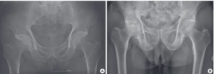
A B
Fig. 3. The initial radiograph of a 76-yr-old male patient shows joint space narrowing of the right hip joint (A). Radiograph five months later shows progression of joint space narrowing and femoral head collapse (B).
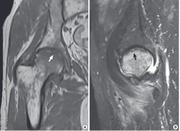
Fig. 4. MR images of the same patient in Fig. 3. (A) T1- and (B) T2-weighted signal intensity images show a subchondral fracture (arrows) and the bone-marrow-edema pattern extending to the subchondral area.
A B
Fig. 5. Gross photograph of the resected femoral head of the same patient in Fig. 3 showed a markedly destroyed articular cartilage.
mes and the extent of initial FHC. However, a significantly larg- er proportion of patients who underwent THA showed JSN and FHC progression compared to the symptom improvement group (P = 0.006 and 0.048, respectively, Table 1). The risk factor sig- nificantly associated with failed conservative treatment was JSN (P = 0.038; OR, 11.8; 95% CI; 1.15-122.26, Table 2). There was a significant difference between the mean age of cases with JSN (71.3 ± 8.5 yr) than those cases without JSN (63.8 ± 6.8 yr;
P = 0.025, Table 3).
DISCUSSION
In the current study, 15 cases (48.4%) of SIFFH underwent THA because the conservative, limited weight-bearing treatment was not satisfactory. The risk for failure of conservative treatment was substantially higher in the patients with JSN.
These results are markedly different from those of fatigue-type subchondral fractures of the femoral head, which is another type of stress fracture of the femoral head that occurs in healthy people with good bone quality, frequently after a sudden in- crease in activity (12, 13). Clinical results of fatigue-type frac- tures have been reported to be very satisfactory with conserva- tive treatment even with some collapse of the femoral head (12- 14). Furthermore, no joint space narrowing has been reported in fatigue-type fractures.
Because of the rare incidence of SIFFH, most of the reported cases have been case reports or small series reports (1-5, 9, 15- 17). Of these limited number of cases, many were reported to be treated by THA. Yamamoto and Bullough (16) reported ten cases of SIFFH on histopathologic basis which had been previ- ously diagnosed as osteonecrosis. All patients had failed initial conservative therapy and they eventually undergone THA be- tween 2 and 13 months from the onset of hip pain. Buttaro et al.
(5) reported four cases of SIFFH. All cases showed progressive femoral head collapse and three of them were treated with THA
Table 1. Univariate analysis comparing symptom improvement group and THA groups*
Variables
Clinical outcome
P value Symptom improvement
(n = 16) THA
(n = 15) Gender
Male
Female 4 (25.0%)
12 (75.0%) 1 (6.5%)
14 (93.5%)
0.333
Patient age (yr) 67.9 ± 7.8 69.9 ± 9.6 0.830
BMI (kg/m2) 23.0 ± 2.0 22.6 ± 2.7 0.682
Singh index 3.1 ± 1.1 2.7 ± 0.8 0.281
CEA (degrees) 28.7 ± 6.4 25.2 ± 6.7 0.175
The extent of initial FHC None
Mild ( < 2 mm) Moderate (2-4 mm) Severe ( > 4 mm)
10 (62.5%) 2 (12.5%) 2 (12.5%) 2 (12.5%)
8 (53.3%) 3 (20.0%) 3 (20.0%) 1 (6.7%)
0.854
FHC progression 4 (25.0%) 9 (60.0%) 0.048
JSN 7 (43.8%) 14 (93.3%) 0.006
*Data represent the mean ± standard deviation. BMI, body mass index; CEA, center- edge angle; FHC, femoral head collapse; JSN, joint space narrowing; THA, total hip arthroplasty.
Table 2. Multiple logistic regression results Parameters Estimate Standard
error Chi-
square P value OR 95% CI for OR
CEA -0.079 0.081 0.954 0.329 0.924 0.789-1.082
FHC progression 1.328 0.946 1.970 0.160 3.773 0.591-24.109
JSN 2.472 1.191 4.309 0.038 11.846 1.148-122.255
OR, odds ratio; CI, confidence interval; CEA, center-edge angle; FHC, femoral head collapse; JSN, joint space narrowing.
Table 3. Univariate analysis comparing the patients with and without joint space nar- rowing*
Variables Joint space narrowing
P value Yes (n = 21) No (n = 10)
Patient age (yr) 71.3 ± 8.5 63.8 ± 6.8 0.025
BMI (kg/m2) 22.7 ± 2.4 23.0 ± 2.2 0.554
Singh index 2.9 ± 0.8 3.1 ± 1.3 0.535
CEA (degrees) 25.7 ± 6.1 29.8 ± 7.3 0.128
*Data represent the mean ± standard deviation. BMI, body mass index; CEA, center- edge angle.
A B
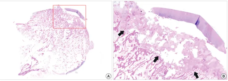
Fig. 6. Histology of the superior portion (dotted read rectangle) of the resected femoral head of the same patient in Fig. 3, showing fracture callus formation (arrows) and granu- lation tissue (H&E, ×40 [A], ×100 [B]).
after 4-12 months of conservative treatment. Recently, Miyani- shi et al. (18) reported the largest case series of 27 patients with SIFFH. Thirteen cases (48%) underwent THA. Femoral head collapse was observed much more frequently in the THA group than in the conservative treatment group, but only age was a statistically significant risk factor for THA with the cut-off value of 71 yr.
In the current study, patient age was not directly associated with failure of conservative treatment, while JSN of the affected hip on plain radiographs compared to the unaffected one was the only significant risk factor for a poor outcome. Joint space narrowing was observed in many cases reported in the litera- ture and some authors suggested that this finding is an early sign of rapidly destructive arthrosis of the hip joint (2, 9, 15, 17, 19). However, there are no studies documenting JSN as a prog- nostic factor of SIFFH. We hypothesized that JSN is a factor for
poor prognosis because it has not been reported in fatigue-type subchondral stress fractures of the femoral head which showed very satisfactory results with conservative treatment, in spite of FHC. And our hypothesis was supported by the results of statis- tical analyses. A univariate analysis proved that a significantly larger proportion of patients that underwent THA showed JSN compared to the symptom improvement group (P = 0.006), and JSN was the significant risk factor for failure of conservative treat- ment according to the result of the multiple logistic regression analysis (P = 0.038; OR, 11.8; 95% CI, 1.15-122.26). The mecha- nism of the development of JSN in SIFFH is unknown. In our study, the mean age of cases with JSN (71.3 ± 8.5 yr) was signifi- cantly older than that of cases without JSN (63.8 ± 6.8 yr; P = 0.025).
In summary, conservative treatment failed in 15 cases (48.4%), and during the average follow-up period of 15.9 months, pain
resolved to a tolerable level in 16 cases, even though there was FHC and JSN on radiographs in some of these cases at last fol- low-up. This may be due to low-demand activity in older pa- tients and the symptoms in these patients may deteriorate with a longer follow-up. Clinical results of conservative treatment for SIFFH in older patients are relatively poor compared to those for fatigue-type subchondral fracture of the femoral head. The patients with JSN are at higher risk of failed conservative treat- ment.
ACKNOWLEDGMENT
We thank Sohee Oh from Seoul National University Boramae Hospital for statistical advice.
DISCLOSURE
The authors declare no potential conflicts of interest.
ORCID
Pil Whan Yoon http://orcid.org/0000-0001-7354-0989 Hong Suk Kwak http://orcid.org/0000-0001-5204-6201 Jeong Joon Yoo http://orcid.org/0000-0002-6304-0101 Kang Sup Yoon http://orcid.org/0000-0001-5917-1881 Hee Joong Kim http://orcid.org/0000-0002-3994-5672 REFERENCES
1. Ikemura S, Yamamoto T, Nakashima Y, Shuto T, Jingushi S, Iwamoto Y.
Bilateral subchondral insufficiency fracture of the femoral head after re- nal transplantation: a case report. Arthritis Rheum 2005; 52: 1293-6.
2. Yamamoto T, Schneider R, Iwamoto Y, Bullough PG. Subchondral in- sufficiency fracture of the femoral head in a patient with systemic lupus erythematosus. Ann Rheum Dis 2006; 65: 837-8.
3. Iwasaki K, Yamamoto T, Nakashima Y, Mawatari T, Motomura G, Ike- mura S, Iwamoto Y. Subchondral insufficiency fracture of the femoral head after liver transplantation. Skeletal Radiol 2009; 38: 925-8.
4. Miyanishi K, Hara T, Hamada T, Maekawa M, Tsurusaki S, Moro-oka TA, Kamo Y, Jingushi S, Torisu T. Co-occurrence of subchondral insuffi- ciency fracture of the femoral head and contralateral femoral neck frac- ture in a rheumatic patient receiving steroid treatment. Mod Rheumatol
2008; 18: 619-22.
5. Buttaro M, Della Valle AG, Morandi A, Sabas M, Pietrani M, Piccaluga F.
Insufficiency subchondral fracture of the femoral head: report of 4 cases and review of the literature. J Arthroplasty 2003; 18: 377-82.
6. Uetani M, Hashmi R, Ito M, Okimoto T, Kawahara Y, Hayashi K, Eno- moto H, Shindo H. Subchondral insufficiency fracture of the femoral head: magnetic resonance imaging findings correlated with micro-com- puted tomography and histopathology. J Comput Assist Tomogr 2003;
27: 189-93.
7. Cabarrus MC, Ambekar A, Lu Y, Link TM. MRI and CT of insufficiency fractures of the pelvis and the proximal femur. AJR Am J Roentgenol 2008;
191: 995-1001.
8. Yamamoto T, Iwamoto Y, Schneider R, Bullough PG. Histopathological prevalence of subchondral insufficiency fracture of the femoral head. Ann Rheum Dis 2008; 67: 150-3.
9. Miyanishi K, Hara T, Kaminomachi S, Maeda H, Watanabe H, Torisu T.
Contrast-enhanced MR imaging of subchondral insufficiency fracture of the femoral head: a preliminary comparison with that of osteonecrosis of the femoral head. Arch Orthop Trauma Surg 2009; 129: 583-9.
10. Yamamoto T. Subchondral insufficiency fractures of the femoral head.
Clin Orthop Surg 2012; 4: 173-80.
11. Yoon PW, Yoo JJ, Koo KH, Yoon KS, Kim HJ. Joint space widening in sy- novial chondromatosis of the hip. J Bone Joint Surg Am 2011; 93: 303-10.
12. Kim JW, Yoo JJ, Min BW, Hong SH, Kim HJ. Subchondral fracture of the femoral head in healthy adults. Clin Orthop Relat Res 2007; 464: 196-204.
13. Song WS, Yoo JJ, Koo KH, Yoon KS, Kim YM, Kim HJ. Subchondral fa- tigue fracture of the femoral head in military recruits. J Bone Joint Surg Am 2004; 86-A: 1917-24.
14. Lee YK, Yoo JJ, Koo KH, Yoon KS, Min BW, Kim HJ. Collapsed subchon- dral fatigue fracture of the femoral head. Orthop Clin North Am 2009;
40: 259-65.
15. Niimi R, Hasegawa M, Sudo A, Uchida A. Rapidly destructive coxopathy after subchondral insufficiency fracture of the femoral head. Arch Orthop Trauma Surg 2005; 125: 410-3.
16. Yamamoto T, Bullough PG. Subchondral insufficiency fracture of the femoral head: a differential diagnosis in acute onset of coxarthrosis in the elderly. Arthritis Rheum 1999; 42: 2719-23.
17. Yamamoto T, Takabatake K, Iwamoto Y. Subchondral insufficiency frac- ture of the femoral head resulting in rapid destruction of the hip joint: a sequential radiographic study. AJR Am J Roentgenol 2002; 178: 435-7.
18. Miyanishi K, Ishihara K, Jingushi S, Torisu T. Risk factors leading to total hip arthroplasty in patients with subchondral insufficiency fractures of the femoral head. J Orthop Surg (Hong Kong) 2010; 18: 271-5.
19. Davies M, Cassar-Pullicino VN, Darby AJ. Subchondral insufficiency fractures of the femoral head. Eur Radiol 2004; 14: 201-7.