의학 석사학위 논문
뇌의 생김새와 자기공명영상을
깨닫는 데 도움이 되는 컴퓨터 풀그림
뇌의 생김새와 자기공명영상을
깨닫는 데 도움이 되는 컴퓨터 풀그림
지도교수 정 민 석
이 논문을 의학 석사학위 논문으로 제출함.
2000
년 12 월
아 주 대 학 교 대 학 원
김이석의 의학 석사학위 논문을 인준함.
심사위원장 정 민 석 인
심사위원 김 선 용 인
심사위원 서 해 영 인
아 주 대 학 교 대 학 원
2000
년 12 월 20 일
감사의
감사의
감사의
감사의 글
글
글
글
이 논문이 나오기까지 지도와 노고를 아끼지 않고 이끌어 주신 정민석 선생님께 감사드리며, 또한 심사를 맡아서 많은 도움을 주신 김선용 선생님과 서해영 선생님께 감사드립니다. 저에게 많은 조언과 격려를 해주신 이영돈 선생님께 감사드리며, 실험에 참여해 서 많은 도움을 주신 황우섭 선생님과 해부학 교실원들께 감사드립니다. 끝으로 사랑과 격려로 저에게 힘을 주신 부모님께 감사드립니다. 2000년 12월 김 이 석- 국문요약국문요약국문요약국문요약 -
뇌의
뇌의
뇌의
뇌의 생김새와
생김새와
생김새와
생김새와 자기공명영상을
자기공명영상을
자기공명영상을 깨닫는
자기공명영상을
깨닫는
깨닫는
깨닫는 데
데 도움이
데
데
도움이
도움이
도움이 되는
되는
되는
되는 컴퓨터
컴퓨터
컴퓨터
컴퓨터 풀그림
풀그림
풀그림
풀그림
뇌에 생긴 질병을 올바르게 진단하고 치료하려면 뇌의 정상 구조가 어떻게 생겼 고, 자기공명영상에서 어떻게 보이는지 깨달아야 한다. 이제까지 뇌의 생김새를 깨닫 기 위해서 시체, 모형, 신경해부학 책과 같은 교육 자료를 써왔고, 뇌의 자기공명영상 을 깨닫기 위해서 자기공명영상 필름, 진단방사선학 책과 같은 교육 자료를 써왔다. 요즘에는 개인용 컴퓨터의 성능이 좋아지면서 이제까지의 교육 자료를 보완하는 컴 퓨터 풀그림을 만들어서 쓰고 있다. 그런데 이제까지 만든 컴퓨터 풀그림은 뇌의 생 김새를 낱낱이 볼 수 없거나 서로 들어맞는 절단표본 영상과 자기공명영상을 함께 볼 수 없다는 단점이 있다. 이 연구에서는 한국 사람의 시체에서 꺼낸 뇌를 가지고 서로 들어맞는 뇌의 절단표본 영상, 자기공명영상, 구역화 영상을 여러 방향으로 절 단해서 볼 수 있고, 아울러 뇌의 해부표본 영상을 볼 수 있는 컴퓨터 풀그림을 만들 었다. 한국 사람의 시체에서 뇌 3개를 꺼냈다. 뇌 1개를 가지고 1.4 mm 간격의 자기공 명영상(122개)과 절단표본(122개)을 만든 다음에 컴퓨터에 입력하였다. 뇌의 구조(10 개)를 구역화하였고, 뇌의 세부 구조(83개)를 가리키고 이름 붙였다. 뇌 2개를 가지고 해부표본(27개)을 만든 다음에 컴퓨터에 입력하였다. 뇌의 세부 구조(102개)를 가리키 고 이름 붙였다. 이러한 영상을 바탕으로 뇌의 생김새와 자기공명영상을 깨닫는 데 도움이 되는 수평절단, 자유절단, 해부 풀그림을 만들었다. 이 연구에서 만든 컴퓨터 풀그림을 쓰면 서로 들어맞는 뇌의 절단표본 영상, 자 기공명영상, 구역화 영상을 여러 방향으로 절단해서 볼 수 있었고, 아울러 뇌의 해부 표본 영상을 볼 수 있었다. 이 컴퓨터 풀그림은 의과대학 학생 또는 의사가 교육 자 료로 쓸 수 있으며, 따라서 씨디 타이틀이나 인터넷을 통해서 널리 퍼뜨릴 계획이다. 핵심되는 말: 뇌, 생김새, 자기공명영상, 컴퓨터 풀그림차례
차례
차례
차례
논문인준서 --- i 감사의 글 --- ii 국문요약 --- iii 차례 --- iv 그림 차례 --- v 표 차례 --- vi I. 서론 --- 1 II. 재료 및 방법 --- 2 III. 결과 --- 7 IV. 고찰 --- 19 V. 결론 --- 22 참고문헌 --- 23 영문요약 --- 25그림
그림
그림
그림 차례
차례
차례
차례
Fig. 1. The process of making computer programs for horizontally-sectioned and
free angle-sectioned as well as conventionally dissected images of brain. --- 3 Fig. 2. The specifications and functions of the computer programs for
horizontally-sectioned and free angle-horizontally-sectioned as well as conventionally dissected images
of brain. --- 8 Fig. 3. The computer program for horizontally-sectioned images of brain. The
window indicates that several levels of horizontally-sectioning can be selected. --- 9 Fig. 4. The computer program for horizontally-sectioned images of brain. The
corresponding images of the horizontally-sectioned, magnetic resonance, segmented, and segmented magnetic resonance images at the level 47 are
shown by selecting in Fig. 3. --- 10 Fig. 5. The computer program for horizontally-sectioned images of brain. The
corresponding images of the horizontally-sectioned, magnetic resonance, segmented, and segmented magnetic resonance images at the level 50 can be
seen by pressing the 'downward' button three times in Fig. 4. --- 11 Fig. 6. The computer program for horizontally-sectioned images of brain. The
corresponding images of the horizontally-sectioned, magnetic resonance, and segmented magnetic resonance images can be seen by pressing the icon for the 'horizontally-sectioned and magnetic resonance images' in Fig. 5. --- 12 Fig. 7. The computer program for horizontally-sectioned images of brain. The names
of detailed structures can be shown in the horizontally-sectioned image by
Fig. 8. The computer program for free angle-sectioned images of brain. The coronal (B) and sagittal (C) images can be visualized by selecting coronal (b) and sagittal (c) planes in the horizontally-sectioned image (A). The oblique images (E, F) can be visualized by selecting oblique planes (e, f) of free angle
in the coronal plane (D). --- 15 Fig. 9. The computer program for free angle-sectioned images of brain. The
sectioned, segmented sectioned, magnetic resonance, and segmented magnetic resonance images are shown by the sagittal image (C) in Fig. 8. --- 16 Fig. 10. The computer program for conventionally dissected images of brain. The
window indicates that several images obtained by conventional dissection
can be selected. --- 17 Fig. 11. The computer program for conventionally dissected images of brain. The
conventionally dissected images with the names of detailed structures are
shown by selecting in Fig. 10. --- 18
표
표
표
표 차례
차례
차례
차례
Table 1. Comparison of the present computer-aided program with other conventional educational tools in understanding brain morphology and magnetic resonance image. --- 19
뇌의
뇌의
뇌의
뇌의 생김새와
생김새와
생김새와
생김새와 자기공명영상을
자기공명영상을
자기공명영상을 깨닫는
자기공명영상을
깨닫는
깨닫는
깨닫는 데
데 도움이
데
데
도움이
도움이
도움이 되는
되는
되는
되는 컴퓨터
컴퓨터
컴퓨터
컴퓨터 풀그림
풀그림
풀그림
풀그림
지도교수
지도교수
지도교수
지도교수 정민석
정민석
정민석
정민석
아주대학교
아주대학교
아주대학교
아주대학교 대학원
대학원
대학원
대학원 의학과
의학과
의학과
의학과
김이석
김이석
김이석
김이석
I.
서론
서론
서론
서론
뇌에 생긴 질병을 올바르게 진단하고 치료하려면 뇌의 정상 구조가 어떻게 생겼 고, 자기공명영상에서 어떻게 보이는지 깨달아야 한다. 보기를 들어서 입체공간외과 수술을 할 때에는 뇌의 구조가 입체적으로 어디에 있고 어떻게 생겼는지 깨달아야 전도자를 올바르게 넣을 수 있다1,2 . 다른 보기를 들어서 자기공명영상을 보고 질병을 진단할 때에는 뇌의 구조가 여러 방향의 자기공명영상에서 어떻게 보이는지 깨달아 야 질병의 위치를 알 수 있다3 . 이제까지 뇌의 생김새를 깨닫기 위해서 시체, 모형, 신경해부학 책과 같은 교육 자료를 써왔고, 뇌의 자기공명영상을 깨닫기 위해서 자기 공명영상 필름, 진단방사선학 책과 같은 교육 자료를 써왔다4-6. 요즘에는 개인용 컴퓨 터의 성능이 좋아지면서 이제까지의 교육 자료를 보완하는 컴퓨터 풀그림을 만들어 서 쓰고 있다7-9 . 그런데 이제까지 만든 컴퓨터 풀그림은 뇌의 생김새를 낱낱이 볼 수 없거나 서로 들어맞는 절단표본 영상과 자기공명영상을 함께 볼 수 없다는 단점이 있다. 이 연구에서는 한국 사람의 시체에서 꺼낸 뇌를 가지고 서로 들어맞는 뇌의 절 단표본 영상, 자기공명영상, 구역화 영상을 여러 방향으로 절단해서 볼 수 있고, 아울 러 뇌의 해부표본 영상을 볼 수 있는 컴퓨터 풀그림을 만들었다.II.
재료
재료
재료
재료 및
및
및
및 방법
방법
방법
방법
뇌의 생김새와 자기공명영상을 깨닫는 데 도움이 되는 컴퓨터 풀그림을 만드는 과정은 조직을 처리하는 과정, 영상을 다듬는 과정, 풀그림을 만드는 과정으로 나눌 수 있다. 각 과정의 내용을 간추리면 그림 1과 같고, 낱낱이 풀이하면 다음과 같다.
3 조직을 처리함. 영상을 다듬음. 풀그림 을 만듦. 그림 1. 뇌의 수평절단, 자유절단, 해부 풀그림을 만드는 과정.
Fig. 1. The process of making computer programs for horizontally-sectioned and free angle-sectioned as well as conventionally dissected images of brain.
14. 해부 풀그림을 만듦. 9. 수평절단 풀그 림을 만듦. 10. 자유절단 풀그 림을 만듦. 8. 절단표본 영상과 자기공명영상을 구역화함. 13. 해부표본 영상 에 이름 붙임. 7. 절단표본 영상을 정렬함. 12. 해부표본을 컴 퓨터에 입력함. 11. 해부함. 1. 시체에서 뇌를 꺼냄. 2. 포매함. 3. 자기공명영상을 찍음. 4. 자기공명영상을 컴퓨터로 옮김. 5. 절단표본을 만듦. 6. 절단표본을 컴퓨터에 입력함.
1. 시체에서 뇌를 꺼냄. 한국 남자의 시체 3구에서 뇌 3개를 꺼냈다. 뇌를 꺼낼 때에는 뇌가 다치지 않게 하였고, 거미막, 뇌신경, 뇌혈관이 뇌에 많이 달려 있게 하였다10. 뇌에 남다른 질병이 없는 것을 맨눈으로 확인하였다. 뇌 1개는 수평절단 풀그림과 자유절단 풀그림을 만 드는 데 썼고, 뇌 2개는 해부 풀그림을 만드는 데 썼다. 2. 포매함. 뇌 1개를 gelatin 용액(14 gm%)으로 포매해서 뇌 블록을 만들었다. 합성수지 판을 silicon으로 붙여서 200 mm × 300 mm × 400 mm 크기의 포매 상자를 만든 다음에, 포매 상자에 뇌와 gelatin 용액(14 gm%)을 넣어서 포매하였다. 이 때 뇌가 해부학자세 를 유지하게 함으로써 수평 방향의 자기공명영상과 절단표본을 마련할 수 있게 하였 다. 포매 상자를 냉동고(-20 ℃)에 넣어서 24시간 동안 얼린 다음에 뇌 블록을 꺼냈다. 뇌 블록을 formalin 용액에 24시간 동안 담가 실온에서 적당히 딱딱해지게 하였다. 3. 자기공명영상을 찍음.
뇌 블록을 자기공명영상 촬영기(G.E. Signa Horizon 1.0 Tesla MRI System)에 놓고 자 기공명영상을 찍었다(proton weighted image). 찍는 방향은 수평이 되게 하였고, 간격은 1.4 mm가 되게 하였다. 이렇게 만든 자기공명영상은 122개였다.
4. 자기공명영상을 컴퓨터로 옮김.
뇌의 자기공명영상을 개인용 컴퓨터로 옮겼다. 이 때 자기공명영상의 DICOM 파 일을 bmp 파일로 바꾸기 위해서 pi-view(Mediface™) 풀그림을 썼다. 개인용 컴퓨터로 옮긴 자기공명영상의 해상도는 300 × 360, 빛깔 개수는 16 bit gray scale, 총 파일 크 기는 15 Mbytes였다.
5 5. 절단표본을 만듦. 뇌 블록을 육절기(HFS-330L, Fuzee™)에 놓고 연속절단하였다. 절단 방향은 수평이 되게 하였고, 절단면은 울퉁불퉁하지 않게 하였고, 절단면과 절단면 사이는 평행이 되게 하였고, 간격은 1.4 mm가 되게 하였다. 이렇게 만든 절단표본 122개는 자기공명 영상 122개와 들어맞았다. 6. 절단표본을 컴퓨터에 입력함.
절단표본을 스캐너(Scanjet 4c, Hewlett Packard™)에 놓고 개인용 컴퓨터에 입력하였 다. 컴퓨터에 입력한 절단표본 영상(bmp 파일)의 해상도는 496 × 582, 빛깔 개수는 224, 총 파일 크기는 15 Mbytes였다. 7. 절단표본 영상을 정렬함. 절단표본 영상을 자기공명영상에 맞추어서 정렬하였다. 8. 절단표본 영상과 자기공명영상을 구역화함. 절단표본 영상과 자기공명영상에서 보이는 뇌의 구조 10개(대뇌, 소뇌, 뇌줄기, 렌즈핵, 꼬리핵, 시상, 시각신경, 뇌활, 뇌동맥, 뇌실)를 수동으로 구역화하였다. 각각 의 구조를 특정한 빛깔로 그려서 구별할 수 있게 하였다. 그 결과, 세 가지 2차원 영 상(절단표본 영상, 자기공명영상, 구역화 영상)을 마련하였다. 또한 절단표본 영상에서 보이는 뇌의 세부 구조 83개를 가리키고 이름 붙였다. 이름 붙일 때에는 해부학 용어 집에 있는 영어 이름과 한글 이름을 함께 썼다11 . 9. 수평절단 풀그림을 만듦. 수평 방향으로 절단한 뇌의 2차원 영상(절단표본 영상, 자기공명영상, 구역화 영 상)을 골라서 볼 수 있는 수평절단 풀그림을 만들었다. 이 풀그림은 Director(version 7.0, Macromedia™)를 써서 만들었다.
10. 자유절단 풀그림을 만듦.
뇌의 2차원 영상(절단표본 영상, 자기공명영상, 구역화 영상)을 쌓아서 3차원 영 상으로 재구성한 다음에, 이 3차원 영상을 여러 방향으로 절단해서 볼 수 있는 자유 절단 풀그림을 만들었다. 이 풀그림은 Visual C++(version 6.0, Microsoft™)을 써서 만들 었다. 11. 해부함. 뇌 1개는 위에서 해부하였고, 다른 뇌 1개는 양쪽으로 나눈 다음에 가쪽과 안쪽 에서 해부하였다. 위에서는 뇌들보, 뇌실, 해마의 순서대로 해부하였고, 가쪽에서는 위세로다발, 바깥섬유막, 렌즈핵, 속섬유막의 순서대로 해부하였고, 안쪽에서는 띠다 발, 꼬리핵, 속섬유막의 순서대로 해부하였다. 12. 해부표본을 컴퓨터에 입력함. 뇌를 순서대로 해부하는 단계마다 사진을 찍어서 해부표본 영상 27개를 마련하였 다. 사진을 찍을 때에는 Nikon FM-2 사진기, Macro 렌즈(55 mm), SB-16 링 플래쉬, 인 화용 칼라 필름(ASA 100)을 썼고, 보통 방법대로 현상, 인화하였다. 인화한 사진을 스 캐너(Scanjet 4c, Hewlett Packard™)에 놓고 개인용 컴퓨터에 입력하였다. 컴퓨터에 입력 한 해부표본 영상(bmp 파일)의 해상도는 460 × 500, 빛깔 개수는 224 , 총 파일 크기 는 5 Mbytes였다. 13. 해부표본 영상에 이름 붙임. 해부표본 영상에서 보이는 뇌의 세부 구조 102개를 가리키고 이름 붙였다. 이름 붙일 때에는 해부학 용어집에 있는 영어 이름과 한글 이름을 함께 썼다11 . 14. 해부 풀그림을 만듦.
7
III.
결과
결과
결과
결과
이 연구에서 만든 컴퓨터 풀그림의 사양과 기능을 간추리면 그림 2와 같다. 수평 절단, 자유절단, 해부 풀그림을 하나의 씨디 타이틀에 담을 수 있었고, 개인용 컴퓨터 의 한글 윈도우즈 환경에서 쓸 수 있었다. 수평절단 풀그림은 파일 크기가 64 Mbytes 였고 하드 디스크에 설치하지 않아도 쓸 수 있었다. 자유절단 풀그림은 파일 크기가 37 Mbytes였고 하드 디스크에 설치해서 압축을 풀어야 쓸 수 있었으며, 이를 위해서 하드 디스크에 150 Mbytes의 여유 공간이 필요하였다. 해부 풀그림은 파일 크기가 127 Mbytes였고 하드 디스크에 설치하지 않아도 쓸 수 있었다. 각 풀그림의 기능을 풀이하면 다음과 같았다.수평절단 풀그림 자유절단 풀그림 해부 풀그림 파일 크기 64 Mbytes 37 Mbytes 127 Mbytes 설치할 필요 없음. 있음. 없음.
하드
디스크 20 Mbytes 150 Mbytes 20 Mbytes 램 32 Mbytes 64 Mbytes 32 Mbytes 해상도 800 × 600 1024 × 768 800 × 600 필요한 컴퓨터 환경 빛깔 개수 2 8 216 28 기능 그림 2. 뇌의 수평절단, 자유절단, 해부 풀그림의 사양과 기능.
Fig. 2. The specifications and functions of the computer programs for horizontally-sectioned and free angle-horizontally-sectioned as well as conventionally dissected images of
수평절단하는 면을 고름. 수평절단면을 봄. 수평절단면에 나타 나는 영상을 고름. 자유절단하는 면을 고름. 자유절단면을 봄. 자유절단면에 나타 나는 영상을 고름. 해부표본 영상 을 고름. 해부표본 영상 을 봄.
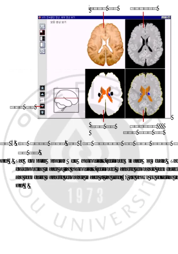
9 수평절단 풀그림의 뇌 그림에서 가장 위가 1번이고 가장 아래가 122번이었다. 47 번에 마우스 포인터를 놓고 딸깍하거나 ‘수평절단’에 47을 입력하면(그림 3) 47번의 네 가지 영상(절단표본 영상, 자기공명영상, 구역화 영상, 자기공명영상 더하기 구역 화 영상)을 볼 수 있었다. ‘아래로 가기’를 세 번 누르면(그림 4) 50번의 영상을 볼 수 있었다. ‘절단표본 영상, 자기공명영상 보기’를 누르면(그림 5) 세 가지 영상(절단표본 영상, 자기공명영상, 자기공명영상 더하기 구역화 영상)을 볼 수 있었다. ‘절단표본 영 상, 풀이 보기’를 누르면(그림 6) 절단표본 영상과 세부 구조의 이름을 함께 볼 수 있 었다(그림 7). 그림 3. 뇌의 수평절단 풀그림. 수평절단하는 면을 고르는 창.
Fig. 3. The computer program for horizontally-sectioned images of brain. The window indicates that several levels of horizontally-sectioning can be selected.
그림 4. 뇌의 수평절단 풀그림. 그림 3에서 수평절단하는 면을 고르면 나타나는 네 가지 영상.
Fig. 4. The computer program for horizontally-sectioned images of brain. The corresponding images of the horizontally-sectioned, magnetic resonance, segmented, and segmented magnetic resonance images at the level 47 are shown by selecting in Fig. 3. 자기공명영상 더하기 구역화 영상 아래로 가기 자기공명영상 구역화 영상 절단표본 영상
11
그림 5. 뇌의 수평절단 풀그림. 그림 4에서 ‘아래로 가기’를 세 번 누르면 나타나는 네 가지 영상.
Fig. 5. The computer program for horizontally-sectioned images of brain. The corresponding images of the horizontally-sectioned, magnetic resonance, segmented, and segmented magnetic resonance images at the level 50 can be seen by pressing the 'downward' button three times in Fig. 4.
자기공명영상 더하기 구역화 영상 절단표본 영상, 자기공명영상 보기 자기공명영상 구역화 영상 절단표본 영상
그림 6. 뇌의 수평절단 풀그림. 그림 5에서 ‘절단표본 영상, 자기공명영상 보기’를 누 르면 나타나는 세 가지 영상.
Fig. 6. The computer program for horizontally-sectioned images of brain. The corresponding images of the horizontally-sectioned, magnetic resonance, and segmented magnetic resonance images can be seen by pressing the icon for the 'horizontally-sectioned and magnetic resonance images' in Fig. 5.
자기공명영상 더하기 구역화 영상 절단표본 영상, 풀이 보기 자기공명영상 절단표본 영상
13
그림 7. 뇌의 수평절단 풀그림. 그림 6에서 ‘절단표본 영상, 풀이 보기’를 누르면 나 타나는 절단표본 영상과 세부 구조의 이름.
Fig. 7. The computer program for horizontally-sectioned images of brain. The names of detailed structures can be shown in the horizontally-sectioned image by pressing the icon for the 'detailed structures' in Fig. 6.
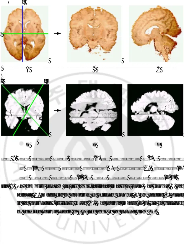
자유절단 풀그림의 수평절단면에서 관상절단하는 면과 시상절단하는 면을 고르면 관상절단면과 시상절단면을 볼 수 있었다. 마찬가지로 관상절단면에서도 수평절단하 는 면과 시상절단하는 면을 고를 수 있었고, 시상절단면에서도 수평절단하는 면과 관 상절단하는 면을 고를 수 있었다. 수평절단면에서 비스듬히 절단하는 면을 고르면 비 스듬한 절단면을 볼 수 있었다. 마찬가지로 관상절단면과 시상절단면에서도 비스듬히 절단하는 면을 고를 수 있었다(그림 8). 자유절단한 다음에 네 가지 영상(절단표본 영 상, 절단표본 영상 더하기 구역화 영상, 자기공명영상, 자기공명영상 더하기 구역화 영상) 중 두 가지 영상을 골라서 함께 볼 수 있었다(그림 9).
15
그림 8. 뇌의 자유절단 풀그림. 수평절단면(A)에서 관상절단하는 면(b)과 시상절단하 는 면(c)을 고르면 나타나는 관상절단면(B)과 시상절단면(C). 관상절단면(D) 에서 비스듬히 절단하는 면(e, f)을 고르면 나타나는 비스듬한 절단면(E, F). Fig. 8. The computer program for free angle-sectioned images of brain. The coronal (B) and
sagittal (C) images can be visualized by selecting coronal (b) and sagittal (c) planes in the horizontally-sectioned image (A). The oblique images (E, F) can be visualized by selecting oblique planes (e, f) of free angle in the coronal plane (D).
A B C
b
c
e f
그림 9. 뇌의 자유절단 풀그림. 그림 8의 시상절단면(C)으로 나타날 수 있는 네 가지 영상.
Fig. 9. The computer program for free angle-sectioned images of brain. The sectioned, segmented sectioned, magnetic resonance, and segmented magnetic resonance images are shown by the sagittal image (C) in Fig. 8.
절단표본 영상 절단표본 영상 더하기 구역화 영상
자기공명영상 자기공명영상 더하기 구역화 영상
17
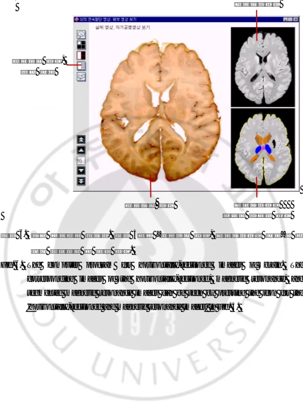
해부 풀그림에서 해부표본 영상 27개 중 하나를 고르면(그림 10) 해부표본 영상 과 세부 구조의 이름을 함께 볼 수 있었다(그림 11).
그림 10. 뇌의 해부 풀그림. 해부표본 영상을 고르는 창.
Fig. 10. The computer program for conventionally dissected images of brain. The window indicates that several images obtained by conventional dissection can be selected.
그림 11. 뇌의 해부 풀그림. 그림 10에서 하나를 고르면 나타나는 해부표본 영상과 세부 구조의 이름.
Fig. 11. The computer program for conventionally dissected images of brain. The conventionally dissected images with the names of detailed structures are shown by selecting in Fig. 10.
19
IV.
고찰
고찰
고찰
고찰
뇌의 생김새와 자기공명영상을 깨닫기 위해서 의사와 의과대학 학생은 다음과 같 은 교육 자료를 써 왔다. 첫째는 시체에서 꺼낸 뇌이다. 그런데 뇌 해부는 아무나 할 수 없고, 과정이 번거롭고, 한 번 하면 돌이킬 수 없다는 단점이 있다5 . 둘째는 종이, 찰흙, 나무, 플라스틱으로 만든 뇌의 모형이다. 그런데 모형은 세밀하지 않고 마음대 로 잘라볼 수 없다는 단점이 있다6 . 셋째는 신경해부학 책과 뇌의 자기공명영상 필름 이다. 그런데 책과 필름에 있는 평면 그림은 마음대로 잘라볼 수 없어서 뇌의 입체 생김새를 깨닫는 데 한계가 있다는 단점이 있다4 . 넷째는 뇌의 컴퓨터 풀그림이다7-9 . 컴퓨터 풀그림은 다른 교육 자료의 단점을 보완할 수 있으며, 특히 요즘에는 개인용 컴퓨터의 성능이 좋아지면서 값어치가 커지고 있다(표 1). 표 1. 각 교육 자료가 뇌의 생김새와 자기공명영상을 깨닫는 데 도움이 되는 정도. Table 1. Comparison of the present computer-aided program with other conventionaleducational tools in understanding brain morphology and magnetic resonance image. 교육 자료 쉽게 되풀이해서 보기 세밀하게 보기 마음대로 잘라보기 시체 – ++ ++ 해부학 모형 ++ – + 책과 자기공명영상 필름 ++ ++ – 컴퓨터 풀그림 ++ ++ ++ –: 도움이 안됨, +: 도움이 조금 됨, ++: 도움이 많이 됨.
그런데 이제까지 만든 뇌의 컴퓨터 풀그림도 몇 가지 단점이 있으며, 이러한 단 점을 보완하기 위하여 이 연구에서는 다음과 같은 기능을 갖춘 컴퓨터 풀그림을 만 들었다. 첫째, 한국 사람의 뇌를 볼 수 있다. 인종과 민족마다 얼굴이 다르게 생긴 것처럼 기관도 다르게 생겼다. 따라서 한국 사람의 뇌로 만든 컴퓨터 풀그림은 한국 사람의 뇌 질병을 진단하고 치료하는 데 도움이 된다. 둘째, 얇게 연속절단한 뇌를 볼 수 있다. 1 cm쯤으로 두껍게 연속절단하면 송과체 처럼 작은 구조를 보지 못할 수 있다12 . 그러나 1.4 mm로 얇게 연속절단하면 뇌의 작 은 구조까지 볼 수 있다. 뇌처럼 큰 기관을 얇게 연속절단하기 위해서 개발한 방법으로는 얼리고 cryomacrotome으로 가는 것13,14 과 celloidin으로 포매하고 polycut으로 절단하는 것15,16 이 있다. 이 연구에서는 gelatin 용액으로 포매하고 육절기로 연속절단하였는데17,18, 이 방 법은 다른 방법에 비해서 시간이 오래 걸리지 않고 값이 싸다는 장점이 있다. 셋째, 서로 들어맞는 자기공명영상, 절단표본 영상, 구역화 영상을 함께 볼 수 있 다. 자기공명영상은 뇌의 빛깔을 그대로 나타내지 못하고 해상도의 한계가 있기 때문 에 어떤 구조의 위치와 생김새를 깨닫기가 어렵다. 따라서 서로 들어맞는 자기공명영 상, 절단표본 영상, 구역화 영상을 함께 보면 뇌의 자기공명영상을 깨닫는 데 도움이 된다7,8,19 . 넷째, 뇌의 3차원 영상을 여러 방향으로 절단해서 볼 수 있다. 수평 방향의 절단 면만 가지고는 뇌의 입체 생김새와 여러 방향의 자기공명영상을 깨닫기가 어렵기 때 문에 여러 방향의 절단면을 볼 필요가 있다5. 다섯째, 뇌의 해부표본 영상을 볼 수 있다. 절단표본 영상만 가지고는 신경해부학 실습을 할 때 뇌를 어떻게 해부해서 어떤 구조를 봐야 하는지 깨닫기가 어렵다20 . 따 라서 뇌를 여러 방향에서 해부하여 만든 풀그림을 쓰면 신경해부학 실습 시간에 뇌
21 여섯째, 개인용 컴퓨터에서 쓸 수 있다. 자기공명영상 촬영기에 딸린 컴퓨터 풀그 림은 자기공명영상 촬영기의 옆에서만 쓸 수 있기 때문에 공간과 시간의 제한이 크 다21. 그러나 개인용 컴퓨터의 한글 윈도우즈 환경에서 쓸 수 있는 풀그림은 이러한 제한이 없다. 이 컴퓨터 풀그림은 의과대학 학생 또는 의사가 교육 자료로 쓸 수 있으며, 따라 서 씨디 타이틀이나 인터넷을 통해서 널리 퍼뜨릴 계획이다. 앞으로 더 도움이 되는 컴퓨터 풀그림을 만들기 위해서는 다음과 같이 연구해야 할 것이다. 첫째, 뇌뿐만 아니라 머리 전체를 연속절단해서 뇌 주변의 구조까지 볼 수 있게 하는 것이다. 이를 위해서는 머리 전체를 얼린 다음에 cryomacrotome으로 가는 것이 바람직하다13,14 . 둘째, 뇌의 구조를 더 세밀하게 구역화하는 것이다. 이를 위해서는 더 얇게 연속 절단한 다음에 절단표본을 더 높은 해상도로 컴퓨터에 입력해야 한다. 셋째, 컴퓨터 풀그림에 새로운 기능을 덧붙이는 것인데, 보기를 들면 뇌를 가상으 로 수술하는 기능이다. 이를 위해서는 이미 만들어진 컴퓨터 풀그림의 기능을 살펴보 고22,23,24 , 의과대학 학생과 의사가 어떤 기능을 바라는지 조사할 필요가 있다.
V.
결론
결론
결론
결론
이 연구에서 만든 수평절단, 자유절단, 해부 풀그림의 기능은 실제로 다음과 같이 도움 된다. 첫째, 뇌의 구조가 어떻게 생겼는지 깨닫는 데 도움이 된다. 수평절단 풀그림을 써서 1.4 mm 간격의 절단표본 영상을 세부 구조의 이름과 함께 이어서 보면 뇌의 구 조가 어떻게 생겼는지 깨닫는 데 도움이 된다. 자유절단 풀그림을 써서 여러 방향의 절단표본 영상과 구역화 영상을 함께 보면 뇌의 구조가 입체적으로 어디에 있고 어 떻게 생겼는지 깨닫는 데 도움이 된다. 해부 풀그림을 써서 해부표본 영상을 세부 구 조의 이름과 함께 보면 신경해부학 실습 시간에 뇌를 해부하는 데 도움이 된다. 둘째, 뇌의 구조가 자기공명영상에서 어떻게 보이는지 깨닫는 데 도움이 된다. 수 평절단 풀그림을 써서 1.4 mm 간격의 자기공명영상을 자기공명영상에 들어맞는 절단 표본 영상, 구역화 영상과 함께 보면 수평 방향의 자기공명영상을 깨닫는 데 도움이 된다. 자유절단 풀그림을 써서 여러 방향의 자기공명영상, 절단표본 영상, 구역화 영 상을 함께 보면 여러 방향의 자기공명영상을 깨닫는 데 도움이 된다.23
VI.
참고문헌
참고문헌
참고문헌
참고문헌
1. Bourgeois G, Magnin M, Morel A, Sartoretti S, Huisman T, Tuncdogan E, Meier D and Jeanmonod D: Accuracy of MRI-guided stereotactic thalamic functional neurosurgery. Neuroradiology 41: 636-645, 1999
2. Smith JR, Hardy TL, Rose DF, Flanigin HF, King DW, Gallagher BB, Murro AM and Fifer A: Comparison of CT- versus MRI-guided, computer-assisted depth electrode implantation. Stereotact Funct Neurosurg 58: 189-193, 1992
3. Le BD, Douek P, Argyropoulou M, Turner R, Patronas N and Fulham M: Diffusion and perfusion magnetic resonance imaging in brain tumors. Top Magn Reson Imaging 5: 25-31, 1993
4. Ackerman MJ: The visible human project. A resource for education. Acad Med 74: 667-670, 1999
5. Hoffman H, Vu D: Virtual reality. Teaching tool of the twenty-first century?. Acad Med 72: 1076-1081, 1997
6. Schubert R, Höhne KH, Pommert A, Reimer M, Schiemann T, Tiede U and Lierse W: A new method for practicing exploration, dissection, and simulation with a complete computerized three-dimensional model of the brain and skull. Acta Anat 150: 69-74, 1994 7. Tiede U, Bomans M, Höhne KH, Pommert A, Riemer M, Schiemann T, Schubert R and
Lierse W: A computerized three-dimensional atlas of the human skull and brain. Am J Neuroradiol 14: 551-559, 1993
8. Toh MY, Falk RB and Main JS: Interactive brain atlas with the visible human project data. Development methods and techniques. Radiographics 16: 1201-1206, 1996
9. Trelease RB: The virtual anatomy practical. A stereoscopic 3D interactive multimedia computer examination program. Clin Anat 11: 89-94, 1998
10. Felle P, Lockman AK and Kellaghan P: Removal of the brain for teaching and examination. Clin Anat 8: 363-365, 1995
12. Opeskin K, Anderson RM: A device for cutting brain slices. Biotech Histochem 69: 253-256, 1994
13. Spitzer VM, Ackerman MJ, Scherzinger AL and Whitlock DG: The visible human male. Technical report. J Am Med Inform Assoc 3: 118-130, 1996
14. Toga AW, Ambach K, Quinn B, Hutchin M and Burton JS: Postmortem anatomy from cryosectioned whole human brain. J Neurosci Methods 54: 239-252, 1994
15. Giles LG, Taylor JR: Histological preparation of large vertebral specimens. Stain Technol 58: 45-49, 1983
16. Qui M: Bone histomorphometric study of iliac crest in normal Chinese subjects. Chung Hua I Hsueh Tsa Chin 70: 685-690, 1990
17. Barnett RI, Lyons GW, Driscoll JD and Forrest WJ: Improved sectioning and Berlin blue staining of whole human brain. Stain Technol 55: 235-239, 1980
18. Roberts M, Hanaway J: Preparation of brain slices for macroscopic study by the copper sulfate-phenol-ferrocyanide technique. Stain Technol 44: 143-146, 1969
19. Toga AW, Goldkorn A, Ambach K, Chao K, Quinn BC and Yao P: Postmortem cryosectioning as an anatomic reference for human brain mapping. Comput Med Imaging Graph 21: 131-141, 1997
20. Lamperti A, Sodicoff M: Computer-based neuroanatomy laboratory for medical students. Anat Rec 249: 422-428, 1997
21. Mankovich NJ, Robertson DR and Cheeseman AM: Three-dimensional image display in medicine. J Digit Imaging 3: 69-80, 1990
22. Hayashi N, Endo S, Shibata T, Ikeda H and Takaku A: Neurosurgical simulation and navigation with three-dimensional computer graphics. Neurol Res 21: 60-66, 1999 23. Kockro RA, Serra L, Tseng TY, Chan C, Yih YS, Gim GC, Lee E, Hoe LY, Hern N and
Nowinski WL: Planning and simulation of neurosurgery in a virtual reality environment. Neurosurgery 46: 118-135, 2000
24. Tanaka H, Nakamura H, Tamaki E, Nariai T and Hirakawa K: Brain surgery simulation system using VR technique and improvement of presence. Stud Health Technol Inform
25
- Abstract -
A Computer Program for Understanding Brain Morphology
and Magnetic Resonance Image
Yi Suk Kim
Department of Medical Sciences The Graduate School, Ajou University (Directed by Assistant Professor Min Suk Chung)
Understanding of brain morphology and magnetic resonance image (MRI) is essential for accurate diagnosis and treatment of the brain diseases. As education tools, the cadaver dissection, plastic models, and neuroanatomy books have been used for understanding brain morphology; and the MRI films and radiology books have been used for understanding brain MRI. Recently, due to the popularization of powerful personal computers, computer programs compensating the conventional education tools have been used. But these computer programs have a disadvantage that it is not possible to visualize the details of brain morphology or to compare the corresponding sectioned specimens and MRI. Therefore, we attempted to make a computer program which could visualize not only the details of brain morphology but also the corresponding sectioned specimens and MRI by using the brains removed from Korean cadavers.
Three brains were removed from Korean cadavers. With a brain, 122 MRI and 122 serially-sectioned specimens with a 1.4 mm interval were acquired and inputted into the computer. Ten brain structures were segmented, and 83 fine structures were designated on the images. With two brains, 27 dissected specimens were acquired and inputted into the computer. One-hundred two fine structures were designated on the images. Based on these images, a computer program for understanding brain morphology and MRI was made.
The computer program, which was made in this study, visualized the corresponding sectioned specimens, MRI, and segmented images after sectioning a brain horizontally or at any angles. In addition, the computer program visualized the images of dissected brain.
This computer program is helpful to understand brain morphology and MRI. This computer program is expected to be used through CD-title or Internet as an educational tool for medical students and doctors.