저작자표시-비영리-동일조건변경허락 2.0 대한민국 이용자는 아래의 조건을 따르는 경우에 한하여 자유롭게 l 이 저작물을 복제, 배포, 전송, 전시, 공연 및 방송할 수 있습니다. l 이차적 저작물을 작성할 수 있습니다. 다음과 같은 조건을 따라야 합니다: l 귀하는, 이 저작물의 재이용이나 배포의 경우, 이 저작물에 적용된 이용허락조건 을 명확하게 나타내어야 합니다. l 저작권자로부터 별도의 허가를 받으면 이러한 조건들은 적용되지 않습니다. 저작권법에 따른 이용자의 권리는 위의 내용에 의하여 영향을 받지 않습니다. 이것은 이용허락규약(Legal Code)을 이해하기 쉽게 요약한 것입니다. Disclaimer 저작자표시. 귀하는 원저작자를 표시하여야 합니다. 비영리. 귀하는 이 저작물을 영리 목적으로 이용할 수 없습니다. 동일조건변경허락. 귀하가 이 저작물을 개작, 변형 또는 가공했을 경우 에는, 이 저작물과 동일한 이용허락조건하에서만 배포할 수 있습니다.
Association between Coronary Artery Calcium
Score and Serum Creatinine concentration
by
Jung Ho Shin
Major in Medicine
Department of Medical Sciences
The Graduate School, Ajou University
Association between Coronary Artery Calcium
Score and Serum Creatinine concentration
by
Jung Ho Shin
A Dissertation Submitted to The Graduate School of
Ajou University in Partial Fulfillment of the Requirements
for the Degree of
Master of Medicine
Supervised by
Bom Taeck Kim, M.D., Ph.D.
Major in Medicine
Department of Medical Sciences
The Graduate School, Ajou University
This certifies that the dissertation
of Jung Ho Shin is approved.
SUPERVISORY COMMITTEE
Bom Taeck Kim
Chang Hee Suh
Sung Ran Cho
The Graduate School, Ajou University
December, 14th, 2012
i -ABSTRACT-
Association between Coronary Artery Calcium Score and
Serum Creatinine concentration
Background: Coronary calcification quantified by coronary multi-detector computed
tomography (MDCT) is a strong predictor for coronary artery disease (CAD). Previous studies have reported that the amplitude of impaired renal clearance is well correlated with the coronary calcification score in patients with chronic kidney disease. We investigated if whether the relationship between coronary calcium score and serum creatinine concentration was still valid in adults with normal serum creatinine.
Methods: We analyzed the data of 1,429 healthy adults, who visited a health promotion
center in Ajou University Hospital from March, 2010 to February, 2012 for physical check-up. We measured Agaston’s coronary artery calcium score (CACS) using coronary MDCT. We divided subjects into quartiles according to serum creatinine level and compared their odds ratios for acquisition of coronary calcification (CACS > 0) or, for severe coronary calcification (CACS ≥ 100) after adjusting for other coronary risk factors.
Results: As serum creatinine level of each quartile incremented, percentages of people with
coronary calcification (p = 0.008) and ratio of people with high CACS (p < 0.001) also incremented. Highest serum creatinine quartile group demonstrated more than double is the risk for acquisition of coronary calcification and high CACS (odds ratio: 2.12 [1.51 - 2.97], 2.83 [1.50 - 5.33], respectively) compared with lowest quartile. This relationship remained significant after adjustment for other CAD risk factors (odds ratio: 2.21 [1.48 - 3.29], 3.91 [1.91-7.99], respectively).
Conclusion: Elevated serum creatinine is an independent predictor for coronary calcification
and its severity even in adults with normal serum creatinine concentration.
Key words: Serum creatinine, coronary artery calcification, multi-detector computed
ii
TABLE OF CONTENTS
ABSTRACT --- - i
TABLE OF CONTENTS --- ii
LIST OF FIGURES --- iii
LIST OF TABLES --- iv
I. INTRODUCTION --- 1
II. MATERIALS AND METHODS --- 2
A. Study Population --- 2
B. Measurements --- 2
1. Coronary artery calcium score (CACS) --- 2
2. Physical measurements and questionnaires --- 2
3. Laboratory study --- 3 C. Statistical analysis --- 3 III.RESULT --- 4 IV. DISCUSSION --- 13 V. CONCLUSION --- 15 REFERENCE --- 16 국문요약 --- 20
iii
LIST OF FIGURES
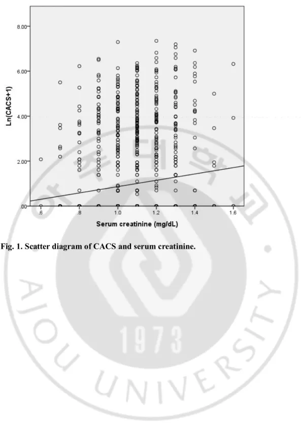
Fig. 1.Scatter diagram of CACS and serum creatinine --- 8 Fig. 2.Means of CACS in the quartiles of serum creatinine --- 9 Fig. 3. Percentages of subjects with CACS exceeding 0 (A) and above 100 (B) according to
iv
LIST OF TABLES
Table 1.Baseline characteristics of subjects --- 6 Table 2.Correlations of CACS with serum creatinine and other risk factors for CAD --- 7 Table 3. Means or percentages of variables according to the quartiles of serum creatinine - 10 Table 4. Odds ratios (95% confidence interval) of the quartiles of serum creatinine for the incidence of CACS exceeding 0 and CACS above 100 by logistic regression analysis --- 12
- 1 -
I. INTRODUCTION
Coronary artery disease (CAD) is the leading cause of death in developed countries and its prevalence is on the rise even in developing countries. Along with traditional risk factors for CAD, coronary calcification is a novel predictor for development of CAD (Margolis et al., 1980). Agaston’s coronary artery calcium score (CACS) measured by coronary multi-detector computed tomography (MDCT) is the most commonly used tool to evaluate the coronary calcification (Agatston et al., 1990). Several guidelines including American Heart Association Guidelines for Cardiovascular Disease Prevention in Women (2004) and European Guidelines on Cardiovascular Disease Prevention in Clinical Practice (2003), advocated the beneficial effects of CACS for risk stratification for CAD and defined coronary calcification as a subclinical CAD (De Backer et al., 2003; Mieres et al., 2005).
Chronic kidney disease (CKD) is defined as decreased glomerular filtration rate (GFR) under 60 mL/min/1.73 m2 for more than three months. The prevalence of CKD has been soaring in aging societies due to high prevalence of hypertension and type 2 diabetes mellitus. CKD patients frequently demonstrate coronary calcification (Ossareh, 2011) and are likely to die from CAD even before impairment of their kidney function progresses to end-stage renal disease (ESRD) (Sarnak et al., 2003). Duration of ESRD is positively correlated with extent of coronary calcification (Mehrotra et al., 2004) and ESRD patients have much higher coronary mortality than age-adjusted general population (Foley et al., 1998; Parfrey and Foley, 1999).
Recent studies demonstrated that minor renal insufficiency like slight reduction of GFR or microalbuminuria is also associated with coronary calcification and increased risk for development of CAD (Culleton et al., 1999; Ritz and McClellan, 2004). Coronary calcification seems to be universal in a population with impaired renal function. This study aimed to investigate that serum creatinine level (Cr) and CACS is still closely related even in adults with normal Cr.
- 2 -
II. MATERIALS AND METHODS
A. Study Population
This study targeted those who underwent a health check-up at the Ajou University Hospital health promotion center between March, 2010 and February, 2012. If one person did a health examination more than once, we analyzed only the first result. We gathered data from subjects who carried out coronary MDCT, blood sampling, physical measurements and questionnaire about medical and smoking histories. We defined Cr below 1.5 mg/dL as normal. Exclusion criteria were subjects with past history of CAD or coronary stent insertion and with Cr over 1.5mg/dL. Finally, we analyzed data from 1,429 adults (1,072 males and 357 females).
B. Measurements
1. Coronary artery calcium score (CACS)
CT scans were performed with 64-slice MDCT scanner (Brilliance 64, Philips Medical Systems, Best, The Netherlands) to calculate the Agaston’s CACS (Agatston et al., 1990). Subjects were required not to take foods containing much caffeine including coffee and soda from previous night and to take metoprolol 50 mg to control purse rate under 65 beats/min 2 hours before examination. We categorized CACS into three levels which represent different risks for development of CAD: 1) CACS = 0, 2) 0 < CACS < 100, 3) CACS ≥ 100 (Rumberger et al., 1999).
2. Physical measurements and questionnaires
Vital signs including systolic blood pressure (BP) and diastolic BP were checked. Height was measured to the nearest 0.1 cm and weight to the nearest 0.1 kg. Body mass index (BMI) was calculated as weight (kg) divided by height (cm2) in meters squared. Waist
circumference was checked on the middle point between lowest sternum and iliac spine with tapeline and measured to the nearest 0.1 cm. We used bioelectrical impedance test to analyze the body composition. We used questionnaires to check the medical histories and current smoking state. As to smoking, subjects were classified into non-current smokers who have
- 3 -
never smoked or quit to smoke and current smokers who smoke in the present.
3. Laboratory study
Blood test was conducted after at least 8-hours of fasting. We gathered laboratory data including serum levels of creatinine, fasting glucose, total cholesterol, triglyceride, low-density lipoprotein (LDL) and high-low-density lipoprotein (HDL). Estimated glomerular filtration ratio (eGFR) was calculated with Modification of Diet in Renal Disease (MDRD) method (Levey et al., 1999). We divided subjects into quartiles based on Cr to compare CACS and other variables.
C. Statistical analysis
SPSS 19.0 (SPSS Ltd, Chicago, IL, USA) was used for all statistical analysis. Data are shown as the mean ± standard deviation (SD) for continuous variables and as number (percentages) for discrete variables, unless otherwise indicated. Frequencies and descriptive analysis were carried out to show the baseline characteristics of subjects. We used log transformed (CACS+1) to reduce skewness. We analyze the correlations of CACS with Cr, age and other CAD risk factors. We also compared CACS and other variables according to the quartiles of Cr with one-way ANOVA test for variables in normal distribution and Kruskal-Wallis test for the others. Bonferroni method was applied for the post hoc analysis. Previous trials have documented that people without coronary calcification have very low chances of development of CAD (Greenland et al., 2004; Arad et al., 2005) and people with CACS above 100 have significantly high risk for development of CAD (Arad et al., 2005). So we carried out logistic regression to calculate the odds ratios for having coronary calcification itself (CACS > 0) and high CACS (CACS ≥ 100) according to the quartiles of Cr with adjustment for other risk factors. P-values those < 0.05 were considered statistically significant.
- 4 -
III. RESULTS
A. General characteristics of study subjects
Table 1 shows the baseline characteristics of 1429 subjects included in this study. Mean age of the subjects was 50.9 ± 8.9 and their average Cr was 1.07 ± 0.17 mg/dL. Mean eGFR was 74.08 ± 10.55 and only 100 subjects (7.5%) had eGFR within optimal range (≥ 90 mL/min/1.73m2). More than 70% of subjects did not have coronary calcification and only 7%
of subjects showed CACS above 100. Traditional CAD risk factors such as age, waist circumference, BMI, blood pressure, fasting glucose, triglyceride and HDL were significantly different between subjects with and without coronary calcification. The subjects with coronary calcification had higher Cr, more muscle mass and smaller eGFR than those without coronary calcification.
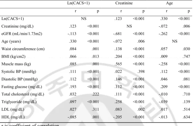
B. Relationship among CACS, Cr and other risk factors
Ln(CACS+1) showed significant correlations with Cr, eGFR and traditional risk factors for CAD including age, waist circumference, BMI, systolic BP, diastolic BP, fasting glucose, triglyceride and HDL (Table 2). Cr also showed positive correlations with most of CAD risk factors except systolic BP and age. Age was negatively correlated with muscle mass, eGFR and Cr. The distribution of CACS and Cr which was in a positive relationship (r=0.123, p<0.001) showed in Figure 1.
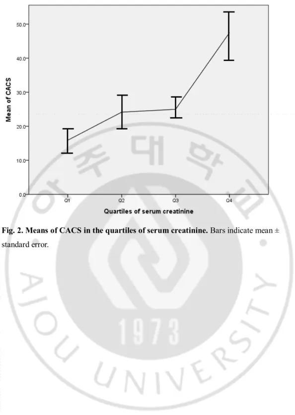
C. CACS and other risk factors according to the quartiles of Cr
As Cr in each quartile incremented, their CACS got higher as well (p < 0.001). Quartile in highest Cr had higher waist circumference, BMI, diastolic BP, total cholesterol, TG, LDL and lower HDL than other quartiles. Age was highest in the lowest Cr quartile group (Table 3).
D. Odds ratios for presence and severity of coronary calcification
according to the quartiles of Cr
- 5 -
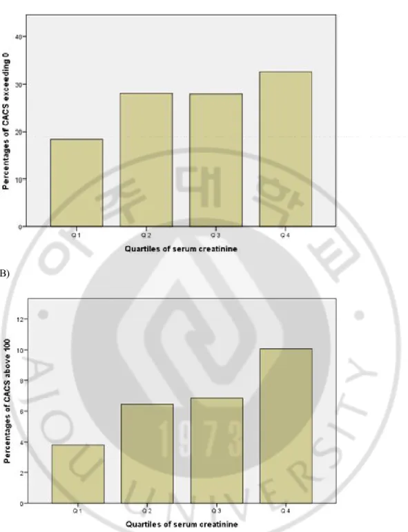
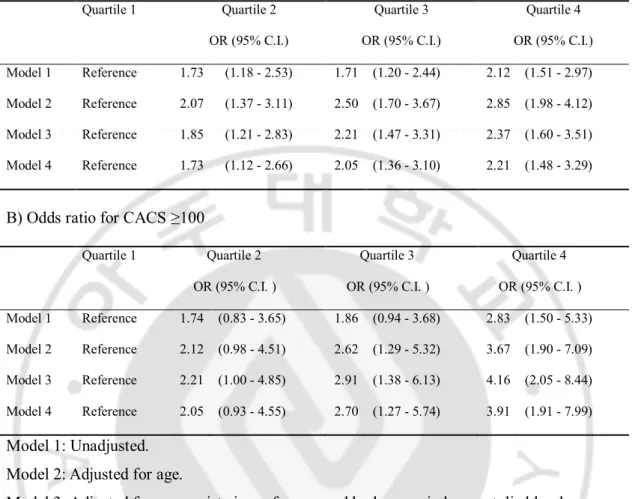
The higher Cr quartile had, the more percentages of subjects with CACS > 0 and CACS ≥ 100 they occupied. Percentages of subjects with CACS > 0 were 18.4%, 28.0%, 27.9% and 32.3% (p <0.001) and CACS ≥ 100 were 3.8%, 6.4%, 6.8% and 9.9% (p =0.010), respectively (Figure 3). Table 4 demonstrates the odds ratios for having CACS > 0 and ≥ 100 according to the quartiles of Cr. The highest Cr quartile group showed more than double is the risk for acquisition of coronary calcification both in unadjusted model (Odds ratio 2.12 [1.51 - 2.97]) and in adjusted model (OR 2.21 [1.48 - 3.29]) for other CAD risk factors. This trend was similar in the odds ratios for having CACS ≥ 100 (Odds ratio 2.83 [1.50 - 5.33]) and 3.91 [1.91 - 7.99]).
- 6 - Table 1. Baseline characteristics of subjects.
Variables Total CACS = 0 CACS > 0
Age(years) 50.9 ± 8.9 49.1 ± 8.5 55.7 ± 8.1* Male 1,073 (75.1%) 742 (71.1%) 330 (85.5%)* Height (cm) 167.4 ± 8.1 167.1 ± 8.3 168.0 ± 7.4* Weight (kg) 69.7 ± 11.8 69.1 ± 12.0 71.2 ± 11.1* Waist circumference (cm) 86.9 ± 8.0 86.5 ± 8.0 88.0 ± 7.9* BMI (kg/cm2) 24.8 ± 3.2 24.6 ± 3.2 25.1 ± 3.1* Muscle Mass (kg) 48.2 ± 8.7 47.7 ± 9.0 49.4 ± 8.0* CACS 29.7 ± 116.2 0 ± 0 108.6 ± 202.3* CACS = 0 1043 (73.0%) 0 < CACS <100 286 (20.0%) CACS >= 100 100 (7.0%) Creatinine (mg/dL) 1.07 ± 0.17 1.06 ± 0.17 1.10 ± 0.16* eGFR (mL/min/1.73m2) 74.04 ± 10.55 74.8 ± 10.3 72.3 ± 10.5* Systolic BP (mmHg) 118.7 ± 14.5 117.9 ± 14.3 121.1 ± 14.6* Diastolic BP (mmHg) 78.0 ± 11.0 77.3 ± 11.0 79.8 ± 11.0* Fasting glucose (mg/dL) 100.8 ± 25.4 98.5 ± 22.0 106.8 ± 32.1* Total cholesterol (mg/dL) 190.9 ± 33.8 189.9 ± 33.0 193.6 ± 35.8 Triglyceride (mg/dL) 137.5 ± 82.0 132.6 ± 79.7 150.2 ± 86.6* LDL (mg/dL) 114.3 ± 31.2 113.7 ± 30.4 116.1 ± 33.2 HDL (mg/dL) 49.1 ± 11.9 49.7 ± 12.0 47.4 ± 11.5* Smoking Non-current 1009 (70.6%) 752 (72.1%) 257 (66.6%)* Current 420 (29.4%) 291 (29.9%) 129 (33.4%)*
Values are presented as mean ± SD or number (%) unless otherwise indicated.
*: Mean is significantly different between CACS=0 and CACS>0
BMI: body mass index, CACS: coronary artery calcium score, eGFR: estimated glomerular filtration ratio, BP: blood pressure, LDL: low-density lipoprotein, HDL: high-density lipoprotein
- 7 -
Table 2. Correlations of CACS with serum creatinine and other risk factors for CAD.
Ln(CACS+1) Creatinine Age
r p r p r p Ln(CACS+1) NS .123 <0.001 .330 <0.001 Creatinine (mg/dL) .123 <0.001 NS -.072 .006 eGFR (mL/min/1.73m2) -.113 <0.001 -.681 <0.001 -.262 <0.001 Age (years) .330 <0.001 -.072 .006 NS Waist circumference (cm) .084 .001 .138 <0.001 .057 .030 BMI (kg/cm2) .066 .013 .204 <0.001 .009 .747 Muscle mass (kg) .085 .001 .565 <0.001 -.258 <0.001 Systolic BP (mmHg) .111 <0.001 .022 .398 .112 <0.001 Diastolic BP (mmHg) .112 <0.001 .146 <0.001 .046 .081 Fasting glucose (mg/dL) .193 <0.001 .112 <0.001 .209 <0.001 Total cholesterol (mg/dL) .032 .222 .111 <0.001 -.010 .710 Triglyceride (mg/dL) .097 <0.001 .258 <0.001 -.039 .139 LDL (mg/dL) .027 .311 .083 .002 .017 .514 HDL (mg/dL) -.085 .001 -.205 <0.001 -.013 .634 r is coefficient of correlation.
CACS: coronary artery calcium score, eGFR: estimated glomerular filtration ratio, BMI: body mass index, BP: blood pressure, LDL: low-density lipoprotein, HDL: high-density lipoprotein.
- 8 -
- 9 -
Fig. 2. Means of CACS in the quartiles of serum creatinine. Bars indicate mean ±
- 10 -
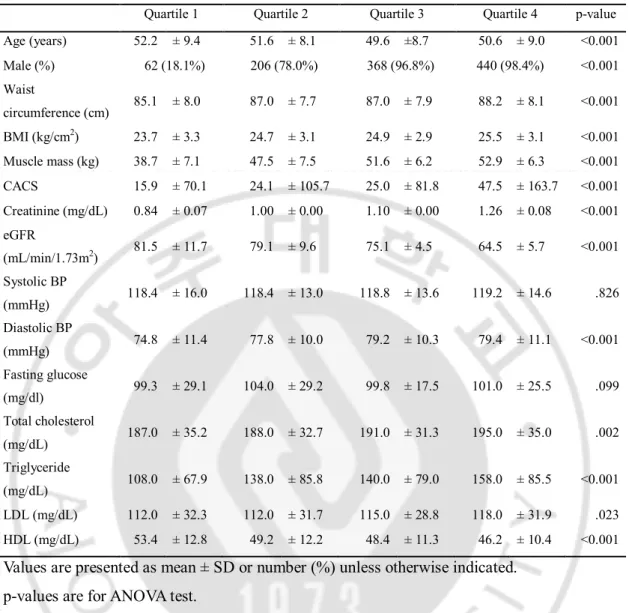
Table 3. Means or percentages of variables according to the quartiles of serum creatinine.
Quartile 1 Quartile 2 Quartile 3 Quartile 4 p-value Age (years) 52.2 ± 9.4 51.6 ± 8.1 49.6 ±8.7 50.6 ± 9.0 <0.001 Male (%) 62 (18.1%) 206 (78.0%) 368 (96.8%) 440 (98.4%) <0.001 Waist circumference (cm) 85.1 ± 8.0 87.0 ± 7.7 87.0 ± 7.9 88.2 ± 8.1 <0.001 BMI (kg/cm2) 23.7 ± 3.3 24.7 ± 3.1 24.9 ± 2.9 25.5 ± 3.1 <0.001 Muscle mass (kg) 38.7 ± 7.1 47.5 ± 7.5 51.6 ± 6.2 52.9 ± 6.3 <0.001 CACS 15.9 ± 70.1 24.1 ± 105.7 25.0 ± 81.8 47.5 ± 163.7 <0.001 Creatinine (mg/dL) 0.84 ± 0.07 1.00 ± 0.00 1.10 ± 0.00 1.26 ± 0.08 <0.001 eGFR (mL/min/1.73m2) 81.5 ± 11.7 79.1 ± 9.6 75.1 ± 4.5 64.5 ± 5.7 <0.001 Systolic BP (mmHg) 118.4 ± 16.0 118.4 ± 13.0 118.8 ± 13.6 119.2 ± 14.6 .826 Diastolic BP (mmHg) 74.8 ± 11.4 77.8 ± 10.0 79.2 ± 10.3 79.4 ± 11.1 <0.001 Fasting glucose (mg/dl) 99.3 ± 29.1 104.0 ± 29.2 99.8 ± 17.5 101.0 ± 25.5 .099 Total cholesterol (mg/dL) 187.0 ± 35.2 188.0 ± 32.7 191.0 ± 31.3 195.0 ± 35.0 .002 Triglyceride (mg/dL) 108.0 ± 67.9 138.0 ± 85.8 140.0 ± 79.0 158.0 ± 85.5 <0.001 LDL (mg/dL) 112.0 ± 32.3 112.0 ± 31.7 115.0 ± 28.8 118.0 ± 31.9 .023 HDL (mg/dL) 53.4 ± 12.8 49.2 ± 12.2 48.4 ± 11.3 46.2 ± 10.4 <0.001 Values are presented as mean ± SD or number (%) unless otherwise indicated.
p-values are for ANOVA test.
BMI: body mass index, CACS: coronary artery calcium score, eGFR: estimated glomerular filtration ratio, BP: blood pressure, LDL: low-density lipoprotein, HDL: high-density lipoprotein.
- 11 - (A)
(B)
Fig. 3. Percentages of subjects with CACS exceeding 0 (A) and above 100 (B) according to the quartiles of serum creatinine.
- 12 -
Table 4. Odds ratios (95% confidence interval) of the quartiles of serum creatinine for the incidence of CACS exceeding 0 and CACS above 100 by logistic regression analysis.
A) Odds ratio for CACS > 0
Quartile 1 Quartile 2 Quartile 3 Quartile 4
OR (95% C.I.) OR (95% C.I.) OR (95% C.I.)
Model 1 Reference 1.73 (1.18 - 2.53) 1.71 (1.20 - 2.44) 2.12 (1.51 - 2.97) Model 2 Reference 2.07 (1.37 - 3.11) 2.50 (1.70 - 3.67) 2.85 (1.98 - 4.12) Model 3 Reference 1.85 (1.21 - 2.83) 2.21 (1.47 - 3.31) 2.37 (1.60 - 3.51) Model 4 Reference 1.73 (1.12 - 2.66) 2.05 (1.36 - 3.10) 2.21 (1.48 - 3.29)
B) Odds ratio for CACS ≥100
Quartile 1 Quartile 2 Quartile 3 Quartile 4
OR (95% C.I. ) OR (95% C.I. ) OR (95% C.I. )
Model 1 Reference 1.74 (0.83 - 3.65) 1.86 (0.94 - 3.68) 2.83 (1.50 - 5.33) Model 2 Reference 2.12 (0.98 - 4.51) 2.62 (1.29 - 5.32) 3.67 (1.90 - 7.09) Model 3 Reference 2.21 (1.00 - 4.85) 2.91 (1.38 - 6.13) 4.16 (2.05 - 8.44) Model 4 Reference 2.05 (0.93 - 4.55) 2.70 (1.27 - 5.74) 3.91 (1.91 - 7.99) Model 1: Unadjusted.
Model 2: Adjusted for age.
Model 3: Adjusted for age, waist circumference and body mass index, systolic blood
pressure, diastolic blood pressure, fasting glucose, total cholesterol, triglyceride, low-density lipoprotein and high-density lipoprotein.
Model 4: Adjusted for age, waist circumference, body mass index, systolic blood pressure, diastolic blood pressure, fasting glucose, total cholesterol, triglyceride, low-density lipoprotein, high-density lipoprotein and smoking.
- 13 -
IV. DISCUSSION
We studied the relationship between CACS and Cr in healthy adults with normal Cr. The results demonstrated thatCr had correlations with traditional CAD risk factors, and when Cr incremented even in reference range, odds ratios for acquisition of coronary calcification and high CACS augmented as well.
Numerous studies reported that the amplitude of reduced renal function predicts the extent of coronary calcification in CKD and ESRD patients (Mitsutake et al., 2008; Russo et al., 2011). Until recent period, abnormal calcium and phosphate metabolism mainly account for coronary calcification in patients with impaired renal function. Renal impairment reduces the renal 1-alpha hydroxylase activity, which decreases production of 1, 25 hydoxycholecalciferol, and causes hypocalcemia and secondary hyperparathyroidism in turn, leading to accelerated bone loss and precipitation of calcium - phosphate complex in coronary artery. However, such mineral abnormalities appear only in severe renal insufficiency with at least GFR < 30 mL/min/1.73m2 (Delmez and Slatopolsky, 1992).
We found that even in the reference range, subtle increase in Cr is associated with acquisition and severity of coronary calcification. Previous studies indicated that traditional CAD risk factors, such as hypertension, diabetes and dyslipidemia, are associated with coronary calcification and renal impairment (Garg et al., 2002). However after adjusting for traditional CAD risk factors, Cr is a independent predictor for coronary calcification. A recent research demonstrated that mild renal insufficiency such as just a slight reduction of GFR or microalbuminuria was also related with coronary calcification. (Roy et al., 2011). Though all subjects in our study showed Cr in normal range, eGFRs of most subjects (1322, 92.5%) were under optimal level (< 90 mL/min/1.73m2). It is suggested that endothelial
dysfunction may play a certain role in the connection between mild renal dysfunction and coronary calcification.(Amann et al., 2006). Several factors lead to endothelial dysfunction, including activation of the renin-angiotensin system, oxidative stress and low-grade inflammation. These are common pathophysiological backgrounds shared by both renal impairment and coronary calcification (Amann et al., 2006). Experimental evidences also supported that endothelial dysfunction results not only in progression of renal disease
- 14 -
(Fujihara et al., 1995) but also in calcium deposition at coronary artery (Schindler et al., 2009).
Strength of our study is that we extend the implication of association between elevated Cr and coronary calcification from CKD patients to the population with normal Cr. Our study has a few limitations. First, our study has cross-sectional setting so that it is difficult to infer cause-result relationship between CACS and Cr. To elucidate causality, a longitudinal study is required in future. Second, we collected data about medical histories from questionnaires, not from medical records or interviews.
- 15 -
V. CONCLUSION
Coronary calcification is a novel risk factor for CAD and its severity is well-represented by Agaston’s CACS measurement using coronary MDCT. Elevated Cr is a predictor for acquisition and severity of coronary calcification, independent of traditional coronary risk factors in a population with normal Cr as it is in CKD and ESRD patients.
- 16 -
REFERENCES
1. Agatston AS, Janowitz WR, Hildner FJ, Zusmer NR, Viamonte M, Jr., Detrano R: Quantification of coronary artery calcium using ultrafast computed tomography. J
Am Coll Cardiol 15: 827-832, 1990
2. Amann K, Wanner C, Ritz E: Cross-talk between the kidney and the cardiovascular system. J Am Soc Nephrol 17: 2112-2119, 2006
3. Arad Y, Goodman KJ, Roth M, Newstein D, Guerci AD: Coronary calcification, coronary disease risk factors, C-reactive protein, and atherosclerotic cardiovascular disease events: the St. Francis Heart Study. J Am Coll Cardiol 46: 158-165, 2005 4. Benjamin EJ, Larson MG, Keyes MJ, Mitchell GF, Vasan RS, Keaney JF, Jr.,
Lehman BT, Fan S, Osypiuk E, Vita JA: Clinical correlates and heritability of flow-mediated dilation in the community: the Framingham Heart Study. Circulation 109: 613-619, 2004
5. Culleton BF, Larson MG, Wilson PW, Evans JC, Parfrey PS, Levy D: Cardiovascular disease and mortality in a community-based cohort with mild renal insufficiency. Kidney Int 56: 2214-2219, 1999
6. De Backer G, Ambrosioni E, Borch-Johnsen K, Brotons C, Cifkova R, Dallongeville J, Ebrahim S, Faergeman O, Graham I, Mancia G, Manger Cats V, Orth-Gomer K, Perk J, Pyorala K, Rodicio JL, Sans S, Sansoy V, Sechtem U, Silber S, Thomsen T, Wood D: European guidelines on cardiovascular disease prevention in clinical practice. Third Joint Task Force of European and Other Societies on Cardiovascular Disease Prevention in Clinical Practice. Eur Heart J 24: 1601-1610, 2003
7. Delmez JA, Slatopolsky E: Hyperphosphatemia: its consequences and treatment in patients with chronic renal disease. Am J Kidney Dis 19: 303-317, 1992
8. Erdogan D, Gullu H, Caliskan M, Yildirim E, Bilgi M, Ulus T, Sezgin N, Muderrisoglu H: Relationship of serum uric acid to measures of endothelial function and atherosclerosis in healthy adults. Int J Clin Pract 59: 1276-1282, 2005
- 17 -
in chronic renal disease. Am J Kidney Dis 32: S112-119, 1998
10. Fujihara CK, De Nucci G, Zatz R: Chronic nitric oxide synthase inhibition aggravates glomerular injury in rats with subtotal nephrectomy. J Am Soc Nephrol 5: 1498-1507, 1995
11. Garg AX, Clark WF, Haynes RB, House AA: Moderate renal insufficiency and the risk of cardiovascular mortality: results from the NHANES I. Kidney Int 61: 1486-1494, 2002
12. Greenland P, LaBree L, Azen SP, Doherty TM, Detrano RC: Coronary artery calcium score combined with Framingham score for risk prediction in asymptomatic individuals. JAMA 291: 210-215, 2004
13. Lee RC, Wang Z, Heo M, Ross R, Janssen I, Heymsfield SB: Total-body skeletal muscle mass: development and cross-validation of anthropometric prediction models.
Am J Clin Nutr 72: 796-803, 2000
14. Levey AS, Bosch JP, Lewis JB, Greene T, Rogers N, Roth D: A more accurate method to estimate glomerular filtration rate from serum creatinine: a new prediction equation. Modification of Diet in Renal Disease Study Group. Ann Intern Med 130: 461-470, 1999
15. Lexell J, Taylor CC, Sjostrom M: What is the cause of the ageing atrophy? Total number, size and proportion of different fiber types studied in whole vastus lateralis muscle from 15- to 83-year-old men. J Neurol Sci 84: 275-294, 1988
16. Macias Nuñez JF, Cameron JS, Oreopoulos DG: The aging kidney in health and disease. New York, Springer, pp.xxii, 553 p., 2008
17. Margolis JR, Chen JT, Kong Y, Peter RH, Behar VS, Kisslo JA: The diagnostic and prognostic significance of coronary artery calcification. A report of 800 cases.
Radiology 137: 609-616, 1980
18. Mehrotra R, Budoff M, Christenson P, Ipp E, Takasu J, Gupta A, Norris K, Adler S: Determinants of coronary artery calcification in diabetics with and without nephropathy. Kidney Int 66: 2022-2031, 2004
- 18 -
Mosca L, Patel AR, Quinones MA, Redberg RF, Taubert KA, Taylor AJ, Thomas GS, Wenger NK: Role of noninvasive testing in the clinical evaluation of women with suspected coronary artery disease: Consensus statement from the Cardiac Imaging Committee, Council on Clinical Cardiology, and the Cardiovascular Imaging and Intervention Committee, Council on Cardiovascular Radiology and Intervention, American Heart Association. Circulation 111: 682-696, 2005
20. Mitsutake R, Miura S, Shiga Y, Kawamura A, Saku K: Is chronic kidney disease associated with coronary artery stenosis or calcification as assessed by multi-detector row computed tomography? Intern Med 47: 1835-1841, 2008
21. Ossareh S: Vascular calcification in chronic kidney disease: mechanisms and clinical implications. Iran J Kidney Dis 5: 285-299, 2011
22. Parfrey PS, Foley RN: The clinical epidemiology of cardiac disease in chronic renal failure. J Am Soc Nephrol 10: 1606-1615, 1999
23. Ritz E, McClellan WM: Overview: increased cardiovascular risk in patients with minor renal dysfunction: an emerging issue with far-reaching consequences. J Am
Soc Nephrol 15: 513-516, 2004
24. Roy SK, Cespedes A, Li D, Choi TY, Budoff MJ: Mild and moderate pre-dialysis chronic kidney disease is associated with increased coronary artery calcium. Vasc
Health Risk Manag 7: 719-724, 2011
25. Rumberger JA, Brundage BH, Rader DJ, Kondos G: Electron beam computed tomographic coronary calcium scanning: a review and guidelines for use in asymptomatic persons. Mayo Clin Proc 74: 243-252, 1999
26. Russo D, Corrao S, Battaglia Y, Andreucci M, Caiazza A, Carlomagno A, Lamberti M, Pezone N, Pota A, Russo L, Sacco M, Scognamiglio B: Progression of coronary artery calcification and cardiac events in patients with chronic renal disease not receiving dialysis. Kidney Int 80: 112-118, 2011
27. Sarnak MJ, Levey AS, Schoolwerth AC, Coresh J, Culleton B, Hamm LL, McCullough PA, Kasiske BL, Kelepouris E, Klag MJ, Parfrey P, Pfeffer M, Raij L, Spinosa DJ, Wilson PW: Kidney disease as a risk factor for development of
- 19 -
cardiovascular disease: a statement from the American Heart Association Councils on Kidney in Cardiovascular Disease, High Blood Pressure Research, Clinical Cardiology, and Epidemiology and Prevention. Hypertension 42: 1050-1065, 2003 28. Schindler TH, Cadenas J, Facta AD, Li Y, Olschewski M, Sayre J, Goldin J,
Schelbert HR: Improvement in coronary endothelial function is independently associated with a slowed progression of coronary artery calcification in type 2 diabetes mellitus. Eur Heart J 30: 3064-3073, 2009
29. Tomiyama H, Matsumoto C, Yamada J, Teramoto T, Abe K, Ohta H, Kiso Y, Kawauchi T, Yamashina A: The relationships of cardiovascular disease risk factors to flow-mediated dilatation in Japanese subjects free of cardiovascular disease.
- 20 - - 국문 요약- 아주대학교 대학원 의학과 신 정 호 (지도교수: 김 범 택)
관상동맥
석회화 점수와 혈중 크레아티닌과의 관계
배경: 다중검출 컴퓨터 단층촬영으로 측정된 관상동맥의 석회화 정도는 관상동맥 질환의 강력한 예측인자이다. 이전 연구들에 의하면, 만성 신질환 환자에서 신기 능 감소는 관상동맥 석회화와 유의한 상관성이 있었다. 이 연구는 정상 혈중 크 레아티닌 농도를 가진 성인에서도 혈중 크레아티닌 농도와 관상동맥 석회화가 서로 관련성이 있는 지를 알아보고자 하였다.. 방법: 2010년 3월부터 2012년 2월까지 아주대 병원 건강검진센터에서 건강검진을 받은 1,429명의 관상동맥 단층촬영 결과와 혈액 검사 결과를 분석하였다. 관상동 맥 석회화 점수는 Agaston 법으로 측정하였다. 관상동맥 질환의 위험인자를 보정 한 후, 혈중 크레아티닌의 사분위수에 따라 관상동맥 석회화 점수를 가지는 경우 (0 초과)와 높은 관상동맥 석회화 점수를 가지는 경우 (100 이상)에 대한 교차비 를 계산하였다. 결과: 혈중 크레아티닌이 더 높은 집단일수록 관상동맥 석회화가 있거나 100 이 상의 석회화 점수를 가지는 대상자의 비율이 높았다. 혈중 크레아티닌 농도가 가 장 높은 사분위수는 가장 낮은 사분위수에 비해 관상동맥 석회화가 나타나거나 높은 관상동맥 점수를 가지는 확률, 교차비가 2 이상이었다 (교차비 2.12 [1.51 – 2.97], 2.83 [1.50 - 5.33]). 이러한 경향은 다른 관상동맥 질환의 위험인자를 보정한 후에도 여전히 유의하였다 (교차비 2.21 [1.48 - 3.29], 3.91 [1.91-7.99]). 결론: 정상 혈중 크레아티닌 농도를 가진 성인에서, 크레아티닌 농도의 증가는- 21 -
관상동맥 석회화의 존재와 중증도에 대한 독립적인 예측인자이다.