Journal of Digestive Cancer Reports 3(2):61-69, 2015
Cancer Cachexia in Pancreatic Cancer Patients: Recent
Advances and New Therapeutic Approach
Sang Hoon Lee1,2, Moon Jae Chung1,2
Department of Internal Medicine, Institute of Gastroenterology, Yonsei University College of Medicine1,
Pancreatobiliary Cancer Center, Yonsei Cancer Hospital2, Seoul, Korea
About 80% of all pancreatic cancer patients suffer from a wasting syndrome defined as the cancer cachexia characte- rized by abnormally low weight, weakness, and loss of skeletal muscle mass, which directly impacts physical activity, quality of life and overall survival. Over the past decades, we have gained new insights into the underlying mechanism of cachexia associated with pancreatic cancer. The aim of this review was to explore recent findings about cancer cache-xia pathophysiology and describe the current pharmacologic approach. Pancreatic cancer cachecache-xia is a multifactorial synd-rome mediated by mechanical factors, inflammatory cytokines, neuropeptides, hormones and tumor-derived factors. The treatment of cancer cachexia remains controversial but is currently an active area of research. Several new targeted drugs are under investigation, and we hope to open a new prospect in the management of cancer cachexia in the future. Key Words: Anorexia-cachexia syndrome, Pancreatic cancer cachexia, Pancreatic adenocarcinoma
Received: December 16, 2015 Accepted: December 21, 2015
Corresponding author: Moon Jae Chung, MD, PhD Department of Internal Medicine, Yonsei University College of Medicine, 50-1 Yonsei-ro, Seodaemun-gu, Seoul 03722, Korea.
Tel: +82-2-2228-1981, Fax: +82-2-393-6884 E-mail: [email protected]
INTRODUCTION
Cachexia is a multifactorial syndrome with ongoing loss of skeletal muscle mass, with or without loss of fat mass that cannot be fully reversed by conventional nutritional support and leads to progressive functional impairment.1 It can occur
in the course of chronic benign disease such as congestive heart failure or human immunodeficiency virus (HIV) infe- ction. However, it is most frequently observed in patients with malignancy, especially in advanced stage of disease. Many pa-tients with advanced cancer suffer from a wasting syndrome characterized by anorexia, loss of weight, sarcopenia, and a poor prognosis, defined as the cancer anorexia-cachexia synd- rome.2
Cachexia is highly prevalent in pancreatic cancer, and up to 80% of pancreatic cancer patients undergo severe cachexia at the time of death.3,4 This wasting syndrome is related with
poor tolerability of cancer treatment, and furthermore, it can
reduce quality of life and expected survival of the patients.5-7
In addition, preoperative existence of cachexia in pancreatic cancer patients has been associated with poor outcome after pancreatoduodenectomy.8
Although new insights into the pathogenesis of cancer cache- xia have been gained over the past decades, the underlying mechanisms are still poorly understood. It is currently to be an active area of research for potential treatment targets of can-cer cachexia. We believed that improvement in overall survival or quality of life in pancreatic cancer patients could be achie- ved from a better management of cachexia. This article revi- ews the current concepts and therapeutic approach of this disabling phenomenon.
1. Definition and classification of cancer cachexia
The consensus diagnostic criteria of cancer cachexia defi- ned as a case of (1) involuntary weight loss more than 5% in the last 6 months if no starvation present; (2) weight loss more than 2% in individuals with body mass index (BMI less than 20 kg/m2; or (3) weight loss more than 2% along with
skeletal muscle index (SMI) consistent with sarcopenia (males <7.26 kg/m2, females <5.45 kg/m2) (Table 1). Any direct mea-
sure of skeletal muscle mass (dual-energy X-ray absorptiom-etry (DEXA), computed tomography (CT), magnetic resona- nce imaging (MRI)) is recommended in case of fluid reten- tion, massive tumor load or obesity.9
Table 1. Diagnosis of cancer cachexia
Weight loss greater than 5% over the past 6 months; or Weight loss greater than 2% in individuals with BMI less than 20 kg/m2; or
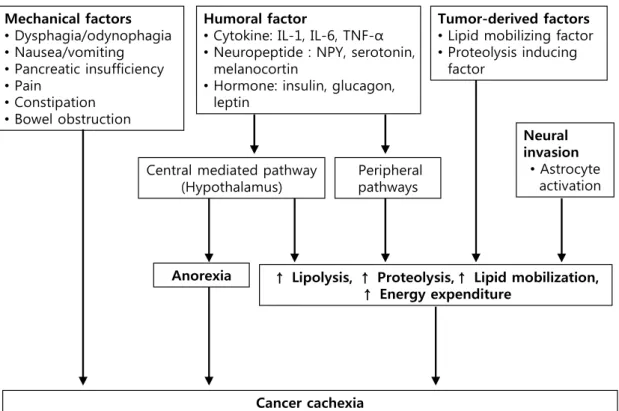
Evidence of sarcopenia*withweightlossgreaterthan 2% *Sarcopenia defined as appendicular skeletal muscle index in males <7.26 kg/m2and infemales <5.45 kg/m2 determined by DEXA. Mechanical factors •Dysphagia/odynophagia •Nausea/vomiting •Pancreatic insufficiency •Pain •Constipation •Bowel obstruction Tumor-derived factors •Lipid mobilizing factor •Proteolysis inducing
factor
Peripheral pathways Humoral factor
•Cytokine: IL-1, IL-6, TNF-α •Neuropeptide : NPY, serotonin,
melanocortin
•Hormone: insulin, glucagon, leptin
Central mediated pathway (Hypothalamus)
↑ Lipolysis, ↑ Proteolysis,↑ Lipid mobilization, ↑ Energy expenditure Anorexia Cancer cachexia Cancer cachexia Neural invasion •Astrocyte activation
Fig. 1. Pathopysiology of cachexia in pancreatic cancer. There are several factors which contribute to develop cachexia in pancreatic cancer, including mechanical factors, tumor-derived factors, humoral factors and neuronal invasion. Several pro-inflammatory cytokines, circulating hormones, neuropeptides, and neurotransmitters result in anorexia and metabolic alteration, such as increased lipolysis, proteolysism lipid mobilization and energy.
*Adapted from Tan et al. Front Physiol 2014;5:88.1 This international consensus also described three stages of cachexia; precachexia, cachexia, and refractory cachexia.9
Severity is based on the degree of depletion of energy store and body protein mass (using BMI) and the rate of ongoing weight loss. In precachexia, patients with early clinical and metabolic signs including anorexia and impaired glucose tol-erance can precede considerable involuntary weight loss. Some patients then have progressive weight loss and meet the crite- ria for cachexia as previously defined. Large retrospective cohort study for pancreatic cancer revealed that a reduction in BMI developed as early as 3 years prior to cancer diagnosis and cachexia-associated symptoms presented at average 2
months before the cancer diagnosis.10,11 Unfortunately, most
patients with pancreatic cancer usually demonstrate in the advanced stage with cachexia symptoms,12 and their cachexia becomes clinically refractory as a result of progressive unres- ponsive to cancer treatment. In refractory cachexia stage, pa-tients have worsening performance status with expected sur-vival less than 3 months.
2. Pathophysiology of cancer cachexia
Cancer cachexia arises from a complex interaction between cancer growth and host response resulting ongoing weight loss, a consequence of a negative protein and energy balance mediated by a combination of reduced food intake and increa- sed metabolism.1,9 The pathophysiology includes a series of
complex metabolic mechanisms directly related to the tumor- host interaction (Fig. 1). There are mechanical factors that contribute to reduced food intake, tumor-derived factors re-leased from the tumor itself and humoral factors generated as the host’s biological response to the tumor. Several pro-in-flammatory cytokines, circulating hormones, neuropeptides,
and neurotransmitters can affect the development of cancer cachexia.13 In addition recent studies have described the other
potentially momentous processes involved in the develop-ment of pancreatic cancer cachexia, including astrocytic acti-vation from neural invasion of pancreatic cancer.14,15
3. Mechanical factors
Mechanical digestive abnormalities that can reduce food intake and result in a lack of appetite include abdominal pain, nausea, dysphagia, odynophagia, pancreatic insufficiency, con- stipation, and intestinal obstruction.16 They can induce and maintain cancer-associated weight loss. These symptoms re-sult from direct cancer invasion to pancreatic duct and/or gast- rointestinal tract, particularly the duodenal second portion. Also, some patients who received the resection of pancreas suffer from pancreatic insufficiency and poor oral intake.
4. Humoral factors
The humoral mediators of cancer cachexia include pro-in-flammatory cytokines (interleukin-1 (IL-1), interleuin-6 (IL-6), interferon-γ (IFN-γ), tumor necrosis factor-α (TNF-α), neu-ropeptides (neuropeptide Y, serotonin, melanocortin) and hormones (insulin, glucagon, leptin). These pathways can be divided into central pathways, which are controlled at brain hypothalamus, and peripheral pathways, which associate with direct lipolysis and proteolysis.
1) Centrally-mediated pathways
Recent evidence suggests that systemic inflammation plays a pivotal role in inducing cancer anorexia by triggering a com- plex neurochemical pathways in hypothalamus.17,18 Increased
cytokine expression prevents the activation of hypothalamus from responding appropriately to peripheral signals by persis-tent stimulation of anorexigenic pathways and inhibition of orexigenic pathways.19,20 Some studies reveal that cancer cache-
xia is associated with hyperactivation of the pro-opiomelno-cortin (POMC)/cocaine and amphetamine-regulated transc- ript (CART) pathways (one of anorexigneic pathways) which may be triggered by IL-1 and other pro-inflammatory cyto- kines.21-24
Leptin is a protein with homeostatic effect released by fatty tissue, which reduces appetite and increase energy expendi- ture through the central nervous system (CNS). In situation of weight loss, leptin release is decreased and this stimulation the appetite in the CNS by activation of neuropeptide Y (NRY)/ Agouti-related peptide (AgRP) pathway (one of orexigenic path-
ways) and reduced activity of anorexigenic neuropeptide, such as corticotropin-releasing factor (CRF) and melanocortin. Cur- rent studies for cancer cachexia suggest that inflammatory cytokine like IL-1 and TNF-α activate the leptin signaling and disturb the orexigenic response to decreased peripheral leptin level.19,25
Serotonin also may play an important role in the develop-ment of cancer anorexia through the melanocortin system. Studies suggested that IL-1 stimulates the release of hypotha- lamic serotonin, which contribute to the persistent activation of POMC/CARD pathway, resulting in decreased appetite and anorexia.26,27
2) Peripheral pathways
Inflammatory cytokines not only contribute to the neuro-chemical changes in the CNS responsible for anorexia, but also have been revealed to induce proteolysis, lipolysis, and the hepatic acute phase protein response (APPR) through the numerous pathways. These processes develop the uncompen- sated loss of muscle and adipose tissue.
TNF-α is one of the first identified as an endogenous cache- xia-inducing factors. TNF-α stimulates protein degradation in the proteasome-ubiquitin system, mediated by transcrip- tion factors, such as nuclear factor kappa B (NF-κB) and MyoD.28-30 Some studies also showed that TNF-α promote
lipolysis in vitro with increase in glycerol release in mouse and human adipocyte, through downregulation of perilipin exp- ression, which subsequently induce hormone-sensitive lipase (HSL), a key regulator of lipolysis, to access the surface of lipid droplets for breakdown.31,32 Additionally, TNF-α has a inhibi-
tory action on adipocyte differentiation, resulting in impaired lipogenesis.33,34
IL-6 is another important cytokine in the development of cachexia in pancreatic cancer, particularly associated with acti- vation of the hepatic APPR. Moses et al found that overpro- duction of IL-6 and elevated APPR (for example; elevated c-reactive protein (CRP) level) have been significantly asso-ciated with decreased survival in patients with pancreatic cancer cachexia.35There is strong relationship between increased IL-6 production of peripheral blood mononuclear cells and the presence of elevated APPR.35-37 The activation of hepatic
APPR promote the mobilization of peripheral amino acid sto- res, mostly from skeletal muscle, contributing to the loss of lean body mass.
5. Tumor factors
hormones and neurotransmitters, tumor-derived factors con-tribute to metabolic dysregulation in pancreatic cancer cache- xia. There are two most well studied factors, lipid mobilizing factor (LMF) and proteolysis-inducing factor (PIF).
LMF was first discovered from a cachexia-inducing murine tumor model and the urine of patients with unresectable pan-creatic cancer with weight loss.38 This material was 43 kDa
and was suggest to be homologous with the plasma protein zinc-α2-glycoprotein (ZAG).38 LMF/ZAG not only induces
lipid mobilization through various signal pathways but also augment substrate utilization and activates mitochondrial oxi-dative pathways in brown adipose tissue. Consequently, LMF/ ZAG causes lipolysis with increased energy expenditure, and catatbolism.39-41 Recently, LMF/ZAG is proposed as a serum bio-
marker in pancreatic cancer cachexia.42
PIF was isolated in 1996 from a mouse tumor model of cachexia, as a 24 kDa glycoprotein inducing skeletal muscle catabolism.43 PIF was detected in the urine of 80% of pan-
creatic cancer patients with cachexia, and rate of weight loss was greater in patients who have PIF in their urine.44 Also,
when PIF from urine of cancer cachexia patients administered intravenously to normal mice, PIF induced significant weight loss without reduction in food and water intake.45 Some studies
suggest that PIF-mediated protein degradation may be media- ted by the ubiquitin-proteasome proteolytic pathway in skel-etal muscle; that process results from activation of NFκB.46-48 Also, PIF inhibits the protein synthesis on skeletal muscle through activation of double-stranded RNA dependent pro-tein kinase (PKR).49
6. Other mechanisms
Recent studies have suggested that neural invasion of pan-creatic cancer is related to astrocyte activation and develop-ment of cachexia in pancreatic cancer patients.14,15 Neuronal
invasion of pancreatic cancer induce the activation of astrocy- tes and microglia in the spinal cord. These activated astrocy- tes can subsequently develop lipolysis and muscle atrophy in pancreatic cancer patients, although more researches is nee- ded to determine the underlying mechanisms involving cache- xia.14
7. Treatment of cancer cachexia
The primary end-points of optimal treatment of cancer cache- xia are improvements in cachexia-associated symptoms such as anorexia and fatigue, lean body mass, resting energy expen- diture, quality of life, and performance status through inhi- bition of effect of pro-inflammatory cytokines.50 Although
there has been recently remarkable advances in preclinical and
clinical research in era of cancer cachexia , the currently avail-able treatment options are still limited.
8. Nutritional support
Nutritional risk is highest among pancreatic cancer patients.51 However, despite several trials of conventional and/or aggre- ssive nutritional support using different feeding techniques, the cachectic state is difficult to be overcomed by nutritional support alone.52 A small multicenter randomized trial for
pa-tients with advanced pancreatic cancer showed a meaningful improvement in weight and body mass composition as well as quality of life with L-Carnitine supplementation.53
9. Pharmacologic treatment
The two major options for pharmacological therapy have been, till now, either progestational agents or corticosteroids. Recently, there are various drugs studied for treatment of can- cer cachexia.
Megestrol acetate is semi-synthetic progesterone widely used as an appetite stimulant. The pharmacologic activity of mege- strol acetate was considered as reduced release of pro-inflam- matory cytokines (IL-1, IL-6, TNF-α) and stimulation of NPY in the hypothalamus.54,55 Several randomized control trials
have demonstrated that megestrol acetate (480-800 mg/ day) significantly improves appetite, nausea, food intake, and weight gain among patients with cancer cachexia, including those with pancreatic cancer.56-59 Megestrol acetate is generally well- tolerated with low incidence of adverse events, such as skin rash, hyperglycemia, adrenal insufficiency, and thromboem-bolic events.57 Since its approval in 1993, several meta-anal-yses have revealed that megestrol acetate has better effect of improved appetite, weight, and quality of life compared than placebo or other drugs (cisapride, dronabinol, corticosteroids, nandrolone).60,61
Corticosteroids, such as dexamethasone, have been studied to treat cancer-associated anorexia and cachexia.62,63 The me-
cha nism of action is likely associated with the inhibition of IL-1, TNF-α, and leptin as well as the stimulation of NPY.64
However, the effects of corticosteroid could not be maintai- ned longer than 4 weeks and related to long-term side effects, such as insulin resistance, fluid retention, steroid-induced myopathy, skin fragility, adrenal insufficiency, and sleep and cognitive disorders.65Owing to their short term symptomatic benefits with long term adverse effects, corticosteroids are just considered as treatment option in patients with short expected survival.
Dronabinol is effective in reducing nausea and increasing appetite with a tendency to weight stabilization. The appe-tite-stimulant effect of dronabinol associated with interaction with endorphin receptors, interference with IL-1 synthesis, activation of cannabinoid receptors involved in the neuronal circuit of leptin and inhibition of prostaglandin synthesis.20 A phase II study showed that dronabinol decreased anorexia in 68% of patients, but 16% of patients had to stop admin-istration due to CNS adverse events, such as euphoria, hallu-cinations, psychosis, and vertigo.66 However, in result of first
clinical trial that compared megestrol acetate with nol, megestrol acetate was appears to be superior to dronabi-nol in aspect of increasing appetite and weight gain.67 Drona-
binol serves as an alternative treatment option as an appetite stimulant and anti-emetic.
Non-steroidal anti-inflammatory drugs (NSAIDs), includ-ing cyclooxygenase-2 (COX-2) inhibitors, ibuprofen, and in-domethacin, reduce release of acute phase proteins and pro- inflammatory cytokines.68,69 NSAIDs are suggested to inhibit
prostaglandin synthesis and thereby prevent downstream ef-fects of systemic inflammation. Preliminary results of some studies for NSAIDs, such as indomethacin and ibuprofen, have been shown to effect to increase weight and muscle mass, improve quality of life, and prolong survival in advanced can-cer patients, especially when combined with progestogens.69-72
However, further large studies are needed to validate the clinical role of NSAIDs in the management of cancer cachexia. Thalidomide have anti-inflammatory and immunomodula- tory properties that downregulate the production of TNF-α and other cytokines, inhibit NF-κB, downregulate COX-2, and inhibit aniogenesis.73 In 2005, Gorden et al. published
the results of a single center, double blinded, placebo-control- led, randomized study aimed at assessing the efficacy and safety of thalidomide in attenuating weight loss in pancreatic cancer patients with cachexia.74 The study population consis-
ted of 50 patients (who had lost at least 10% of their body weight) randomized to administer thalidomide 200 mg/day or a placebo for 24 weeks. The conclusion of the study strongly suggested that thalidomide was effective for attenuating loss of weight and lean body mass in patients with cancer cache- xia. Thalidomide was typically well-tolerated. Adverse events included peripheral neuropathy, dizziness, somnolence, con-stipation, rash, and possible increased risk of venous throm- boembolism. These results are significant but further large- scale clinical trials are needed to validate the efficacy of thali-domide in treating pancreatic cancer cachexia.
Cyproheptadine is an antiserotoninergic agent with anti-histamine properties. Despite some promising results of pilot
studies, controlled clinical trials have not yet proved its effi-cacy in cancer cachexia patients.75,76 Pizotifen is an antisero-
toninergic drug studied in the treatment of anorexia asso-ciated with other benign causes, which has not been inves-tigated in cancer patients.
Eicosapentaenoic acid (EPA) and docosahexaenoic acid (DHA), a long-chain polyunsaturated fatty acid of the ome-ga-3 family, are have been revealed to suppress production of pro-inflammatory cytokines, including IL-1, TNF-α, and IL-6.77,78 EPA can also inhibit the downstream effect of LMF
and PIF.79-81 Their efficacy in cancer cachexia has not been
fully validated in well-organized clinical trials.82-84 Two
sys-tematic literature reviews conclude the EPA and DHA in monotherapy show no significant improvement in appetite, fat free mass, survival and quality of life compared with pla- cebo.85,86 The efficacy of EPA in cancer cachexia treatment
remains uncertain although recent study suggest that EPA supplementation may not be effective as a single agent or even in combination with megestrol acetate in patients with cancer cachexia.
Current studies are investigating an approach of drug com-binations to attenuate cancer cachexia. A recent data from a large multicenter trial with 332 patients comparing medroxy- progesterone, megestrol acetate, and oral supplementation with EPA, L-carnitine, and thalidomide found that the combi-nation therapy was significantly effective in improving lean body mass and appetite than any other treatment arms with single drug treatment.87
10. New therapeutic targets
Due to lack of the available drugs that have shown sustai- ned effects on weight stabilization and improvement in sur-vival, various researches have continued to explore new ther-apeutic targets and to develop new drugs.
OHR/AVR 118 is a recently developed, broad-spectrum peptide-nucleic acid immunomodulator that target both TNF-α and IL-6. A phase II study including patients with advanced cancer and cachexia presented an improvement in anorexia, dyspepsia, strength anddepression.88 A phase IIb study is cur-
rently underway to evaluate the safety and efficacy of OHDR/ AVR118(NCT01206335).
ALD518 (also known as BMS-945429), a humanized mon-oclonal IL-6 antibody, showed promising beneficial results in phase II randomized, double-blinded, placebo-controlled trials with non-small cell lung cancer (NSCLC) patients with cachexia.89,90 This agent was safe and well tolerated. ALD518
of lean body mass with significant improvement of fatigue score.89,90
Ghrelin is the endogenous ligand of the growth hormone receptor that produces the release of growth hormone and NPY.91 In addition, Ghrelin induces the release of anti-inflam-
matory cytokine, IL-10, which suppressed the production of pro-inflammatory cytokines, including IL-1β, Il-6 and TNF-α.92-94 Several controlled clinical trials using oral ghrelin
mim-etic named RC-1291 or anamorelin, demonstrated an impro- vement in increasing appetite and weight in cancer cachexia patients.95-97 Macimorelin is a novel oral ghrelin compound,
with a good oral availability and stability, which binds the growth hormone secretagogue receptor (GHSR) 1a with simi- lar affinity to ghrelin.98 These findings promoted further inves
tigation for ghrelin analogs andmore phase II trials are ongo- ing (NCT01505764, NCT01614990).
MT-102, a novel anabolic/catabolic transforming agent, has a multi-targeting effect on three potential pharmacological pathways in cancer cachexia, primarily reduced catabolism through nonselective β-blockade, improve fatigue and ther-mogenesis through blocking central 5-HT 1a receptor, and increased anabolism through partial activation of β-2 recep- tor.99 Two phase II studies in stage III/IV colorectal cancer and NSCLC are under investigation (ACT-ONE and ACT-TWO; NCT01238107).99
BYM338 (bimagrumab) is a fully humanized monoclonal antibody blocking the activin II receptor type IIB (ActRIIB) and preventing receptor occupation by myostatin. Myostatin, a member of the TGF-β superfamily, is expressed almost exclu- sively in skeletal muscle and acts as a negative regulator of muscle growth, through binding to the ActRIIB by activating multiple downstream pathways.100 Inhibition of muscle diffe-
rentiation by myostatin is mediated, in part, through Smad 2/3 phosphorylation-dependent inhibition of the Akt/mTOR pathway.100 In preclinical study, ActRIIB blockade prevented
muscle loss and prolonged survival in C-26 tumor-bearing mice.101 A multicenter, randomized, double-blind, placebo-
controlled phase II trials to investigate the efficacy of BYM 338 in attenuating loss of body mass in cachectic patients with stage IV NSCLC or stage III/IV pancreatic cancer has been comple- ted and in preparation to report the results (NCT01433263). LY2495655 is another humanized anti-myostatin antibody currently under investigation. A phase II study in patients with locally advanced or metastatic pancreatic cancer is ongoing to evaluate two different doses of LY2495655 in combination with standard of care chemotherapy in improving survival, lean body mass and physical performance (NCT01505530).
CONCLUSION
Although cachexia is a major problem in cancer and many chronic diseases, cachexia treatment is still largely ignored even for inpatient management or limited to dietary counsel-ing to treat weight loss with poor efficacy. Regardcounsel-ing that approximately 80% of pancreatic cancer suffer from cachec-tic symptoms and up to 30% die from cachexia-related com-plications, importance of treatment for cancer cachexia, espe-cially in pancreatic cancer patients, should be acquired in the future.1,102 Although pancreatic cancer cachexia is
consid-ered as multifactorial syndrome mediated by mechanical fac-tors, pro-inflammatory cytokines, neuropeptides, hormones and tumor-derived factors, further researches to understand the basic mechanism involved in induction and maintenance of pancreatic cancer cachexia are further needed. Recently, some preliminary studies for targeted agents have been shown promising results. We hoped that new therapeutic strategies will be developed to improve the quality of lifer and prolong the survival of pancreatic cancer patients in the future.
REFERENCES
1. Fearon KC, Voss AC, Hustead DS, et al. Definition of cancer cachexia: effect of weight loss, reduced food intake, and sys-temic inflammation on functional status and prognosis. Am J Clin Nutr 2006;83:1345-1350.
2. Inui A. Cancer anorexia-cachexia syndrome: current issues in research and management. CA Cancer J Clin 2002;52:72-91. 3. Wigmore SJ, Plester CE, Richardson RA, et al. Changes in
nutritional status associated with unresectable pancreatic can- cer. Br J Cancer 1997;75:106-109.
4. Dewys WD, Begg C, Lavin PT, et al. Prognostic effect of weight loss prior to chemotherapy in cancer patients. Eastern Cooperative Oncology Group. Am J Med 1980;69:491-497. 5. Mueller TC, Burmeister MA, Bachmann J, et al. Cachexia
and pancreatic cancer: are there treatment options? World J Gastroenterol 2014;20:9361-9373.
6. Fearon KC. Cancer cachexia: developing multimodal therapy for a multidimensional problem. Eur J Cancer 2008;44:1124- 1132.
7. Ozola Zalite I, Zykus R, Francisco Gonzalez M, et al. Infl- uence of cachexia and sarcopenia on survival in pancreatic ductal adenocarcinoma: a systematic review. Pancreatology 2015;15:19-24.
8. Pausch T, Hartwig W, Hinz U, et al. Cachexia but not obesity worsens the postoperative outcome after pancreatoduodene- ctomy in pancreatic cancer. Surgery 2012;152:S81-S88.
9. Fearon K, Strasser F, Anker SD, et al. Definition and classifi- cation of cancer cachexia: an international consensus. Lancet Oncol 2011;12:489-495.
10. Pannala R, Leibson CL, Rabe KG, et al. Temporal association of changes in fasting blood glucose and body mass index with diagnosis of pancreatic cancer. Am J Gastroenterol 2009;104: 2318-2325.
11. Chari ST, Leibson CL, Rabe KG, et al. Pancreatic cancer-asso-ciated diabetes mellitus: prevalence and temporal association with diagnosis of cancer. Gastroenterology 2008;134:95-101. 12. Sah RP, Nagpal SJ, Mukhopadhyay D, et al. New insights into
pancreatic cancer-induced paraneoplastic diabetes. Nat Rev Gastroenterol Hepatol 2013;10:423-433.
13. Tisdale MJ. Cachexia in cancer patients. Nat Rev Cancer 2002; 2:862-871.
14. Imoto A, Mitsunaga S, Inagaki M, et al. Neural invasion in-duces cachexia via astrocytic activation of neural route in pan-creatic cancer. Int J Cancer 2012;131:2795-2807.
15. Mitsunaga S, Kinoshita T, Hasebe T, et al. Low serum level of cholinesterase at recurrence of pancreatic cancer is a poor prognostic factor and relates to systemic disorder and nerve plexus invasion. Pancreas 2008;36:241-248.
16. Deutsch J, Kolhouse JF. Assessment of gastrointestinal func-tion and response to megesterol acetate in subjects with gas-trointestinal cancers and weight loss. Support Care Cancer 2004;12:503-510.
17. Laviano A, Meguid MM, Rossi-Fanelli F. Cancer anorexia: clinical implications, pathogenesis, and therapeutic strategies. Lancet Oncol 2003;4:686-694.
18. Fearon K, Arends J, Baracos V. Understanding the mecha-nisms and treatment options in cancer cachexia. Nat Rev Clin Oncol 2013;10:90-99.
19. Suzuki H, Asakawa A, Amitani H, et al. Cancer cachexia path-ophysiology and translational aspect of herbal medicine. Jpn J Clin Oncol 2013;43:695-705.
20. Tuca A, Jimenez-Fonseca P, Gascon P. Clinical evaluation and optimal management of cancer cachexia. Crit Rev Oncol Hematol 2013;88:625-636.
21. Wisse BE, Frayo RS, Schwartz MW, et al. Reversal of cancer anorexia by blockade of central melanocortin receptors in rats. Endocrinology 2001;142:3292-3301.
22. Marks D, Cone RD. The role of the melanocortin-3 receptor in cachexia. Ann N Y Acad Sci 2003;994:258-266. 23. Marks DL, Butler AA, Turner R, et al. Differential role of
melanocortin receptor subtypes in cachexia. Endocrinology 2003;144:1513-1523.
24. Scarlett JM, Jobst EE, Enriori PJ, et al. Regulation of central melanocortin signaling by interleukin-1 beta. Endocrinology 2007;148:4217-4225.
25. Inui A. Cancer anorexia-cachexia syndrome: are neuropep- tides the key? Cancer Res 1999;59:4493-4501.
26. Shintani F, Kanba S, Nakaki T, et al. Interleukin-1 beta aug-ments release of norepinephrine, dopamine, and serotonin in the rat anterior hypothalamus. J Neurosci 1993;13:3574- 3581.
27. Heisler LK, Cowley MA, Tecott LH, et al. Activation of cent-
ral melanocortin pathways by fenfluramine. Science 2002;297: 609-611.
28. Llovera M, Carbo N, Lopez-Soriano J, et al. Different cyto-kines modulate ubiquitin gene expression in rat skeletal mus- cle. Cancer Lett 1998;133:83-87.
29. Li YP, Reid MB. NF-kappaB mediates the protein loss indu- ced by TNF-alpha in differentiated skeletal muscle myotubes. Am J Physiol Regul Integr Comp Physiol 2000;279:1165- 1170.
30. Guttridge DC, Mayo MW, Madrid LV, et al. NF-kappaB-in-duced loss of MyoD messenger RNA: possible role in muscle decay and cachexia. Science 2000;289:2363-2366. 31. Zhang HH, Halbleib M, Ahmad F, et al. Tumor necrosis
fac-tor-alpha stimulates lipolysis in differentiated human adipo-cytes through activation of extracellular signal-related kinase and elevation of intracellular cAMP. Diabetes 2002;51:2929- 2935.
32. Ryden M, Arvidsson E, Blomqvist L, et al. Targets for TNF- alpha-induced lipolysis in human adipocytes. Biochem Bio- phys Res Commun 2004;318:168-175.
33. Cawthorn WP, Heyd F, Hegyi K, et al. Tumour necrosis fac-tor-alpha inhibits adipogenesis via a beta-catenin/TCF4(TCF7L2)- dependent pathway. Cell Death Differ 2007;14:1361-1373. 34. Hammarstedt A, Isakson P, Gustafson B, et al. Wnt-signaling
is maintained and adipogenesis inhibited by TNFalpha but not MCP-1 and resistin. Biochem Biophys Res Commun 2007; 357:700-706.
35. Moses AG, Maingay J, Sangster K, et al. Pro-inflammatory cytokine release by peripheral blood mononuclear cells from patients with advanced pancreatic cancer: relationship to acute phase response and survival. Oncol Rep 2009;21:1091-1095. 36. Martignoni ME, Dimitriu C, Bachmann J, et al. Liver macro-phages contribute to pancreatic cancer-related cachexia. Oncol Rep 2009;21:363-369.
37. Martignoni ME, Kunze P, Hildebrandt W, et al. Role of mon-onuclear cells and inflammatory cytokines in pancreatic can-cer-related cachexia. Clin Cancer Res 2005;11:5802-5808. 38. Todorov PT, McDevitt TM, Meyer DJ, et al. Purification
and characterization of a tumor lipid-mobilizing factor. Can- cer Res 1998;58:2353-2358.
39. Hirai K, Hussey HJ, Barber MD, et al. Biological evaluation of a lipid-mobilizing factor isolated from the urine of cancer patients. Cancer Res 1998;58:2359-2365.
40. Russell ST, Hirai K, Tisdale MJ. Role of beta3-adrenergic receptors in the action of a tumour lipid mobilizing factor. Br J Cancer 2002;86:424-428.
41. Tan CR, Yaffee PM, Jamil LH, et al. Pancreatic cancer cache- xia: a review of mechanisms and therapeutics. Front Physiol 2014;5:88.
42. Felix K, Fakelman F, Hartmann D, et al. Identification of serum proteins involved in pancreatic cancer cachexia. Life Sci 2011;88:218-225.
43. Todorov P, Cariuk P, McDevitt T, et al. Characterization of a cancer cachectic factor. Nature 1996;379:739-742. 44. Wigmore SJ, Todorov PT, Barber MD, et al. Characteristics
cachectic factor. Br J Surg 2000;87:53-58.
45. Cariuk P, Lorite MJ, Todorov PT, et al. Induction of cachexia in mice by a product isolated from the urine of cachectic cancer patients. Br J Cancer 1997;76:606-613.
46. Belizario JE, Lorite MJ, Tisdale MJ. Cleavage of caspases-1, -3, -6, -8 and -9 substrates by proteases in skeletal muscles from mice undergoing cancer cachexia. Br J Cancer 2001;84: 1135-1140.
47. Whitehouse AS, Tisdale MJ. Increased expression of the ubiq-uitin-proteasome pathway in murine myotubes by proteol-ysis-inducing factor (PIF) is associated with activation of the transcription factor NF-kappaB. Br J Cancer 2003;89:1116- 1122.
48. Wyke SM, Tisdale MJ. NF-kappaB mediates proteolysis-in-ducing factor induced protein degradation and expression of the ubiquitin-proteasome system in skeletal muscle. Br J Can- cer 2005;92:711-721.
49. Eley HL, Tisdale MJ. Skeletal muscle atrophy, a link between depression of protein synthesis and increase in degradation. J Biol Chem 2007;282:7087-7097.
50. Donohoe CL, Ryan AM, Reynolds JV. Cancer cachexia: mecha- nisms and clinical implications. Gastroenterol Res Pract 2011; 2011:601434.
51. Bozzetti F, Group SW. Screening the nutritional status in on-cology: a preliminary report on 1,000 outpatients. Support Care Cancer 2009;17:279-284.
52. Evans WJ, Morley JE, Argiles J, et al. Cachexia: a new defini- tion. Clin Nutr 2008;27:793-799.
53. Kraft M, Kraft K, Gartner S, et al. L-Carnitine-supplementa-tion in advanced pancreatic cancer (CARPAN)--a randomized multicentre trial. Nutr J 2012;11:52.
54. McCarthy HD, Crowder RE, Dryden S, et al. Megestrol ace-tate stimulates food and water intake in the rat: effects on regional hypothalamic neuropeptide Y concentrations. Eur J Pharmacol 1994;265:99-102.
55. Mantovani G, Maccio A, Lai P, et al. Cytokine activity in can- cer-related anorexia/cachexia: role of megestrol acetate and medroxyprogesterone acetate. Semin Oncol 1998;25:45-52. 56. Bruera E, Macmillan K, Kuehn N, et al. A controlled trial
of megestrol acetate on appetite, caloric intake, nutritional status, and other symptoms in patients with advanced cancer. Cancer 1990;66:1279-1282.
57. Loprinzi CL, Ellison NM, Schaid DJ, et al. Controlled trial of megestrol acetate for the treatment of cancer anorexia and cachexia. J Natl Cancer Inst 1990;82:1127-1132.
58. Loprinzi CL, Michalak JC, Schaid DJ, et al. Phase III evalua-tion of four doses of megestrol acetate as therapy for patients with cancer anorexia and/or cachexia. J Clin Oncol 1993;11: 762-767.
59. Westman G, Bergman B, Albertsson M, et al. Megestrol ace-tate in advanced, progressive, hormone-insensitive cancer. Effects on the quality of life: a placebo-controlled, randomi- sed, multicentre trial. Eur J Cancer 1999;35:586-595. 60. Pascual Lopez A, Roquei Figuls M, Urrutia Cuchi G, et al.
Systematic review of megestrol acetate in the treatment of anorexia-cachexia syndrome. J Pain Symptom Manage 2004;
27:360-369.
61. Lesniak W, Bala M, Jaeschke R, et al. Effects of megestrol acetate in patients with cancer anorexia-cachexia syndrome--a systematic review and meta-analysis. Pol Arch Med Wewn 2008;118:636-644.
62. Willox JC, Corr J, Shaw J, et al. Prednisolone as an appetite stimulant in patients with cancer. Br Med J (Clin Res Ed) 1984; 288:27.
63. Bruera E, Roca E, Cedaro L, et al. Action of oral methyl-prednisolone in terminal cancer patients: a prospective rando- mized double-blind study. Cancer Treat Rep 1985;69:751- 754.
64. Plata-Salaman CR. Dexamethasone inhibits food intake sup-pression induced by low doses of interleukin-1 beta adminis- tered intracerebroventricularly. Brain Res Bull 1991;27:737- 738.
65. Loprinzi CL, Kugler JW, Sloan JA, et al. Randomized compar-ison of megestrol acetate versus dexamethasone versus fluox-ymesterone for the treatment of cancer anorexia/cachexia. J Clin Oncol 1999;17:3299-3306.
66. Nelson K, Walsh D, Deeter P, et al. A phase II study of delta- 9-tetrahydrocannabinol for appetite stimulation in cancer-asso- ciated anorexia. J Palliat Care 1994;10:14-18.
67. Jatoi A, Windschitl HE, Loprinzi CL, et al. Dronabinol versus megestrol acetate versus combination therapy for cancer-asso-ciated anorexia: a North Central Cancer Treatment Group study. J Clin Oncol 2002;20:567-573.
68. Preston T, Fearon KC, McMillan DC, et al. Effect of ibupro-fen on the acute-phase response and protein metabolism in patients with cancer and weight loss. Br J Surg 1995;82:229- 234.
69. Wigmore SJ, Falconer JS, Plester CE, et al. Ibuprofen reduces energy expenditure and acute-phase protein production com-pared with placebo in pancreatic cancer patients. Br J Cancer 1995;72:185-8.
70. Lundholm K, Gelin J, Hyltander A, et al. Anti-inflammatory treatment may prolong survival in undernourished patients with metastatic solid tumors. Cancer Res 1994;54:5602-5606. 71. McMillan DC, O'Gorman P, Fearon KC, et al. A pilot study
of megestrol acetate and ibuprofen in the treatment of ca-chexia in gastrointestinal cancer patients. Br J Cancer 1997; 76:788-790.
72. McMillan DC, Wigmore SJ, Fearon KC, et al. A prospective randomized study of megestrol acetate and ibuprofen in gas-trointestinal cancer patients with weight loss. Br J Cancer 1999; 79:495-500.
73. Sampaio EP, Sarno EN, Galilly R, et al. Thalidomide selecti- vely inhibits tumor necrosis factor alpha production by stimu-lated human monocytes. J Exp Med 1991;173:699-703. 74. Gordon JN, Trebble TM, Ellis RD, et al. Thalidomide in the
treatment of cancer cachexia: a randomised placebo control- led trial. Gut 2005;54:540-545.
75. Couluris M, Mayer JL, Freyer DR, et al. The effect of cyprohe- ptadine hydrochloride (periactin) and megestrol acetate (megace) on weight in children with cancer/treatment-related cachexia. J Pediatr Hematol Oncol 2008;30:791-797.
76. Kardinal CG, Loprinzi CL, Schaid DJ, et al. A controlled trial of cyproheptadine in cancer patients with anorexia and/or cachexia. Cancer 1990;65:2657-2662.
77. Meydani SN, Lichtenstein AH, Cornwall S, et al. Immunolo- gic effects of national cholesterol education panel step-2 diets with and without fish-derived N-3 fatty acid enrichment. J Clin Invest 1993;92:105-113.
78. Wigmore SJ, Fearon KC, Maingay JP, et al. Down-regulation of the acute-phase response in patients with pancreatic cancer cachexia receiving oral eicosapentaenoic acid is mediated via suppression of interleukin-6. Clin Sci (Lond) 1997;92:215- 221.
79. Tisdale MJ, Beck SA. Inhibition of tumour-induced lipolysis in vitro and cachexia and tumour growth in vivo by eicosa-pentaenoic acid. Biochem Pharmacol 1991;41:103-107. 80. Tisdale MJ. Inhibition of lipolysis and muscle protein
degra-dation by EPA in cancer cachexia. Nutrition 1996;12:S31- S33.
81. Hussey HJ, Tisdale MJ. Effect of a cachectic factor on carbo-hydrate metabolism and attenuation by eicosapentaenoic acid. Br J Cancer 1999;80:1231-1235.
82. Bruera E, Strasser F, Palmer JL, et al. Effect of fish oil on appetite and other symptoms in patients with advanced can-cer and anorexia/cachexia: a double-blind, placebo-controlled study. J Clin Oncol 2003;21:129-134.
83. Fearon KC, Barber MD, Moses AG, et al. Double-blind, pla-cebo-controlled, randomized study of eicosapentaenoic acid diester in patients with cancer cachexia. J Clin Oncol 2006; 24:3401-3407.
84. Persson C, Glimelius B, Ronnelid J, et al. Impact of fish oil and melatonin on cachexia in patients with advanced gastro-intestinal cancer: a randomized pilot study. Nutrition 2005; 21:170-178.
85. Dewey A, Baughan C, Dean T, et al. Eicosapentaenoic acid (EPA, an omega-3 fatty acid from fish oils) for the treatment of cancer cachexia. Cochrane Database Syst Rev 2007: CD 004597.
86. Mazzotta P, Jeney CM. Anorexia-cachexia syndrome: a sys-tematic review of the role of dietary polyunsaturated Fatty acids in the management of symptoms, survival, and quality of life. J Pain Symptom Manage 2009;37:1069-1077. 87. Mantovani G, Maccio A, Madeddu C, et al. Randomized
phase III clinical trial of five different arms of treatment in 332 patients with cancer cachexia. Oncologist 2010;15:200-211. 88. Chasen M, Hirschman SZ, Bhargava R. Phase II study of the
novel peptide-nucleic acid OHR118 in the management of cancer-related anorexia/cachexia. J Am Med Dir Assoc 2011; 12:62-67.
89. Rigas JR, Schuster M, Orlov SV, et al. Efect of ALD518, a humanized anti-IL-6 antibody, on lean body mass loss and symptoms in patients with advanced non-small cell lung can-cer (NSCLC): Results of a phase II randomized double-blind
safety and efficacy trial.J Clin Oncol 2010;28s.Abstracts 7622. 90. Schuster M, Rigas JR, Orlov SV, et al. ALD518, a humanized
anti-IL-6 antibody, treats anemia in patients with advanced non-small cell lung cancer (NSCLC): Results of a phase II, ran- domized, double-blind, placebo-controlled trial. J Clin Oncol 2010;28s.Abstracts 7631.
91. Kojima M, Hosoda H, Date Y, et al. Ghrelin is a growth-hor-mone-releasing acylated peptide from stomach. Nature 1999; 402:656-660.
92. Gonzalez PV, Cragnolini AB, Schioth HB, et al. Interleukin-1 beta-induced anorexia is reversed by ghrelin. Peptides 2006; 27:3220-3225.
93. Waseem T, Duxbury M, Ito H, et al. Exogenous ghrelin mod-ulates release of pro-inflammatory and anti-inflammatory cyto- kines in LPS-stimulated macrophages through distinct signal-ing pathways. Surgery 2008;143:334-342.
94. Dixit VD, Schaffer EM, Pyle RS, et al. Ghrelin inhibits leptin- and activation-induced proinflammatory cytokine expression by human monocytes and T cells. J Clin Invest 2004;114:57-66. 95. Garcia JM, Polvino WJ. Effect on body weight and safety of
RC-1291, a novel, orally available ghrelin mimetic and gro- wth hormone secretagogue: results of a phase I, randomized, placebo-controlled, multiple-dose study in healthy volunteers. Oncologist 2007;12:594-600.
96. Neary NM, Small CJ, Wren AM, et al. Ghrelin increases energy intake in cancer patients with impaired appetite: acute, ran- domized, placebo-controlled trial. J Clin Endocrinol Metab 2004;89:2832-2836.
97. Garcia JM, Friend J, Allen S. Therapeutic potential of anamo- relin, a novel, oral ghrelin mimetic, in patients with cancer- related cachexia: a multicenter, randomized, double-blind, crossover, pilot study. Support Care Cancer 2013;21:129-137. 98. Broglio F, Boutignon F, Benso A, et al. EP1572: a novel peptido-
mimetic GH secretagogue with potent and selective GH-relea- sing activity in man. J Endocrinol Invest 2002;25:RC26-28. 99. Stewart Coats AJ, Srinivasan V, Surendran J, et al. The ACT-
ONE trial, a multicentre, randomised, double-blind, place-bo-controlled, dose-finding study of the anabolic/catabolic transforming agent, MT-102 in subjects with cachexia related to stage III and IV non-small cell lung cancer and colorectal can- cer: study design. J Cachexia Sarcopenia Muscle 2011;2:201- 207.
100. Ma JD, Heavey SF, Revta C, et al. Novel investigational biologics for the treatment of cancer cachexia. Expert Opin Biol Ther 2014;14:1113-1120.
101. Zhou X, Wang JL, Lu J, et al. Reversal of cancer cachexia and muscle wasting by ActRIIB antagonism leads to prolon- ged survival. Cell 2010;142:531-543.
102. Bachmann J, Ketterer K, Marsch C, et al. Pancreatic cancer related cachexia: influence on metabolism and correlation to weight loss and pulmonary function. BMC Cancer 2009; 9:255.