INTRODUCTION
Valvular involvement in heart disease results in stenosis, insufficiency, or both, and the most common cause of mitral stenosis is chronic rheumatic heart disease (1, 2). Rheumatic fever (RF) is the consequence of a pharyngeal infection with group A streptococcus in a susceptible host, leading to autoim-mune disease induced by antigen mimicry of human cardiac myosin with the streptococcal glycoprotein (3). While most disease manifestations are transient and leave no residua, rheumatic carditis can lead to chronic rheumatic heart dis-ease (CRHD), which is characterized by fibrotic valvular deformity and can produce permanent and severe cardiac dys-function even decades later (1).
The current hypothesis of CRHD is that fibrotic valvular deformity evolves through organization of acute inflamma-tion induced by RF, with subsequent thickening and retrac-tion of valvular leaflets, and with secondary damage result-ing from turbulent flow induced by valvular dysfunction (1, 4). There are some evidences that CRHD is associated with ongoing inflammation of valvular leaflets. The plasma con-centrations of TNF- and high sensitive C-reactive protein (CRP) are significantly higher in CRHD than in controls
(5-7). It has been shown that lymphocytes obtained from CRHD patients exhibit T helper 2 (Th2) type cytokine response whereas those from RF patients show T helper 1 (Th1) cyto-kine profile in cell culture with streptococcal superantigen (8). The Th2 immune response is known to promote fibrot-ic process by activating fibroblast proliferation, myofibrob-last differentiation, extracellular matrix deposition, and trans-forming growth factor-beta 1 (TGF- 1) production, where-as Th1 type cytokines such where-as interferon- suppress these processes (9-11).
TGF- 1 plays a critical role in matrix remodeling and in enhancing collagen synthesis (12). Upon tissue injury, inflam-mation occurs and many cytokines are secreted and TGF- 1 is released, promotes myofibroblast differentiation, and stim-ulates fibroblasts and other reparative cells to proliferate and to synthesize extracellular matrix components. Under normal conditions this leads to provisional repair; however, with repeat-ed injury, the increase in TGF- 1 production is sustainrepeat-ed, lead-ing to tissue fibrosis (13, 14). Recent studies have investigat-ed TGF- 1 expression in diseasinvestigat-ed heart valves (15-17). Sero-tonin, which is associated with carcinoid heart disease (18), has been demonstrated to up-regulate TGF- 1 (15). Other reports indicate that TGF- 1 is present within calcific aor-41
Lucia Kim, Do Kyun Kim*, Woo Ick Yang�, Dong Hwan Shin�,
Ick Mo Jung�, Han Ki Park
*, Byung Chul Chang*
Department of Pathology, Inha University College of Medicine, Incheon; Departments of Cardiovascular Surgery*and Pathology�
, Yonsei University College of Medicine, Seoul; Department of Internal Medicine�
, Ehwa University College of Medicine, Seoul, Korea
Address for correspondence Lucia Kim, M.D.
Department of Pathology, Inha University Hospital, 7-206, 3rd st, Shinheung-dong, Jung-gu, Incheon 400-711, Korea
Tel : +82.32-890-3984, Fax : +82.32-890-3464 E-mail : [email protected]
*This work was supported by Inha University Research Grant (INHA-31448).
DOI: 10.3346/jkms.2008.23.1.41
Overexpression of Transforming Growth Factor- 1 in the Valvular
Fibrosis of Chronic Rheumatic Heart Disease
For the purpose of determining the pathogenic role of transforming growth factor-1 (TGF- factor-1) in the mechanism of chronic rheumatic heart disease, we evaluated the expression of TGF- 1, proliferation of myofibroblasts, and changes in extracel-lular matrix components including collagen and proteoglycan in 30 rheumatic mitral valves and in 15 control valves. High TGF- 1 expression was identified in 21 cases (70%) of rheumatic mitral valves, whereas only 3 cases (20%) of the control group showed high TGF- 1 expression (p<0.001). Additionally, increased proliferation of myofibroblasts was observed in the rheumatic valves. High TGF- 1 expression pos-itively correlated with the proliferation of myofibroblasts (p=0.004), valvular fibrosis (p< 0.001), inflammatory cell infiltration (p=0.004), neovascularization (p=0.007), and calcification (p<0.001) in the valvular leaflets. The ratio of proteoglycan to collagen deposition inversely correlated with TGF- 1 expression in mitral valves (p=0.040). In conclusion, an ongoing inflammatory process, the expression of TGF- 1, and proliferation of myofibroblasts within the valves have a potential role in the valvular fibrosis, calcification, and changes in the extracellular matrix that lead to the scar-ring sequelae of rheumatic heart disease.
Key Words : Rheumatic Heart Disease; Transforming Growth Factor- 1; Heart Valves; Fibrosis
Received : 29 January 2007 Accepted : 13 June 2007
tic stenosis cusps and that it mediates the calcification of aor-tic valve interstitial cells in cell culture via apoptosis (16). Addi-tionally, in CRHD, the possibility that TGF- 1 gene poly-morphism might play a role in determining the susceptibil-ity to CRHD was suggested (17).
In this study, we investigated the expression of TGF- 1 and changes in extracellular matrix components in mitral valves affected by CRHD in order to determine the role of TGF- 1 in the delayed valvular fibrosis and deformity caused by CRHD.
MATERIALS AND METHODS
Patients and specimens
Rheumatic mitral valves were obtained from pathology slides of 30 patients with CRHD who underwent valve remo-val and prosthetic remo-valve replacement surgery carried out from 2002 to 2003 in Severance Hospital, Yonsei University Col-lege of Medicine, Seoul, Korea. These patients showed typi-cal echocardiographic and gross findings of CRHD. Cases complicated with infectious endocarditis or collagen vascu-lar disease were excluded. The age of the patients ranged from 25 to 74 yr (mean, 49.7 yr old), with 13 males and 17 females. The number of patients having mitral stenosis was 15, those with mitral regurgitation were 5, and those with both mitral stenosis and regurgitation were 10. Twenty-three patients demonstrated multiple valve involvement.
Normal heart valves were obtained from autopsies (14 cases) and from the removed heart of a transplantation recip-ient (1 case), with the diagnosis of dilated cardiomyopathy with Marfan syndrome but without definite valvular abnor-mality. The age of the patients ranged from 12 to 73 yr (mean, 39.9 yr), with 12 males and 3 females. The hearts obtained at autopsy were devoid of any abnormal findings except mild thickening of valvular leaflets in three cases. The causes of death were not heart-related. Specimens were fixed in 10% buffered formalin and embedded in paraffin. Serial sections were stained with hematoxylin and eosin for general mor-phology.
Pathologic examination
Pathologic examination of sections was carried out blind-ed to the clinical diagnosis. The status of valvular architec-ture was evaluated in comparison to the well established histology offered by the normal valves. The extent of fibro-sis, the degree of inflammatory infiltrates and neovascular-ization, and the presence of patchy necrosis, calcification, and thrombus attachment in valvular leaflets were examined. The extent of fibrosis was semiquantitatively graded as follows: no change (-); mild fibrosis without architectural distortion: (1+); moderate fibrosis with architectural distortion: (2+);
severe fibrosis obliterating the valvular architecture (3+). The inflammatory infiltrates was graded as follows: no inflamma-tion: (-); scanty amount of inflammatory cell infiltration in the stroma: (1+); and moderate inflammatory cell infiltration: (2+). The cases showing severe inflammation in the stroma and endo-cardial surface of the valvular leaflets were excluded in this study. The degree of neovascularization was graded as: no change: (-); presence of several thin-walled vessels: (1+); presence of a few muscularized vessels: (2+); and presence of aggregates of muscularized and ectatic thin-walled vessels: (3+). Myxoid degeneration of valvular leaflets was scored as follows: <5%: (-), 6-25%: (1+), 25-50%: (2+), and >50% of areas showing myxoid degeneration in any low-power field: (3+).
Characterization of extracellular matrix deposition in valvular leaflets
Histochemical evaluation was performed for connective tissue elements with Movat pentachrome stain, which differ-entially stains collagen, elastin, and proteoglycans, to evalu-ate the architectural distortion, alcian blue (pH 2.0) to stain for proteoglycans, and Sirius red to stain for collagen, accord-ing to previously described methods. The slides stained with Movat pentachrome and alcian blue were examined under light microscope, and the sections stained with Sirius red were observed under polarizing microscope. Analysis of the extent of alcian blue and Sirius red staining was performed with a personal computer-based quantitative 24-bit (16.2 million unique combinations) color image analysis system (KS400, Ver. 2.0, Zeiss, Germany). The slides were scanned, negative background (black) was chosen for the threshold and the positive area was calculated by subtraction. The same threshold was applied to all specimens. The percentage of the total area with positive color for each section was recorded. Immunohistochemistry
Paraffin-embedded tissues were sectioned in 4- m slices. Sections were preincubated with 3% hydrogen peroxide, and antibodies against TGF- 1 (Serotec, Oxford, U.K.), vimentin (M7020, Dako Cytomation, Glostrup, Denmark), alpha-sm-ooth muscle actin (SMA) (Dako Cytomation), CD3 (Novo-castra, Newcastle, U.K.), CD20 (BD Pharmingen, San Diego, CA, U.S.A.), and CD68 (Dako Cytomation) were applied and incubated for 60 min at room temperature. Sections were incu-bated with biotinylated secondary antibody (Cap-plus kit, Zymed, San Francisco, CA, U.S.A.) for 30 min and then incu-bated with horseradish peroxidase-labelled streptavidin solu-tion (Cap-plus kit, Zymed) for 30 min. Slides were rinsed in phosphate-buffered saline (pH 7.4) after each incubation step. Peroxidase activity was revealed by diaminobenzidine. Slides were counterstained with hematoxylin and mounted. Posi-tive reaction for TGF- 1 was semi-quantitaPosi-tively evaluated according to the extent of positive stromal area in any
low-power field (X40) as follows: <5%: negative, 5 to 25%: low, and >25%: high expression. The proliferation of myofibrob-lasts was graded according to the extent of SMA-positive myofi-broblasts among valvular interstitial cells as: <5%: negative, 5 to 25%: low, and >25%: high degree.
Statistical analysis
Statistical analysis was performed using the PC-SPSS pro-gram (Ver 11.0, IL, U.S.A.). The data were calculated as mean±standard deviation when available. Fisher’s exact test was used to analyze the data obtained from immunohis-tochemical findings, and the non-parametric Mann-Whit-ney U test was employed to evaluate the association of TGF-1 expression with the deposition of proteoglycan and col-lagen. A p value less than 0.05 was considered to be statisti-cally significant.
RESULTS
Histologic findings of rheumatic mitral valves
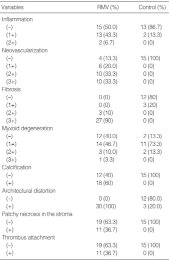
Histologic findings of the rheumatic mitral valves and con-trol valves were summarized in Table 1. All concon-trol valvular leaflets showed architectural preservation of three layers includ-ing spongiosa, fibrosa, and ventricularis (Fig. 1A). Mild fibro-sis was seen in three of them, mild to moderate myxoid degen-eration in thirteen, and scanty lymphocytic infiltrates in two. Several CD68-positive histiocytes were present in the stroma of valve leaflets. Neovascularization, calcification, thrombus attachment, and necrosis of the leaflets were not observed.
By contrast, in all rheumatic mitral valves, valvular archi-tectures were markedly distorted due to moderate-to-severe fibrosis (Fig. 2A). Neovascularization was seen in the central portion of the valvular leaflet in 26 cases. Six cases showed
thin-walled small vascular structures and others contained thick muscularized vessels. Inflammatory cells infiltrated into the perivascular stroma in 15 cases (Fig. 2B) and were
RMV, rheumatic mitral valve; (-), negative; (+), positive.
Variables RMV (%) Control (%) Inflammation (-) 15 (50.0) 13 (86.7) (1+) 13 (43.3) 2 (13.3) (2+) 2 (6.7) 0 (0) Neovascularization (-) 4 (13.3) 15 (100) (1+) 6 (20.0) 0 (0) (2+) 10 (33.3) 0 (0) (3+) 10 (33.3) 0 (0) Fibrosis (-) 0 (0) 12 (80) (1+) 0 (0) 3 (20) (2+) 3 (10) 0 (0) (3+) 27 (90) 0 (0) Myxoid degeneration (-) 12 (40.0) 2 (13.3) (1+) 14 (46.7) 11 (73.3) (2+) 3 (10.0) 2 (13.3) (3+) 1 (3.3) 0 (0) Calcification (-) 12 (40) 15 (100) (+) 18 (60) 0 (0) Architectural distortion (-) 0 (0) 12 (80.0) (+) 30 (100) 3 (20.0)
Patchy necrosis in the stroma
(-) 19 (63.3) 15 (100)
(+) 11 (36.7) 0 (0)
Thrombus attachment
(-) 19 (63.3) 15 (100)
(+) 11 (36.7) 0 (0)
Table 1. Histologic features of rheumatic mitral valves and con-trol valves
Fig. 1. Histologic and immunohistochemical findings of control valves. (A) Valvular leaflets obtained from the control group showed well-pre-served leaflet architecture without fibrosis or inflammatory cell infiltration (H&E, ×40). (B) Immunohistochemical staining for TGF- 1 demon-strated positivity in the subendothelial stroma of valvular leaflets (TGF- 1, ×40).
predominantly composed of CD3+ T lymphocytes. Throm-bi were attached on the valvular leaflets in 11 cases, calcifica-tion was noted in 18 cases, and focal necrosis was found in the fibrotic stroma in 11 cases. Myxoid degeneration of valvu-lar stroma was seen in 18 cases.
In those cases showing extensive stromal fibrosis in the vavlu-ar leaflets, more pronounced neovasculvavlu-arization (p<0.001), increased inflammatory infiltrates (p=0.033), calcification (p< 0.001), stromal necrosis (p=0.004), and thrombus attachment (p=0.009) were observed. Myxoid degeneration was seen in areas where fibrosis or calcification was not intense.
Immunohistochemical expression of TGF- 1 and SMA in control and rheumatic mitral valves
In CRHD, 21 cases of rheumatic valves (70%) showed high TGF- 1 expression, whereas only three cases of control valves (20%) demonstrated high TGF- 1 expression (p<0.001) (Table 2). Control valves exhibited TGF- 1 positivity on the subendothelial stroma of valvular leaflets (Fig. 1B). On the other hand, in CRHD, TGF- 1 was expressed in the endothe-lial cells and smooth muscle cells of the blood vessels (13 cases), Fig. 2. Histologic and immunohistochemical findings of the rheumatic mitral valves. (A) Rheumatic mitral valves showed severe fibrosis and distorted architecture (H&E, ×40). (B) A high-power view demonstrated small thin-walled vessels and perivascular lymphocytic infiltra-tion (H&E, ×200). (C) High TGF- 1 expression was seen in the endothelial cells and smooth muscle cells of the vessels, in the perivas-cular interstitial cells, and stroma of the valves (×200). (D) Myofibroblasts that were positive for SMA immunostaining were present in the subendothelial densely fibrotic area (×40).
A B
C D
RMV, rheumatic mitral valve.
TGF- 1 expression
Negative Low High
Total p
value
RMV 0 (0%) 9 (30.0%) 21 (63.3%) 30 (100%) <0.001
Control 4 (26.7%) 8 (53.3%) 3 (20.0%) 15 (100%) Table 2. Comparison of TGF- 1 expression between rheumatic mitral valves and control group valves
SMA, alpha-smooth muscle actin. Proliferation of SMA-positive
myofibroblasts
Negative Low High
Total p
value
RMV 2 (6.7%) 9 (30.0%) 19 (63.3%) 30 (100%) <0.001 Control 6 (40.0%) 8 (53.3%) 1 (6.7%) 15 (100%) Table 3. Proliferation of SMA-positive myofibroblasts in rheumatic mitral valves and control group valves
the interstitial cells and perivascular stroma (3 cases), or both (14 cases) (Fig. 2C). High TGF- 1 expression correlated with severe valvular fibrosis (p<0.001), inflammatory cell infiltra-tion (p=0.004), neovascularizainfiltra-tion (p=0.007) and calcifica-tion (p<0.001), but no correlacalcifica-tion was observed between TGF- 1 expression and myxoid degeneration (p=0.173).
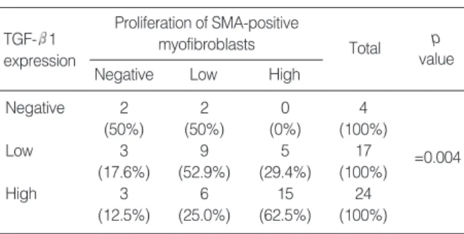
The number of SMA-positive myofibroblasts was signifi-cantly increased in valvular leaflets of CRHD compared to control valves (Table 3), and the proliferation of SMA-positive myofibroblasts positively correlated with TGF- 1
expres-sion (p=0.004) (Table 4) and fibrosis of valvular leaflets (p< 0.001). SMA-positive myofibroblasts were usually seen in the areas showing increased collagen deposition, and in the subendocardial stroma (Fig. 2D).
Deposition of collagen and proteoglycan in control and rheumatic mitral valves
Extracellular matrix deposition of proteoglycan and colla-gen was markedly different in the CRHD and control groups. In control valves, the proteoglycan was present in the spon-giosa and collagen was found in the fibrosa. Whereas, in CRHD, proteoglycan was deposited in the perivascular stro-ma or in areas showing myxoid degeneration, and the colla-gen was in the stroma showing dense fibrosis or myofibrob-lastic proliferation. The proportion of alcian blue-positive areas quantitatively calculated by image analyzer occupied a mean of 3.75% (range 0-23.5%) of valvular matrix in CRHD, compared to the mean of 13.2% (range 0-41.9%) in the con-trol group (p=0.014) (Fig. 3A). The proportion of Sirius red-positive areas, which represent type I and III collagen deposi-tion, averaged 23% (range 8.1-51.4%) in CRHD and 11.2% (range 5.9-22.0%) in the control group (p<0.001) (Fig. 3B). Additionally, the ratio of proteoglycan to collagen deposition
Fig. 3. Proteoglycan and collagen deposition in rheumatic mitral valves and control valves. (A) The proportion of proteoglycan deposited areas was lower in rheumatic mitral valve group (RM) than in the control group (black bars represented mean values: 3.75% vs. 13.2%) (p=0.014). (B) The rheumatic mitral valve group (RM) showed extensive collagen deposition compared to the con-trol group (black bars represented mean values: 23% vs. 11.2%) (p<0.001). (C) The ratio of proteoglycan to collagen deposited areas was lower in the TGF- 1 positive group than in the TGF- 1 negative group (black bars represented mean values: 0.38 vs. 0.66) (p=0.040). Proteoglycan deposition 50 40 30 20 10 0 -10 RM group (N=30) Control (N=14) A Collagen deposition 60 50 40 30 20 10 0 RM group (N=30) Control (N=15)
Ratio of proteoglycan to collagen
3.0 2.5 2.0 1.5 1.0 0.5 0.0 -0.5 Negative (N=20) Positive (N=23) C TGF- 1 expression B
SMA, alpha-smooth muscle actin.
Proliferation of SMA-positive myofibroblasts TGF- 1
expression
Negative Low High
Total p value Negative 2 2 0 4 =0.004 (50%) (50%) (0%) (100%) Low 3 9 5 17 (17.6%) (52.9%) (29.4%) (100%) High 3 6 15 24 (12.5%) (25.0%) (62.5%) (100%)
Table 4. Proliferation of SMA-positive myofibroblasts in valvular leaflets according to TGF- 1 expression
was lower in the rheumatic mitral valve group than in the con-trol valve group (p=0.001). The proportion of collagen and proteoglycan deposition was not associated with TGF- 1 ex-pression (p=0.34 and 0.087), but the ratio of proteoglycan to collagen deposition was lower in the TGF- 1 positive group than in the negative group (p=0.04) (Fig. 3C).
DISCUSSION
CRHD, the most significant complication of RF, demon-strates severe fibrosis and distortion of valvular leaflets (1, 3). The factors leading to continued fibrosis and subsequent valvu-lar disease remain incompletely defined. There are consider-able evidences to indicate that TGF- 1 overproduction plays some role in fibrosis and dysfunction in heart valves. TGF- 1 expression is increased in carcinoid valve cusps (15), calcific aortic valve cusps (16), and prolapsed valves in a mouse model of Marfan syndrome (19). In cases of CRHD, TGF- 1 poly-morphisms may predispose to valvular disease. Chou et al. de-monstrated that patients with CRHD have a lower frequency of the TGF- 1 C509T CC genotype and a higher frequen-cy of the T869C T allele (17). They proposed the possibility that TGF- 1 gene polymorphisms might play a role in deter-mining susceptibility to CRHD. However, there has been no study showing localized overproduction of TGF- 1 in valvu-lar leaflets of CRHD. Our study has demonstrated high TGF-1 expression in valvular leaflets of CRHD and might sup-port the hypothesis that TGF- 1 overproduction would play a role in the valvular fibrosis of CRHD.
It may be difficult to appreciate that, in valvular leaflets of CRHD, fibrosis and remodeling is going on even several de-cades after initial attack. There are some studies demonstrat-ing persistent inflammation in valvular leaflets of CRHD (4, 20, 21). Lymphocytic infiltrations have been identified in valves removed 10-20 yr after initial attacks of RF, and the predom-inant cellular infiltrate was T lymphocytes (4). Prolonged per-sistence of group A streptococcal carbohydrate antibody was demonstrated in CRHD even decades later (20). Additional-ly, the plasma concentrations of TNF- and highly sensitive CRP are significantly higher in CRHD (5, 6, 7, 21). Accord-ing to the study by Bhatnagar et al. (8), lymphocytes derived from CRHD patients exhibit the Th2 type cytokine response whereas those from RF patients show the Th1 cytokine pro-file with response to the streptococcal superantigen. The Th2 immune response is associated with fibrotic process including TGF- 1 secretion, whereas Th1 type cytokines suppresses diverse fibrotic activities (9-11). Guilherme et al. (22) demon-strated that CD4+ T cells derived from heart lesions predom-inantly secrete Th1 type cytokines in both RF and CRHD patients. The different results observed in these two studies are probably due to the different characterization of CRHD patients analysed. The study by Guilherme et al. included the early stage of CRHD patients with age range from 10 to 20
yr, whereas the age of the patients in the study by Bhatnagar et al. ranged from 22 to 54 yr (8, 22). Therefore, the cytokine profiles of early stage CRHD might be similar to that of RF. It is possible that Th2 type cytokine response seen in the ad-vanced CRHD (8) might be induce the TGF- 1 overexpres-sion and the fibrotic process as well as prolonged inflammation seen in the distorted rheumatic valves. Our report confirmed the fact that ongoing inflammation mainly composed of T lymphocytes was present in the apparently resting valves of advanced CRHD, and TGF- 1 expression correlated with the degree of inflammatory infiltrates. Additionally, TGF-1 expression was present predominantly in the perivascular stroma where inflammatory cells mainly infiltrated. These results support the hypothesis that a continued inflammato-ry process and overproduction of TGF- 1 within the valves of the susceptible hosts would lead to the prolonged fibrosis of valvular leaflets in advanced CRHD.
TGF- 1 induces the differentiation of cardiac fibroblasts to myofibroblasts (24), which can produce up to twice as much collagen as their fibroblast precursors (25). The emer-gence of these cells has been closely correlated with valvular fibrosis and degenerative lesions (26). Our study demonstrat-ed that increasdemonstrat-ed proliferation of myofibroblasts was observdemonstrat-ed in the rheumatic valves without alteration of cellularity of valvular interstitial cells and that high TGF- 1 expression was related to the proliferation of myofibroblasts and calcifica-tion of valvular leaflets. Calcific aortic stenosis cusps demon-strate higher concentrations of TGF- 1 within the extracel-lular matrix compared with noncalcified cusps (16). When cultured interstitial cells are exposed to TGF- 1 for a long period of time, aggregates of cells evolve into nodules that become apoptotic, express alkaline phosphatase, and then calcify (16). Therefore, increased TGF- 1 expression in rheu-matic heart valves may induce the differentiation of valvular interstitial cells to the myofibroblasts and the overproduction of extracellular matrix, resulting in progressive fibrosis and valvular distortion, and prolonged exposure to TGF- 1 prob-ably induces calcification of valvular leaflets seen in CRHD.
TGF- 1 is also known to play a critical role in matrix remod-eling by enhancing collagen synthesis (12). The normal heart valve is composed of collagen, proteoglycan and elastin, and approximately 85% of the total collagen in the heart is type I collagen (27). In rheumatic valves, the total amounts of col-lagen, proteoglycan, and elastin are significantly increased (27). Our result demonstrated that the proportion of collagen depo-sition was increased, and proteoglycan depodepo-sition was decreased in rheumatic valvular leaflets compared to control valves, but that it did not correlate with TGF- 1 overexpression. Because only the percentage of collagen and proteoglycan deposited areas in the valvular leaflets was evaluated and the total amount of collagen was not calculated, we were unable to investigate the accurate relationship of collagen deposition to TGF- 1 expression. However, the ratio of proteoglycan to collagen deposited areas was lower in the TGF- 1 positive group than
in the negative group. Therefore TGF- 1 expression might be related to the conversion of extracellular matrix from pro-teoglycan to collagen rather than to the percentage of colla-gen or that of proteoglycan.
There are a number of efforts that have aimed to reduce fibro-sis in cardiovascular disease, such as post-ischemic fibrofibro-sis, and dilated and hypertrophic cardiomyopathies, and targeting the TGF- 1 may be effective in attenuating fibrosis and hypertro-phy in the heart (26). Our study indicated that TGF- 1 may play some role in valvular fibrosis and deformity in CRHD, and therefore, TGF- 1 might be an appropriate target for prevention of subsequent valvular dysfunction in patients having suffered from RF.
In conclusion, TGF- 1 expression is increased in rheumatic mitral valves, and higher TGF- 1 expression is associated with valvular fibrosis, inflammatory cell infiltration, differ-entiation of valvular fibroblasts to myofibroblasts, calcifica-tion, and changes in the extracellular matrix in the valves of CRHD. These results support the hypothesis that TGF- 1 expression might play some role in the prolonged fibrosis and severe valvular deformity typical of CRHD.
ACKNOWLEDGMENTS
We thank Kyung Moo Yang, Pathologist in National Insti-tute of Scientific Investigation for his contribution of collec-tion of control valves, and members of the Department of Pathology, Inha University Hospital and Yonsei University College of Medicine for their supports. We express our sor-row over the death of Dr. Dong Hwan Shin, one of the co-authors of this article.
REFERENCES
1. Schoen FJ. The heart. In: Cotran RS, Kumar V, Collins T, editors.
Robbins pathologic basis of disease. 6th ed. Philadelphia: W.B. Saun-ders 1999: 543-99.
2. Rosai J. Cardiovascular system. In: Rosai J, editor. Ackerman’s
sur-gical pathology. 8th ed. St. Louis: Mosby 1996: 2173-226.
3. Veasy LG, Hill HR. Immunologic and clinical correlations in
rheu-matic fever and rheurheu-matic heart disease. Pediatr Infect Dis J 1997; 16: 400-7.
4. Kemeny E, Grieve T, Marcus R, Sareli P, Zabriskie JB.
Identifica-tion of mononuclear cells and T cell subsets in rheumatic valvulitis. Clin Immunol Immunopathol 1989; 52: 225-37.
5. Chen MC, Chang HW, Wu CJ, Yang CH, Yu TH, Chen CJ, Hung WC. Balance between plasma levels of tumor necrosis factor-alpha
and interleukin-10 in rheumatic mitral stenosis. Cardiology 2005; 104: 171-5.
6. Chiu-Braga YY, Hayashi SY, Schafranski M, Messias-Reason IJ.
Further evidence of inflammation in chronic rheumatic valve dis-ease (CRVD): high levels of advanced oxidation protein products
(AOPP) and high sensitive C-reactive protein (hs-CRP). Int J Car-diol 2006; 109: 275-6.
7. Davutoglu V, Celik A, Aksoy M. Contribution of selected serum
inflammatory mediators to the progression of chronic rheumatic valve disease, subsequent valve calcification and NYHA functional class. J Heart Valve Dis 2005; 14: 251-6.
8. Bhatnagar A, grover A, ganguly NK. Superantigen-induced T cell
responses in acute rheumatic fever and chronic rheumatic heart disease patients Clin Exp Immunol 1999; 116: 100-6.
9. Lakos G, Melichian D, Wu M, Varga J. Increased
bleomycin-induc-ed skin fibrosis in mice lacking the Th1-specific transcription factor T-bet. Pathobiology 2006; 73: 224-37.
10. Kimura T, Ishii Y, Yoh K, Morishima Y, Iizuka T, Kiwamoto T, Matsuno Y, Homma S, Nomura A, Sakamoto T, Takahashi S, Sek-izawa K. Overexpression of the transcription factor GATA-3
enhan-ces the development of pulmonary fibrosis. Am J Pathol 2006; 169: 96-104.
11. Wen FQ, Liu X, Kobayashi T, Abe S, Fang Q, Kohyama T, Ertl R, Terasaki Y, Manouilova L, Rennard SI. Interferon-gamma inhibits
transforming growth factor-beta production in human airway epithe-lial cells by targeting Smads. Am J Respir Cell Mol Biol 2004; 30: 816-22.
12. Border WA, Noble NA. Transforming growth factor beta in tissue
fibrosis. N Eng J Med 1994; 331: 1286-92.
13. O’Kane S, Ferguson MW. Transforming growth factor betas and
wound healing. Int J Biochem Cell Biol 1997; 29: 63-78.
14. Mutsaers SE, Bishop JE, McGrouther G, Laurent GJ. Mechanisms
of tissue repair: from wound healing to fibrosis. Int J Biochem Cell Biol 1997; 29: 5-17.
15. Jian B, Connolly J. Savani RC, Narula N, Liang B, Levy R. Serotonin
mechanisms in heart valve disease I: Serotonin-induced up-regulation of transforming growth factor-beta1 via G-protein signal transduc-tion in aortic valve interstitial cells. Am J Pathol 2002; 161: 2111-21.
16. Jian B, Narula N, Li QY, Mohler ER 3rd, Levy RJ. Progression of
aortic valve stenosis: TGF- 1 is present in calcified aortic valve cusps and promotes aortic valve interstitial cell calcification via apop-tosis. Ann Thorac Surg 2003; 75: 457-65.
17. Chou HT, Chen CH, Tsai CH, Tsai FJ. Association between
transform-ing growth factor-beta1 gene C-509T and T869C polymorphisms and rheumatic heart disease. Am Heart J 2004; 148: 181-6.
18. Robiolio PA, Rigolin VH, Wilson JS, Harrison JK, Sanders LL, Ba-shore TM, Feldman JM. Carcinoid heart disease: Correlation of high
serotonin levels with valvular abnormalities detected by cardiac cathe-terization and echocardiography. Circulation 1995; 92: 790-5.
19. Ng CM, Cheng A, Myers LA, Martinez-Murillo F, Jie C, Bedja D, Gabrielson KL, Hausladen JM, Mecham RP, Judge DP, Dietz HC.
TGF-beta-dependent pathogenesis of mitral valve prolapse in a mouse model of Marfan syndrome. J Clin Invest 2004; 114: 1586-92.
20. Ayoub EM, Toranta A, Bartley TD. Effect of valvular surgery on
antibody to the group A streptococcal carbohydrate. Circulation 1974; 50: 144-50.
21. Golbasi Z, Ucar O, Keles T, Sahin A, Cagli K, Camsari A, Diker E, Aydogdu S. Increased levels of high sensitive C-reactive protein in
inflammation. Eur J Heart Fail 2002; 4: 593-5.
22. Guilherme L, Cury P, Demarchi LM, Coelho V, Abel L, Lopez AP, Oshiro SE, Aliotti S, Cunha-Neto E, Pomerantzeff PM, Tanaka AC, Kalil J. Rheumatic heart disease: proinflammatory cytokines play a
role in the progression and maintenance of valvular lesions. Am J Pathol 2004; 165: 1583-91.
23. Gorelik L, Flavell RA. Transforming growth factor-beta in T-cell
biology. Nat Rev Immunol 2002; 2: 46-53.
24. Walker GA, Masters KS, Shah DN, Anseth KS, Leinwand LA.
Valvu-lar myofibroblast activation by transforming growth factor-beta: implications for pathological extracellular matrix remodeling in
heart valve disease. Circ Res 2004; 95: 253-60.
25. Lijnen P, Petrov V. Transforming growth factor-beta 1-induced
col-lagen production in cultures of cardiac fibroblasts is the result of the appearance of myofibroblasts. Methods Find Exp Clin Pharma-col 2002; 24: 333-44.
26. Khan R, Sheppard R. Fibrosis in heart disease: understanding the
role of transforming growth factor- 1 in cardiomyopathy, valvular disease and arrhythmia. Immunology 2006; 118: 10-24.
27. Lis Y, Burleigh MC, Parker DJ, Child AH, Hogg J, Davies MJ.
Bio-chemical characterization of individual normal, floppy and rheumatic human mitral valves. Biochem J 1987; 244: 597-603.