저작자표시-비영리-변경금지 2.0 대한민국 이용자는 아래의 조건을 따르는 경우에 한하여 자유롭게 l 이 저작물을 복제, 배포, 전송, 전시, 공연 및 방송할 수 있습니다. 다음과 같은 조건을 따라야 합니다: l 귀하는, 이 저작물의 재이용이나 배포의 경우, 이 저작물에 적용된 이용허락조건 을 명확하게 나타내어야 합니다. l 저작권자로부터 별도의 허가를 받으면 이러한 조건들은 적용되지 않습니다. 저작권법에 따른 이용자의 권리는 위의 내용에 의하여 영향을 받지 않습니다. 이것은 이용허락규약(Legal Code)을 이해하기 쉽게 요약한 것입니다. Disclaimer 저작자표시. 귀하는 원저작자를 표시하여야 합니다. 비영리. 귀하는 이 저작물을 영리 목적으로 이용할 수 없습니다. 변경금지. 귀하는 이 저작물을 개작, 변형 또는 가공할 수 없습니다.
A Dissertation
for the Degree of Masters of Sciences
Enhancement of immunogenicity through conjugation of
complement fragment C4d to porcine epidemic diarrhea virus
surface protein antigen, S0
보체 절편 C4d 의 돼지유행성설사 바이러스 표면 항원 S0 에 결합을
통한 면역증진
February 2019
By
Young-Saeng Kim Cho
Department of Agricultural Biotechnology
Graduate School
I
Summary
The current landscape for vaccines is colored with concerns about the safety of vaccines in the wake of live attenuated or inactivated vaccines. From these concerns, subunit vaccines have risen into the next hot topic for vaccine production. The main advantage of a subunit vaccine is its inherent characteristic of consisting of the antigen-neutralizing subunit of the whole virus, being an inherently safer alternative to other types of vaccines. However, the biggest disadvantage a subunit vaccine must overcome is its low immunogenicity compared to live attenuated vaccines. In order to increase the immunogenicity of the subunit vaccine consisting of the S0 spike protein from PEDV’s (Porcine Epidemic Diarrhea Virus) coronavirus, complement fragment C4d was used as a fusion adjuvant.
Cloning by using isocaudomers was successful and was confirmed by fragment size distribution through agarose gel electrophoresis and Sanger sequencing. It is important to express recombinant proteins in a soluble way, so three factors were optimized to increase solubility. The first factor was a chaperone, the second was optical density, and the third was IPTG (isopropyl β-D-1-thiogalactopyranoside) concentration. After optimization experiments, it was determined that trigger factor chaperone must be co-expressed with target proteins. In addition, the optimal optical density for inducing proteins is A600 = 0.6, which is during the
mid-log phase of growth, using 0.1 mM IPTG. The new, optimized protocol was used for expressing the recombinant proteins S0, S0-mC4d1, S0-mC4d2, and
II
mC4d in a large scale 500 mL flask. The proteins were purified using Ni-NTA (Nickel-nitrilotriacetic acid) resin and dialyzed in PBS (phosphate buffered saline) before being concentrated down.
After finding the concentration of the proteins, 6 groups of mice were immunized using the same molar concentration of proteins. The groups were, PBS control, S0 control, the two fusion protein groups, and two mixture groups. After immunization, serum and spleens were taken from the mice and used for ELISA (enzyme-linked immunosorbent assay) and ELISpot (enzyme-linked immunospot), respectively. ELISA shows that end point titers of IgG increased significant in S0-mC4d2 and two mixture groups after 4 weeks and in all mC4d treatment groups after 6 weeks. ELISpot shows the presence of mC4d-specific Th2 cells in the two fusion protein groups but not in the S0 or the mixture groups.
In conclusion, recombinant proteins were produced well with relatively high yield and purity and immunization results show that S0-mC4d1 and S0-mC4d2 increases humoral immunity through C4d-specific auto-reactive Th2 cells.
Keywords: Porcine Epidemic Diarrhea. Porcine Epidemic Diarrhea Virus. Adjuvant. Subunit vaccine. C4d. Fusion Adjuvant. Spike protein. Recombinant protein. Optimization. Solubility
III
Contents
Summary ... I Contents ... III List of Tables and Figures ... VI Tables ... VI Figures ...VII List of Abbreviations ... IX
I. Introduction ...1
II. Review of Literature ...3
1. Porcine Epidemic Diarrhea Virus ...3
1) Overview of Porcine Epidemic Diarrhea ...3
2) Porcine Epidemic Diarrhea Virus ...4
3) Infection of the host cell ...7
4) Porcine Epidemic Diarrhea Virus vaccines...9
2. Complement fragment C4d ...10
1) Overview of complement fragment C4d ...10
2) C3d’s mechanism as an auto-reactive helper T-cell epitope donor ...12
3) In silico analysis of broad binding serum proteins ...15
IV
4. Adjuvant system ...22
1) Vaccine adjuvants ...22
2) Fusion proteins as adjuvants ...23
5. Solubility of recombinant proteins ...24
1) Inclusion bodies ...24
2) Trigger factor chaperone co-expression ...27
3) Other physiological factors ...29
III. Materials and Methods ...31
1. Preparation of protein ...31
1) Plasmids and strains ...31
2) Recombinant protein expression ...34
3) Ni-NTA affinity chromatography ...35
4) SDS-PAGE ...37
5) Quantification of recombinant proteins ...38
2. Optimization of protein expression conditions ...38
1) Sample preparation ...38
2) Western Blot analysis ...39
3) Densitometer analysis ...40
3. In vivo immunization ...40
V
2) Blood extraction ...44
3) S0-specific indirect ELISA ...44
4) Spleen isolation and detection of auto-reactive helper T-cells ...45
5) ELISpot analysis ...45
4. Statistical analysis ...46
IV . Results and Discussion ...47
1. Production of soluble recombinant fusion protein in E. coli ...47
1) Vector construction ...47
2) Production of soluble recombinant proteins ...50
3) Expression, purification, and quantification of soluble recombinant proteins ...67
2. In vivo immunization for development of PEDV recombinant subunit vaccine...73
1) S0-specific humoral immune response ...73
2) S0 and C4d-specific cellular immune response ...75
V. Conclusion ...83
Works Cited ...85
Summary in Korean ...95
VI
List of Tables and Figures
Tables
Table 1. Broad binding serum protein epitope content. ...20
Table 2. Roles of adjuvants ...23
Table 3. Buffer compositions for Ni-NTA affinity chromatography ...37
Table 4. Immunization scheme ...43
Table 5. Trigger factor chaperone’s effect on recombinant protein solubility as a percent. ...54
Table 6. Effects of changes to expression condition variables for S0-mC4d1. ...62
Table 7. Effects of changes to expression condition variables for S0-mC4d2. ...66 Table 8. Purity levels and pure protein yields for recombinant, target proteins. .72
VII
Figures
Figure 1. Genome organization of and the structure of PEDV. ...5
Figure 2. PEDV infection via interaction of spike protein with pAPN. ...8
Figure 3. Complement activation and formation of C4d. ... 11
Figure 4. Hypothesized mechanism of action for C4d fused with antigen of interest ...14
Figure 5. Biological role of C3d. ...15
Figure 6. Premise of IEDB analysis tool. ...18
Figure 7. Broad binding serum protein MHC-II epitopes. ...19
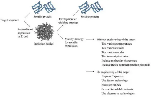
Figure 8. Downstream strategies to obtain soluble recombinant protein ...26
Figure 9. The role of trigger factor (tig) chaperone in folding nascent protein polypeptides ...28
Figure 10. Vector construction scheme containing characteristics of the expression vector, placement of genetic regions of interest, and expected sizes of the target protein sizes. ...33
Figure 11. Schematics of the pET28a (+) – S0- mC4d recombinant protein vector map and overall construction design ...34
Figure 12. In vivo immunization schedule. ...42
Figure 13. Confirmation of cloning of mouse C4d into pET28a (+)–S0 in a series through restriction enzyme digestion (NheI and XhoI) run in 1% agarose gel.. ...49
VIII
Figure 14. Trigger factor (tig) chaperone’s effect on solubility of recombinant
proteins ...53
Figure 15. Western blot confirmation of soluble recombinant protein expression with or without chaperone co-expression in E. coli ...55
Figure 16. Optimized expression of recombinant S0-mC4d1 ...59
Figure 17. Densitometer analysis of S0-mC4d1 solubility ratios. ...61
Figure 18. Optimized expression of recombinant S0-mC4d2 ...63
Figure 19. Densitometer analysis S0-mC4d2 solubility ratios ...65
Figure 20. Relative expression and purification of recombinant S0-mC4d1 by Ni-NTA column ...68
Figure 21. Relative expression and purification of recombinant S0-mC4d2 by Ni-NTA column. ...69
Figure 22. Relative expression and purification of recombinant mC4d by Ni-NTA column...71
Figure 23. Quantification of pure recombinant proteins ...72
Figure 24 Indirect ELISA for the in vitro detection of serum antibodies ...75
Figure 25. IL-4 secretion in splenocytes ...79
Figure 26 T cell release of IL-4 in response to protein stimulation ...80
Figure 27. Correlation between mC4d specific IL-4 secreting T cells and S0 specific IL-4 secreting T cells. ...81
IX
List of Abbreviations
A600 = Optical density at a wavelength of 600 nmACK lysis buffer: Ammonium-Chloride-Potassium lysis buffer APC: Antigen presenting cell
BLAST: Basic Local Alignment Search Tool BSA: Bovine serum albumin
CFA: Complete Freund’s Adjuvant DNA: Deoxyribonucleic acid ECL: Enhance chemiluminescence
ELISA: Enzyme-linked immunosorbent assay ELISpot: Enzyme-linked immunospot assay FBS: Fetal bovine serum
Flagellin-EGFP: Flagellin enhanced fluorescent protein HRP: Horseradish peroxidase
IB: Inclusion bodies
IEDB: Immune Epitope Database IFA: Incomplete Freund’s Adjuvant IL-18: Interleukin-18
IMD: Imidazole
IPTG: Isopropyl β-D-1-thiogalactopyranoside LB: Lysogeny broth
MBL: Mannose binding lectin mC4d: Mouse C4d
X
Ni-NTA: Nickel-nitrilotriacetic acid pAPN: Porcine aminopeptidase N PBS: Phosphate Buffer Saline PED: Porcine Epidemic Diarrhea PEDV: Porcine Epidemic Diarrhea Virus P/S: Penicillin/Streptomycin
RNA: Ribonucleic acid ssRNA: single strand RNA RT: Room temperature
RPMI 1640: Roswell Park Memorial Institute medium
SDS-PAGE: Sodium dodecyl sulfate – polyacrylamide gel electrophoresis SPF: Specific-pathogen-free
TBST: Tris Buffered Saline with Tween 20
1
I. Introduction
Porcine epidemic diarrhea (PED) is an economically significant disease within the pig farming industry due to its devastating effects on the pork industry. PEDV is the etiological agent of PED. The disease mainly affects mostly piglets and has and can completely destroy a farmer’s yield, with mortality rates of up to 100% (Sun, Cai et al. 2012). Current vaccination strategies in Korea consist of live attenuated vaccines that have been developed to vaccinate pregnant sows and protect piglets through passive immunity (Song and Park 2012). The use of vaccines based on live organisms can produce a powerful immune response and thus, a large portion of licensed vaccines fall into the live attenuated vaccine or inactivated vaccine category (Perrie, Kirby et al. 2007). In fact, most commercial PED vaccines come from attenuated live or killed viruses. These vaccines can be effective; however, safety concerns have been raised regarding their application. Antigenic variation and safety concerns make it difficult for generic commercial live attenuated or inactivated PEDV to protect animals from disease. Recombinant subunit vaccines are an alternative vaccine candidate to ameliorate this safety issue as subunit vaccines are safer, easier to produce, and more cost-effective than live attenuated vaccines.
Due to the low immunogenicity of subunit vaccines, fusion adjuvant strategy (Perrie, Mohammed et al. 2008) was deemed to be the most prudent plan of action. By fusing the adjuvant directly onto the antigen, the chance of being engulfed
2
together increases. C4d was the fusion adjuvant of choice. C4d’s mechanism may follow that of C3d’s wherein, T-cell epitopes of C4d may be presented to auto-reactive T helper cells with the ability to activate B cells that have recognized the spike protein. The end result is the differentiation of B cells into plasma cells.
In this study an E. coli expression system was used to produce recombinant proteins that consist of S0 fused with tandem repeats of mC4d linked together with glycine-serine linkers. In the first part of this study recombinant protein plasmids were constructed by using restriction enzymes to cut the vector, pET28a (+), and the insert, mC4d, and ligate them together. Then, classical cloning techniques were utilized to produce the final products as recombinant plasmids. In the next part, the recombinant plasmids were extracted from their cloning hosts and transformed expression hosts with the recombinant plasmids. The bioreactor most commonly used to produce recombinant protein-based subunit vaccines are
E. coli due to their relatively high production yield. However, expression of
recombinant proteins in E. coli often results in the formation of insoluble aggregates known as inclusion bodies (IB). Chaperone co-expression strategies have been proposed to facilitate soluble expression of recombinant proteins. In this section of the study, the focus was to optimize the production of the proteins by utilizing a protein chaperone and adjusting physiological aspects of the protein production procedure. Finally, the effects on humoral and cellular immunity of the recombinant proteins were evaluated in immunized mice through indirect ELISA and ELISPOT assay, respectively.
3
II. Review of Literature
1. Porcine Epidemic Diarrhea Virus
1) Overview of Porcine Epidemic Diarrhea
Porcine epidemic diarrhea is an economically devastating viral enteritis in pigs. The causative agent is PEDV (Song, Moon et al. 2015). Characterized mainly by watery diarrhea and vomiting, the severe water loss eventually leads to dehydration, depression, and death (Lee 2015). PEDV infects pigs of all stages of development and the most important age group is in neonatal piglets as the dehydration and diarrhea commonly leads to death rates of 80% to 100% (Sun, Cai et al. 2012). PEDV’s rapid spreading across various countries of the world not only brings illness and loss of pig lives, but also poses a threat to the economic ventures of the swing farming industry and public health as a whole (Huang, Dickerman et al. 2013).
First observed in 1971 as an unrecognized enteric disease in pigs, the etiological agent that was identified as the main cause was the PEDV prototype strain CV777 (Huang, Dickerman et al. 2013). The disease spread across Europe affecting pigs of all ages until the 1990s when PED’s effects subsided in Europe but had since spread across Asia (Lee 2015), jumping continents. Since its emergence in South Korea in 1992, PEDV has been a constant occurrence each year until 2010 (Lee and Lee 2014). From the 1980s to present day, PED has caused higher rates of
4
mortality than when previously encountered in European countries becoming an endemic in Asian pig farming countries (KUSANAGI, KUWAHARA et al. 1992, Song and Park 2012). In 2013, PEDV suddenly experienced a resurgence when the first case of PEDV was recorded in the United States and quickly spread across North America. Recent strains of PEDV in South Korea appear to be the most genetically like the strains of PEDV from the 2013 outbreak in the United States marking 2013 as a landmark year for PEDV research (Lee and Lee 2014, Lin, Chung et al. 2014).
2) Porcine Epidemic Diarrhea Virus
The etiological agent of PED is the enveloped RNA virus porcine epidemic diarrhea virus (Lin, Chung et al. 2014). The taxonomical classification of PEDV is of the genus Alphacoronavirus and within the Coronaviridae family. The genome protein size is 28 kb long and contains at least 7 open reading frames, 4 of which encode for the structural proteins including the glycosylated spike protein (S), nucleocapsid protein (N), membrane protein (M), and the envelope protein (E) (Lee 2015).
5
Figure 1. Genome organization and the structure of PEDV. (A) Structure of PEDV genomic RNA (B) Model of PEDV structure (Lee 2015)
6
As noted above, the structural proteins of PEDV consist of the S, E, M, and N proteins. The spike (S) protein interacts with the pAPN (porcine aminopeptidase N) receptor during virus entry and is often used to stimulate neutralizing antibodies in the host. For example, the N-terminal domain of the S1 domain of the spike protein has been of interest. The reason for this is because some have reported that the N-terminal domain has some inactions with a sugar co-receptor, Neu5Ac, which can help PEDV bind to the enterocytes of pigs (Piao, Lee et al. 2016). Due to the antigenicity of the spike protein, it is considered as a main player in developing a defense against PED (Oh, Song et al. 2003, Li, Li et al. 2017).
Typically, PEDV strains are categorized into either S-INDEL or non-INDEL strains. By studying the phylogenic differences in the spike (S) gene of PEDV, researchers found that there are high amounts of differences between strains (Masuda, Murakami et al. 2015). Strains with either insertions or deletions, which indicate overall variance, are considered S-INDEL (Wang, Guo et al. 2016) while strains without insertions or deletions in the S gene are called non-INDEL. S-INDEL and non-INDEL strains are also different from one another as they also can have different amounts of danger to the pigs. As antibodies
reactive against S0 from one strain will have an effect against S0 from other non-INDEL strains (Li, Li et al. 2017), S0 was selected for as the subunit candidate in this study. In addition, non-INDEL strains, which S0 has cross-reactive activity over, are considered to be more dangerous than their S-INDEL
7
counterparts, consist of the strains in current circulation (Jarvis, Lam et al. 2016).
The spike protein contains a wide variety of domains with each domain having their own function. It was also discovered that each domain is unique enough that antibodies that bind to one domain may not bind to a different domain within the same spike protein. Despite this, monoclonal antibodies that bind to a specific domain may bind to the same domain of a different strain as long as they are both S-INDEL or both non-INDEL (Li, Li et al. 2017).
3) Infection of the host cell
Like many animal viruses, PEDV has the ability to enter animal cells and “hijack” the cell’s machinery in order to replicate and proliferate, greatly increasing its numbers after infection of the host. For this reason, PEDV is an ssRNA (single strand RNA) virus. Due to the restricted tissue tropism of PEDV, the virus seems to replicate with the most efficiency in porcine intestinal villous enterocytes. The targeted cellular receptor for PEDV’s spike protein is the pAPN receptor which exists on the surface of porcine enterocytes (Nam and Lee 2010). As seen in figure 2, PEDV infects the host cell after its spike protein binds to pAPN. Once the virus has been internalized, the viral genome is released into the cytosol for genome replication and hijacking is complete (Lee 2015).
8
Figure 2. PEDV replication cycle and infection route via interaction of spike protein with pAPN (Lee 2015).
9
4) Porcine Epidemic Diarrhea Virus vaccines
In countries where PED is considered an endemic, vaccination is the best plan of action in order to contain and prevent any additional damage caused by the disease. The majority of commercially available PED vaccines fall into the category of inactivated or attenuated vaccines. (Song et al 2007). A Vero cell-attenuated PEDV PV-5 vaccine was developed first in Japan and has also become available in South Korea (Lee, 2015). Additionally, there are other cell-attenuated strains that originated from the field known as PEDV SM-98-1 and DR-13 which are used in Korean mainly (Lee, 2015). While these vaccines have staying power due to their current market use along with positive results in testing conditions, their overall effectiveness and concerns with the well-being and safety of the animals provides an outlet for the exploration of different options, especially in different types of vaccines. In addition, the efficacy and safety of commercial PEDV vaccines used in Korea has been called into question. It seems that immunity from strains that were previously considered to be endemic fail to produce cross-protection to recent PEDV strains. This may be due to recent Korean PEDV field isolates differing genetically from vaccine strains used in Korea (Ayudhya, Assavacheep et al. 2012).
Currently, there is the development of other types of vaccines as new candidates of PED. One type of vaccine is RNA/DNA vaccines. There was a report that using the DNA of the spike protein combined with IL-18 elicited humoral and cytotoxic T cell immune responses (Suo, Ren et al. 2012). A US company, Harris Vaccine
10
Inc., produced an RNA vaccine (Kim, Lee et al. 2016) and in 2014, Harris Vaccine Inc., was able to attain a US Department of Agriculture license and could distribute its vaccine into use. Another type comes from a South Korean research group where they inserted the core neutralizing epitope of PEDV into tobacco plants using a tobacco-mosaic virus-based vector. This resulted in a plant-based vaccine (Kang, Kim et al. 2005). Another venture is into using recombinant protein vaccines. An attempt at a subunit vaccine using porcine kidney cells to produce the PED virus spike protein has been relatively successful at producing yields (Oh, Lee et al. 2014).
2. Complement fragment C4d
1) Overview of complement fragment C4d
Complement fragment C4d is a split product of the C4 complement protein from the innate immune system’s complement system. The complement system’s main role is to complement the ability of antibodies and phagocytic cells to clear microbes and damaged cells, promote inflammation, and directly attack pathogens by puncturing the pathogen’s cell membrane (Janeway, Travers et al. 2005). As seen in figure 3, C4, when activated by the classical pathway, is triggered by either IgM or IgG sticking to the surface of the pathogen. After complement protein complex C1qrs binds to target-bound antibodies to cleave C4 and C2. C4b sticks to C2a to form C3 convertase. C3 convertase’s main role is to continue the complement cascade cleaving C3. C3b complexes with C4bC3a to form C5
11
convertase which can begin the membrane attack complex which will lyse the pathogenic cell (Murata and Baldwin III 2009). Formation of C3 convertase on the surface of cells amplifies the activation of complement however, chronic amplification of C3 convertase may lead to injury to the host or lysis of bystander cells.
Figure 3. Formation of C4d through complement activation.
The lectin pathway is the second pathway in which C4d is also present. The lectin pathway’s analog to C1q, MBL (mannose binding lectin), bind to mannose or N-acetyl glucosamine both of which are commonly found on surfaces of bacteria. After MBL binds, it complexes with C1r and C1s and the process continues like that of the classical pathway (Janeway, Travers et al. 2005).
12
Regardless of the pathway, C3 convertase present on the surface of cells will amplify the activation of complement. However, chronic amplification of C3 convertase may lead to injury of the host or lysis of innocent bystander cells. To put the brakes on the chronic activation of the complement cascade, C3 convertase is mediated by plasma factor I which eventually generates C4d (Platt 2002).
While bound C4d is considered an orphan ligand, it’s mainstream use is as a marker of classical complement activation which can reveal antibody-based attacks against a host’s cells (Feucht 2003), even after the complement system has finished the cascade. For this reason, many have used the detection of C4d to determine if the body has rejected a grafted kidney or other organ (Herzenberg, Gill et al. 2002). The exact reason for why C4d is prevalent on a rejected graft, or what its overall biological role is remains unknown (Nickeleit, Zeiler et al. 2002).
2) C3d’s mechanism as an auto-reactive helper T-cell epitope donor
The proposed mechanism of C4d was extrapolated from the mechanism of the adjuvant effect from complement fragment C3d. C3d is often used as a fusion adjuvant and originally C3d was thought to be the ligand for a co-receptor on B cells that would help activate them (Carroll 2008). However, new evidence has been found that suggests that the mechanism of adjuvanticity is different. C3d has been discovered to be rich in T-cell epitopes thus creating a different, plausible narrative for how C3d acts as an adjuvant (De Groot, Ross et al. 2015). Typically, C3d would be prevalent in the wake of a complement cascade. When C3d has
13
adhered onto the surface of an antigen, B cells that recognize the antigen engulf both the antigen and the C3d fragment. This results in the display of both the lysed fragment of the antigen and T-cell epitopes of C3d. Pre-existing autoreactive T cells, which recognize fragments originating from complement, may recognize C3d displayed in MHC II and activate the naïve B cell immediately, resulting in an antibody producing plasma cell.
Two curiosities that arise from this situation are the characteristic of a complement fragment having an remarkable high T cell epitope content (De Groot, Ross et al. 2015) and the second oddity is the abnormal existence of C3d-specitifc auto-reactive T cells. Auto-reactive T cells, on principle, should not exist due to positive selection of naïve T cells during T cell education in the thymus (Starr, Jameson et al. 2003). These two pieces of information together suggest the existence of possible biological role of C3d along the crux of a C3d-specific autoreactive T cell axis.
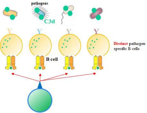
Should the existence of C3d-specific autoreactive T cells be confirmed, their application opens up a new possibility of cellular immunity shown in figure 4. Hypothetically, bacteria that are linked with C3d, from the complement pathway, is phagocytized by B cells, C3d, with its inherently high content of T cell epitopes, would be fragmented and presented on the MHC-II of B cells. The MHC-II and peptide complexes can be recognized by C3d specific T cells and provide activation signals to B cells. As a characteristic of C3d is the ability to bind to a wide range of pathogens, if pathogen-specific B cells all present T cells
14
originating from C3d, C3d-specific T cells can still react to pathogen-specific B cells. This eliminates the need to wait for a pathogen-specific T cell to come along and find it’s “perfect-match” B cell. Along with this, if C3d-specific autoreactive T cells are already primed and prepared to act, they can provide aid to naïve B cells quickly. Maturation of T cells is the rate-limiting step for antibody production (Gershon 1974) and quickening this step may speed up the entire antibody production process.
15
Figure 5. Proposed biological role of C3d.
3) In silico analysis of broad binding serum proteins
The existence of other serum proteins with similar characteristics of C3d is currently unknown. These characteristics would include the ability to attach itself to a broad range of pathogens and, perhaps more importantly, have high T cell epitope content. If both of these characteristics are satisfied, theoretically, this protein could attach itself to a pathogen and be engulfed by the pathogen-specific B cell. Presentation of the T cell epitopes within the serum protein would encourage the T cell to activate the B cell to produce antibodies. In order to find the existence of such serum proteins, in-silico analysis was performed.
16
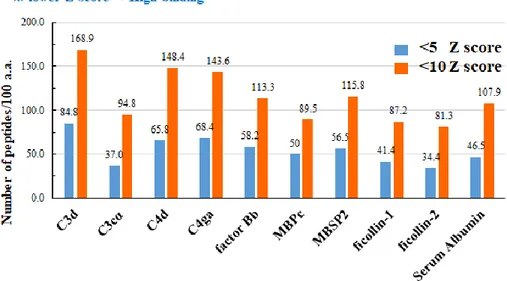
Blood serum candidates were analyzed for T-cell epitopes using the IEDB (Immune Epitope Database) tool for MHC II (major histocompatibility complex II) binding prediction. Protein sequences for various blood serum proteins were collected using BLAST (Basic Local Alignment Search Tool), which contains a database of many DNA sequences, from the National Center for Biotechnology Information (NCBI, USA) and entered into the IEDB tool. IEDB may only make binding predictions for humans and mice and does not include the target animal, pigs, therefore human MHC II was selected along with the appropriate MHC alleles to cover about 90% of the human population (DRB1*01:01, 03:01, 07:01, 09:01, 11:01, 15:01). Despite the target animal being swine, humans were selected due to further applications possibly venturing into vaccines for humans. The tool selected every possible amino acid fragment through chopping the entire protein sequence and assigned a Z-score with lower Z-scores representing good binding ability to each of the chopped protein sequences.
The data from the tool was compiled and only fragments with Z-scores below 10 or below 5 were considered T-cell epitopes with high binding affinity for MHC II.
The rationale for selecting broadly binding serum proteins is due to the working hypothesis that autoreactive T cells have an evolutionary origin. Their ability to broadly bind to various pathogens and the existence of multiple T cell epitopes within their protein structure may be for the purpose of seeking out pathogens and creating an immune response against them.
17
In addition, there is an inherent advantage of having copious amounts of epitopes as increasing the probability of a B cell presenting any given epitope to a T cell in turn increases the likelihood of B cell activation. Finally, any given individual’s MHC will be slightly different from another individual’s due to genetic shuffling and the variability of DNA. As a result, having a diverse and broad repertoire of epitopes is an advantage because the different epitopes can activate MHC-II from different individuals. Thus, the formulated hypothesis states that broad binding serum proteins that also double as epitope donors may act in a manner similar to C3d and thus have an adjuvant-like effect for the activation of B cells through autoreactive T-helper cells.
Serum proteins were selected from common knowledge of which serum proteins bind broadly to a wide range of pathogens derived from immunology textbooks. To validate their use, an in-silico experiment was conducted. Protein sequences of various serum proteins were used in the IEDB database to
determine their respective T-cell epitope content. The IEDB analysis tool chops up the protein sequences and measure their ability to bind with MHC-II.
Fragments with high binding ability to MHC-II were given a low Z-score (below 1, 5, or 10). Using the scores the database provided, the number of low Z-scores, shown in figure 7, either below 5 or below 10, for each serum protein, were graphed to produce a visualization of the amount of low Z-score fragments amongst the broad pathogen binding serum proteins.
18 Figure 6. Premise of IEDB analysis tool.
19
Figure 7. The number of T cell epitopes per 100 amino acids from within broad binding serum proteins.
The positive control was C3d, as it has been proven to contain a high T cell epitope content (De Groot, Ross et al. 2015) and the negative control was serum albumin. After compiling the z-score data from all the serum protein candidates, the number of fragments containing z-scores below 10 or below 5 per 100 amino acids were graphed. According to figure 7 and table 1, serum proteins containing the greatest number of low scoring peptides (z-scores below 5 or below 10) within 100 amino acids were C3d, and C4d. The negative control, serum albumin, contained 108 fragments that scored below 10 and 47 fragments per 100 amino acids that scored below 5. C4d had 149 peptides with z-scores below 10 per 100 amino acids and 66 fragments below 5 per 100 amino acids,
20
negative control. In addition, C3d had 169 peptides with z-scores below 10 per 100 amino acids and 85 peptides with z-scores below 5 per 100 amino acids, meaning C4d’s high binding peptide count is comparable with the positive control, C3d.
Table 1. Tabulated number of T cell epitopes per 100 amino acids for broad binding
serum protein candidates
Interpretation of these data leads to the conclusion that C3d and C4d contain the greatest number of MHC-II high binding peptides, or in other words, the greatest number of T-cell epitopes.
3. Recombinant subunit vaccine
Recombinant subunit vaccines are a subtype of inactivated whole vaccines in which, only the essential epitopes of the antigen are used to inoculate the host. As such they can be an alternative vaccine type for live attenuated vaccines or traditional whole, inactivated vaccines. Various live organism-based vaccines exhibit high efficacy but can cause adverse reactions to the recipient. Generally, vaccines greatly benefit from satisfying certain criterium such as having the ability to elicit an immune response, be reasonably safe to the host, and should have wide
21
spread coverage, for example, a single dose administration (Perrie, Mohammed et al. 2008). Regarding the safety to the administered, subunit vaccines have several positive aspects about them that make them attractive as an alternative vaccine candidate. For the most part, subunit vaccines are widely considered to be the safer alternative to other types of vaccines because subunit vaccines do not use a virus and thus, viral replication is completely prevented. As a testament to this, many subunit vaccines are preferentially used in children and elders (Tristram, Welliver et al. 1993, Falsey and Walsh 1997). In addition to this, subunit vaccines are very high productive efficiency as the design and large-scale production process of a subunit vaccine can be quickly done and is relatively simple to complete. The completed process will yield highly-purified recombinant proteins that can induce an antigen specific immune response in the host (Coller, Clements et al. 2011).
Despite the several advantages of using subunit vaccines, there are several disadvantages as well. Namely, that subunit vaccines have low immunogenicity and low stability within the host. These issues cause high incidence of degradation and a need for multiple injections. Work arounds for this include the addition of adjuvants. It has been stated that peptides can become more immunogenic when coupled with synthesized T-cell epitopes for better binding with MHC class II (Burnette 1991).
22
4. Adjuvant system
1) Vaccine adjuvants
As stated above, a subunit vaccine’s ability to induce an immune response is considered as low compared to other types of vaccines. Therefore, the use of adjuvants can enhance the immunogenicity of subunit vaccines (Perrie, Mohammed et al. 2008).
Adjuvants are components that may enhance or shape an antigen-specific immune response. They may be used to enhance the efficacy of weak antigens. Because subunit vaccines do not generate a large humoral response and likely no T cell response at all, multiple immunizations may be necessary to generate the desired humoral response (Reed, Orr et al. 2013). Commonly used adjuvants include the insoluble aluminum salts adjuvant and the water in oil emulsions, introduced by Freund. Both adjuvants are designed to extend the duration of antigen persistence at the injection site while increasing inflammation to recruit antigen presenting cells (O’Hagan and De Gregorio 2009). A summary of their roles has been provided in Table 1.
23
Table 2. Roles of adjuvants (modified from (Reed, Orr et al. 2013) Essential roles of adjuvants
1. Dose sparing
2. Enabling a more rapid immune response
3. Immune response broadening (via cross-reactivity)
4. Developing vaccines for effective T cell responses
Adjuvants have been grouped in classes to provide a clearer description of how they work. These classes include ‘delivery systems’, and ‘immune potentiators’. Delivery systems refer to adjuvants whose main purpose is to deliver antigens to immune cells. Immune potentiators are the alternative group that has direct effects on the immune cells themselves, providing them means of activation (O’Hagan and De Gregorio 2009).
2) Fusion proteins as adjuvants
There is an understanding that many adjuvants activate the host immune system via TLR-mediated signaling through PAMP recognition (Janeway 1989). Therefore, it was surmised that the physical linking of a PAMP to an antigen would increase the immunogenicity of said antigen. A common example of a recombinant fusion protein used as an adjuvant is flagellin. Genetically fusing an
24
antigen of interest to flagellin increases immunogenicity and the protective capacity of the antigen (Huleatt, Nakaar et al. 2008). In vitro, proteins fused with bacterial flagellum were able to mature APCs (antigen presenting cells) and mature APCS were able to produce cytokines capable of inducing inflammation (Cuadros, Lopez-Hernandez et al. 2004). In vivo, FlaB was shown to boost mucosal and systemic immunity in mice (Lee, Kim et al. 2006). In light of this discovery, other proteins could also be genetically fused and physically linked to antigens of interest to bolster their immunogenicity.
5. Solubility of recombinant proteins
1) Inclusion bodies
Recombinant proteins do not require post-translational modification and thus, an expression system using a bioreactor such as E. coli or yeast are commonly used in the production of such proteins. However, an overall problem in recombinant protein production is the aggregation of proteins into inclusion bodies (Fahnert, Lilie et al. 2004), a major flaw in utilizing E. coli as bioreactors. Because inclusion bodies are considered to have little biological activity and the refolding process is time-consuming and requires additional labor, soluble recombinant protein expression must be prioritized. This aggregation of recombinant protein appears to be due to the high expression levels seen in E. coli along with the nature of the recombinant protein not originating from the expression host and being considered “non-native” (Lilie, Schwarz et al. 1998).
25
Thus, recombinant proteins of mammalian origin can be a waste of time and effort to purify in high yield as a soluble form (Galloway, Sowden et al. 2003). The production of inclusion bodies has some positive attributes like high levels of protein expression and protection against degradation (Singh, Panda et al. 2005). However, the main and most important detracting point from inclusion body formation is that inclusion bodies do not have biological activity. In order to reestablish the lost biological activity, inclusion bodies require solubilization, refolding, and purification procedures (Singh, Panda et al. 2005). However, the best-case scenario is to produce a high ratio of soluble to insoluble proteins and avoiding refolding steps altogether.
26
Figure 8. Downstream strategies to obtain soluble recombinant protein (Sørensen and Mortensen 2005)
27
2) Trigger factor chaperone co-expression
The role of chaperone proteins is to bind to nascent polypeptide chains, prevent inappropriate interactions or aggregations and to help them fold into their native structure. This in turn, reduces inclusion bodies while increasing soluble proteins. There are many kinds of chaperone plasmids which express their respective chaperone protein. Amongst these, pTf16, which expresses trigger factor, was selected in this study to reduce inclusion body production.
Trigger factor (tig) is the only ribosome-associated chaperone in bacteria found constitutively expressed in the cytosol and is plentifully found, as it outnumbers ribosomes 5:2 (Crooke, Guthrie et al. 1988). When in action, tig transiently associates directly to the ribosome and is available to “catch” the growing polypeptide chains that leave the ribosome itself (Hoffmann, Bukau et al. 2010). The unique shape of tig prevents inappropriate interactions along with delaying the folding of the protein. Upon release of tig the nascent chains fold spontaneously or must require further folding assistance from other downstream chaperones such as DnaK and its chaperone DnaJ or GroEL with its co-chaperone GroES (Hoffmann, Bukau et al. 2010).
28
Figure 9. The role of trigger factor (tig) chaperone in folding nascent protein polypeptides (Hoffmann, Bukau et al. 2010)
29
3) Other physiological factors
Besides the use of a chaperone to aid in producing soluble recombinant proteins, there are other physiological factors that play a role in preventing inclusion bodies. These other physiological factors include growth and induction temperatures, growth phase at expression induction, and IPTG induction
concentrations.
Aggregates of recombinant proteins were observed in E. coli that were grown at higher temperatures, such as at 37 °C, but not in E. coli that were grown at 23-30 °C. In E.coli the formation of soluble recombinant proteins is favored by lower growth temperatures (Schein 1989). Expression of soluble protein can be enhanced greatly by reducing temperatures of induction as well (Galloway, Sowden et al. 2003).
Besides temperature-based factors, the yield of expressed soluble recombinant proteins can be affected by manipulating the growth phase of induction.
Recombinant protein expression protocols call for induction during the mid-log phase with the reasoning that the culture’s growth is rapid and thus the protein translation would be at its height (Galloway, Sowden et al. 2003). It appears that formation of aggregated proteins are due to an accumulation of high
concentrations of folding intermediates or the chaperones being oversaturated (Sørensen and Mortensen 2005).
30
IPTG is often used to induce the expression of recombinant proteins with most agreeing that induction with lower concentrations decreases the recovery of soluble protein. Typically, low concentrations of inducers are used in order to avoid oversaturation of the cell’s machinery and the chaperone as oversaturation would lead to mis-folded aggregates, otherwise known as inclusion bodies (Kaur, Kumar et al. 2018). In fact, induction with 0.1 mM IPTG was shown to maximize the yield of soluble recombinant protein while induction with 1.0 mM IPTG reduced the total yield (Sørensen and Mortensen 2005).
31
III.
Materials and Methods
1. Preparation of protein
1) Plasmids and strains
S0 was selected as the representative subunit for PEDV due to its cross-reactivity across non-INDEL PEDV strains (Li, Li et al. 2017). As such, the recombinant proteins produced in this study were S0, S0 fused with one mC4d (S0-mC4d1), S0 fused with two tandem repeats of mC4d (S0-mC4d2) and free mC4d. The final recombinant protein plasmid maps can be seen in figure 10. S0-mC4d1, S0-mC4d2, and free mC4d were cloned into the pET28a (+) (Novagen, USA) vector by using the Nhe I and Xho I restriction enzyme sites, respectively. The pET28a (+) vectors and inserts were digested with BamHI and Xho I and
Bgl II and Xho I, respectively, overnight at 37 °C. S0 was inserted into pET28a
(+) using the NheI and XhoI restriction enzyme sites, respectively. To create the fusion proteins S0-mC4d1 and S0-mC4d2, pET28a (+) containing S0 was first opened with BamHI and XhoI. mC4d inserts contained the restriction enzyme sites for BglII and XhoI. BamHI and BglII are isocaudomers and thus may stick together, as seen in figure 10. Within the mC4d insert a second restriction enzyme site for BamHI down-stream from BglII was included to repeat the process for the mC4d tandem repeat to create S0-mC4d2. The cloning scheme is represented in figure 11.
32
After digestion, genes were cloned into pET28a (+) vectors using T4 DNA ligase (NEB, USA). The target genes transferred into pET28a (+) vectors were confirmed via size confirmation after gel electrophoresis (2% agarose gel) and visualized with ChemiDoc™, along with Sanger sequencing. No mutations were induced.
The bioreactor selected for this study was the expression host E. coli BL21 (DE3). E. coli are useful to produce recombinant proteins due to their easy expression system, cheapness, ability to reach high densities of growth very quickly, and the fact that E. coli have well-studied genetics with tools that are already available for the use in E. coli (Sørensen and Mortensen 2005). In addition, the BL21(DE3) competent cell was selected as it is a T7 expression strain from the lysogens of bacteriophage DE3. As such, they carry the
chromosomal copy of T7 RNA polymerase under the control of lacUV5, which expression can be easily induced through IPTG. Expression vectors were transformed into E. coli BL21 (DE3) competent cells (Invitrogen, USA) through heat shock transformation at 42°C for 90 seconds. The transformation was confirmed via sequencing and no mutations were induced. Proteins were expressed from recombinant E. coli under the control of the Lac operon induced by IPTG (Calbiochem, USA). The primers for confirmation of the target gene were the T7 promoter (5’-TAATACGACTCACTATAGGG-3’) and the T7 terminator (5’-GCTAGTTATTGCTCAGCGG-3’).
33
Figure 10. Vector construction scheme containing characteristics of the expression vector, placement of genetic regions of interest, and expected sizes of the target protein sizes.
34
Figure 11. pET28a (+) – S0 – mC4d recombinant protein vector construction scheme
The chaperone plasmid used was pTf16 (TaKaRa, Japan). Plasmids were transformed into the E. coli BL21 (DE3)/pTf16 competent cell, which is designed to produce tig (trigger factor chaperone), under control of the L-arabinose inducible promoter, araB.
2) Recombinant protein expression
A seed culture was prepared by inoculation with a single colony of
transformed recombinant E.coli and grown overnight in Lysogeny Broth (LB) (BD, USA) containing kanamycin (1000 μg/mL ) and chloroamphenicol (2000 μg/mL). 500 mL of LB broth containing 1000 μg/mL of kanamycin was
inoculated with 0.1% of the seed culture. For chaperone co-expression, the seed culture was prepared by inoculating a single colony of recombinant E. coli and grown overnight in LB broth (BD, USA) containing kanamycin (1000 μg/mL) and chloroamphenicol (2000 μg/mL) followed by adding 1000x L-arabinose for the induction of chaperone proteins. Cells were grown in a shaking incubator at
35
37 °C and 230 rpm and with growth monitored by spectrophotometer. Once A600
of the culture reached the set point, target proteins were induced by adding 0.1 mM IPTG at 15 °C and 150 rpm for 20 hours.
Cells were harvested and washed once with 1x phosphate buffered saline (PBS) before they were lysed with 200x lysozyme on a rotator for 30 minutes at 37 °C. Cells were then disrupted on ice by sonication (VCX 750, SONICS, USA) for 3 minutes of 2 seconds on/5 seconds off, amp 40%. Following centrifugation at 12,000 rpm for 15 minutes, supernatants were collected as a soluble fraction, while pellets were solubilized using 50 mM Tris-Cl, 2 M Urea, pH 12.5 buffer and collected as an insoluble fraction.
3) Ni-NTA affinity chromatography
The clear supernatant from the previous sonication step of the 500 mL culture was purified by using His-tag affinity chromatography. Before purification crude protein was filtered with a 0.45 μm syringe filter (Sigma, USA) to remove cell debris.
The composition of the various buffers used for Ni-NTA affinity
chromatography is shown in Table 2. The purification process followed the generalized protocol for Ni-NTA affinity chromatography (Crowe, Masone et al. 1995). Briefly, His-bound resin (Thermo Scientific, USA; 3 mL) was packed into a column for use with the soluble protein. His-bound resin was equilibrated with 2 volumes of binding buffer and charged with 5 volumes of charging buffer.
36
After washing with 5 volumes to remove uncharged nickel ions, the column was loaded with crude protein. Then, the column was washed with washing buffer containing different imidazole (IMD) concentrations to remove non-specific proteins. The 6x His-tag bearing recombinant protein was eluted with elution buffer. Each fraction was analyzed via SDS-PAGE (sodium dodecyl sulfate-polyacrylamide gel electrophoresis) for purification quality and relative purity of the eluted protein.
Next, eluted protein was dialyzed against 3 L of 1x PBS at 4 °C for 20 hours with 3 fresh PBS changes to remove elution buffer salts. Following dialysis, samples were concentrated using Amicon ultra-15 centrifugal filters (Merck, Germany).
37
Table 3. Buffer compositions for Ni-NTA affinity chromatography
* Volume = resin volume
4) SDS-PAGE
Protein expression was analyzed using band intensity and expression purity levels were determined using the percentages of target bands within the total bands of each lane on SDS-PAGE. SDS-PAGE consisted of 5% stacking gels and 12% resolving gels under a reducing condition and run in a Mini-PROTEAN electrophoresis system (BioRad, USA). Gels were stained with Coomassie Blue R250 (AMRESCO, USA). A representative sample of each expression step was collected for SDS-PAGE (Stacking at 80 V for 30 min, Resolving at 120 V for 120 min). Relative band intensity was displayed and quantified by the Image Lab statistical software (Bio-Rad, USA).
38
5) Quantification of recombinant proteins
Concentrated proteins were quantified by at first producing a standard curve using Bovine Serum Albumin (BSA). After the standard curve was created, 20μL of each recombinant protein was loaded in SDS-PAGE. The band densities of the recombinant proteins were analyzed and plotted based on the concentration and associated band densities of the BSA standard curve.
2. Optimization of protein expression conditions
1) Sample preparation
A seed culture was prepared by inoculation with a single colony of
transformed recombinant E. coli BL21 (DE3) and grown overnight in LB broth (BD, USA) containing kanamycin (1000 μg/mL) and chloroamphenicol (2000 μg/mL). A total of 8 100 mL Erlenmeyer flasks with 25 mL of LB broth containing kanamycin and chloroamphenicol was inoculated with 0.1 % of the seed culture. For tig chaperone optimization, addition of 1000x L-arabinose was applied to half of the flasks. Each recombinant protein was added to two of the flasks, one with L-arabinose and one without L-arabinose induction. Cells were grown in a shaking incubator at 37 °C and 230 rpm and optical densities were monitored by spectrophotometer. Once A600 = 0.4 target proteins were induced
by adding 0.1 mM IPTG in each flask. Subsequent to IPTG induction, flasks were incubated at 15 °C and 150 rpm for 20 hours. Cells were harvested and
39
washed once with 1 x PBS before they were lysed with 200x lysosome on a rotator for 30 minutes at 37 °C. Cells were then disrupted on ice by sonication (VCX 750, SONICS, USA) for 1 minute of 2 seconds on/5 seconds off at amp 23%. The inclusion body pellet and soluble supernatant were separated by centrifugation at 12,000 rpm for 15 minutes.
For determining of optimal A600 and IPTG induction concentrations, a similar
protocol was performed with a total of 6 100 mL Erlenmeyer flasks with 25 mL of LB broth containing kanamycin and chloroamphenicol, inoculated with 0.1% of the seed culture. For chaperone co-expression, 1000x L-arabinose was added for the induction of chaperone proteins. Once A600 of the culture reached either
0.4~0.6, for three flasks, or 2.0 for the remaining three flasks, target proteins were induced by adding 0.1 mM, 0.5 mM, and 1.0 mM IPTG in each of the different flasks. At this point, the cell disruption and separation of inclusion bodies with soluble supernatant is as was previously described.
2) Western Blot analysis
Western blot assays were performed by first running proteins in SDS-PAGE and then transferring them to a nitrocellulose membrane (Whatman, USA) in an XCell II™ Blot Module (Invitrogen, USA). After blocking the membrane with Tris buffered saline with Tween 20 (TBST) and 5% skim milk (BD/Difco, USA) overnight at 4°C, the membrane was probed with rabbit anti-His monoclonal antibodies (abcam, UK) in TBST at a dilution factor of 1:2,000 for 1 hour at RT.
40
The membrane was washed with TBST 3 times and the second antibody, HRP-conjugated goat anti-rabbit IgG (abcam, UK) diluted at a factor of 1:5,000 in TBST and 2.5% skim milk, was rotated with the membrane for 1 hour at room temperature (RT). After washing for 3 times, detection of horseradish peroxidase (HRP) was done through an ECL (enhanced chemiluminescence) detection kit (GE Healthcare, Sweden).
3) Densitometer analysis
Measurements of SDS-PAGE band volumes were conducted using the ImageLab™ Software (Bio-Rad, USA). SDS-PAGE gels were visualized using the ChemiDoc™ XRS+ (Bio-Rad, USA) molecular imager® and the
ImageLab™ program that was installed together. Volumes were generated by manually selecting each lane and the band of interest before analyzing the band.
3. In vivo immunization
1) Mouse immunization
To evaluate the adjuvanticity of mouse C4d, mice were immunized with S0, S0-mC4d1, S0-mC4d2, and two different S0 and mC4d mixture groups with CFA (complete Freund’s adjuvant) and IFA (incomplete Freund’s adjuvant) via the intramuscular route for 6 weeks. The immunization schedule consisted of a primary injection using CFA and two booster injections each 2 weeks apart (at 2 weeks and 4 weeks post primary injection) with IFA. Both the humoral immune
41
response, seen through indirect IgG ELISA, and the cellular immune response, seen through splenocytes on ELISpot assay, were studied to determine relative immunogenicity of target proteins. Indirect ELISA for S0-specific IgG end point antibody titers was conducted using antisera at 7 days, 2 weeks, 4 weeks, and 6 weeks. In addition, T cells from the spleen were removed from mice after 6 weeks post injection and analyzed.
Mice were purchased from KoaTech (Korea) and the experiment was performed in accordance with the guidelines for the care and use of laboratory animals under the approval of the animal ethics committee at Seoul National University (SNU-161114-8).
4 female BALB/c mice that were 6 weeks old were provided with free access to food and water under standard pathogen-free conditions. The detailed
immunization schedule can be seen in figure 12. After 1 week of acclimatization, mice were immunized intramuscularly through the right gastrocnemius muscle with recombinant protein suspended in 1x PBS three times at two-week intervals. CFA and IFA were mixed with antigens for the priming and boosting of mice respectively. General information of the mouse immunization such as organization of groups and dosages are provided in Table 3.
42 Figure 12. In vivo immunization schedule and overall design.
43 Table 4. Immunization scheme
No. Group Mice
Dose Note nmol/mouse μg/mouse 1 Untreated 4 --- --- Negative control 2 S0 4 0.12 3.0 S0 antigen alone 3 S0-mC4d1 4 8.0 S0 fused with one mC4d 4 S0-mC4d2 4 12.9 S0 fused with two mC4d tandem repeats 5 S0 + mC4d 4 0.12 + 0.12 3.0 + 5.2 S0 mixed with mC4d 6 S0 + mC4d (x2) 4 0.12 + 0.24 3.0 + 10.4 S0 mixed with mC4d two times
44
2) Blood extraction
Blood samples of immunized were collected at 1, 2, 4, and 6 weeks after immunization. Each blood sample was collected before immunization when applicable. Blood sampling was conducted by piercing the cheek with a syringe and allowing blood to drip into microtainers (BD, USA) followed by isolation of serum from blood via centrifugation at 14,000 rpm for 3 minutes, serum was stored at 4 °C until later use.
3) S0-specific indirect ELISA
The induction of antigen specific serum IgG level was measured by indirect ELISA. 96 well immunoplates (SPL life science, Korea) were coated with 10 μg/mL of purified recombinant S0 in 0.05 M carbonate-bicarbonate buffer (pH 9.6) (Sigma, USA) (100 μg/well) at 37 °C for 2 hours. After 1 wash with PBS (200 μg/well), plates were blocked with 0.5% skim milk in 1x PBST (100 μL/well) at room temperature for 1 hour. After washing with PBST (200 μL/well) once, serum originally diluted at 1:50 was serially diluted down the plate rows and incubated at 37 °C for 2 hours. After 3 washes with PBST (200 μL/well), HRP conjugated goat anti-mouse IgG 1:4000 was used as the secondary antibody (100 μL/well). After incubation at room temperature for 1 hour, the 96 well immunoplates were washed with PBST 3 times (200 μL/well). Then, plates were incubated with a TMB substrate solution (Sigma, USA) (100 μL/well) at room temperature for 30 minutes without light interference and the
45
reaction was stopped by added stop solution (1 M H2SO4; 100 μL/well).
Absorbance at 450 nm was measured in an Infinite 200 PRO microplate reader (TECAN, Switzerland). ELISA results were expressed as the endpoint titer.
4) Spleen isolation and detection of auto-reactive helper T-cells
Spleens were isolated from 2 mice per group at 6 weeks after immunization. The spleen was stored in 5 mL of pre-warmed RPMI 1640 medium (Gibco, USA) containing 10% FBS (GenDEPOT, USA) and 5% P/S until use. The spleen was placed on an empty petri dish and ground using the plunger end of a syringe. Cells were then filtered through a cell strainer (Falcon, USA) with an extra 5 mL medium. After centrifugation at 1,000 rpm for 5 minutes, supernatant was removed by suction and 1 mL ACK lysis buffer (Gibco, USA) was added. After incubation on ice for 10 minutes, 5 mL of medium was added followed by centrifugation at 1,000 rpm for 5 minutes. After the supernatant was removed, cells were resuspended in cRPMI.
5) ELISpot analysis
IL-4 + ELISpot plate purchased from RnD SYSTEMS® kit from R&D Systems, USA. S0, mouse C4d, and mouse C3d was diluted in cRPMI 1640. With 100μL added to each well, each well contained 5 μg as a final
concentration after 100μL cell suspensions at a final density of 1 x 10⁶ was
added. Plates were then incubated at 37 °C in a 5% CO2 humidified incubator for
46
instructed by the kits. Briefly, the wells were aspirated then each well was washed twice with dH20 and then washed three times with wash buffer provided
with the kit. After the prepared Detection Antibody Solution was added, the lid was replaced and incubated for 2 hours at RT. After washed again with wash buffer, the HRP was added and then incubated once more. After incubation and more washes, the substrate solution was added, and spotting was monitored.
ELISpot wells were punched onto Eli.Foil (A.EL.VIS, USA) using Eli.Punch tool (A.EL.VIS, USA) and spots on each well were photographed using the A.EL.VIS ELISPOT software called Eli Analyze (A.EL.VIS, USA). Using the images from Eli Analyze, exact spot measurements were counted using the naked eye.
4. Statistical analysis
All results are expressed as mean + standard deviation (SD). Statistical significance was assessed using paired samples Student’s T-test and Bonferroni post hoc test. All statistical analysis was performed using GraphPad PRISM software (GraphPad Software, Inc.). All statistical significance is denoted by *P < 0.05, **P < 0.01, and ***P < 0.001.
47
IV . Results and Discussion
1. Production of soluble recombinant fusion protein in E.
coli
1) Vector construction
From the cell epitope database analysis, C4d had become a candidate as a T-cell epitope donor. Thus, a recombinant plasmid was designed using C4d as the adjuvant which would be fused to the S0 spike protein of PEDV.
Briefly, all genetic inserts of the recombinant proteins were inserted within the
NheI and XhoI restriction enzyme sites of the pET28a (+) plasmid, a suitable T7
expression plasmid. C4d tandem repeats were added behind the S0 sequence by taking advantage of the same sticky ends of BamHI and BglII. Each recombinant gene has a 6x his tag on both ends and each mC4d insert contains a GS (glycine serine) linker. For a more detailed description of the cloning process, refer to the corresponding methods and materials section.
The cloning process was confirmed through two methods, restriction enzyme digestion and through sequencing. After digestion with NheI and XhoI, the digested plasmids were run through a 1% agarose gel with electrophoresis, as seen in figure 13. The cut pET28a (+) vector’s expected size is 5.4 kb and each
48
of the inserts’ sizes are 0.6 kb for S0, 1.2 kb for mC4d, 1.8 kb for S0-mC4d1, and 3.0 kb for S0-mC4d2.
Visualization of the bands show that each of the recombinant plasmids were cut to the correct size indicating that the cloning proceeded as planned and was a success as shown in figure 13. Sequencing was performed through Sanger sequencing at the NICEM facility at Seoul National University. The sequencing primers were the universal primers T7 and SP6, respectively. Sequenced obtained through Sanger sequencing were compared with template sequences and analyzed through BLAST (Basic Local Alignment Search Tool, NCBI, USA) (data not shown). Sanger sequencing revealed that the identity of the samples matched the template sequences for S0-mC4d1, S0-mC4d2, and mC4d alone at a match rate of 100%.
49
Figure 13. Confirmation of cloning of mouse C4d into pET28a (+)–S0 in a series through restriction enzyme digestion (NheI and XhoI) run in 1% agarose gel. (A) S0 is separated from pET28a (+) with digestion with NheI and XhoI, (B) restriction enzyme digestion (NheI and XhoI) of the recombinant plasmid pET28a (+)–S0– mC4d1, (C) restriction enzyme digestion (NheI and XhoI) of the recombinant plasmid pET28a (+)–S0–mC4d2, and (D) restriction enzyme digestion (NheI and
XhoI) of the recombinant plasmid pET28a (+)-mC4d alone (A) Lanes: M, 1kb
DNA ladder; 1, pET28a (+)-S0 (B) Lanes: M, 1kb DNA ladder, 1, pET28a (+)– S0–mC4d1; (C) Lanes: M, 1kb DNA ladder; 1, pET28a (+)–S0–mC4d2; (D) Lanes: M, 1kb DNA ladder; 1, pET28a (+) -mC4d alone Band size of mC4d alone, 1.2 kb; pET28a (+), 5.3 kb, S0-mC4d1, 1.8 kb, S0-mC4d2, 3.0 kb.
Gel image reveals the presence of “ghost bands”. Upon further inspection of the electrophoresis gels, the un-cut plasmids had a pronounced band volume. Rather than simply being un-cut plasmids, as they were given sufficient time for digestion, these bands could very well be “ghost bands”. Ghost bands may often be caused by supercoiled DNA during the plasmid preparation. As they are in supercoiled form, the restriction enzymes sites are obscured, and they are not cut
50
by the restriction enzymes. The existence of the ghost bands does not impact the overall study as the point was to see the correct sizes for the inserts and vectors. In the end, three correctly sized bands were visualized and confirmed.
Following the cloning of the recombinant plasmids, each recombinant plasmid was transformed to an expression host, the competent cell E. coli BL21 (DE3) (TaKaRa, Japan) through heat-shock transformation.
2) Production of soluble recombinant proteins
After the expression vectors were cloned to the competent cells, the generic protocol for protein expression was followed. Briefly, cells were cultivated until they reached the mid-log phase of growth (A600 = 0.4-0.6) and then 0.1 mM
IPTG was added to the culture to induce protein expression. After incubation at 15° for 24 hours, cells were disrupted, and the disruption products were
separated for SDS-PAGE analysis. As shown in figure 14A, following this procedure caused all recombinant proteins to be expressed within the inclusion body. Densitometer analysis revealed that S0 was expressed completely in the inclusion body, about 99% insoluble. On the other hand, mC4d by itself was able to be expressed 85% in a soluble form. In addition, adding sequentially
increasing tandem repeats of S0-mC4d1 and S0-mC4d2 increases the relative solubility of S0, 41% and 62%, respectively. In conclusion, expression of proteins using a generic, plain expression condition produces insoluble S0. The
51
joining of mC4d to S0 increases the solubility however, even with mC4d conjugation, recombinant proteins remain somewhat insoluble.
The overall goal of recombinant protein expression is to express the proteins as soluble as possible. With this goal in mind, there were three factors that were considered for soluble protein expression. The first factor was the use of a chaperone.
In order to test for the utility of tig chaperone, two protein expression conditions were analyzed using SDS-PAGE gels in figure 14A and 14B. Band volumes were quantified, and relative solubility was determined. As shown in figure 14C and 14D, there is a markedly apparent difference in solubility of expressed proteins when the tig chaperone is used, versus when the chaperone is not co-expressed with target proteins. S0-mC4d1’s solubility increases from 41% to 75% with chaperone assistance. S0-mC4d2 solubility also benefits from chaperone co-expression as evident from its relative solubility increasing from 62% to 98%. Even S0, which is incredibly insoluble, saw an increase of 1% to 60% solubility with chaperone assistance. mC4d was very soluble without chaperone assistance and with chaperone co-expression the solubility of mC4d experienced a slight decrease from 85% to 78%, however the solubility remained relatively high, despite the drop. A compiled table containing all of the
percentages of solubility may be referenced in table 5. From this data, it was determined that when tig is co-expressed with the recombinant proteins, the solubility of each of the proteins drastically increases regardless of which
52
recombinant protein is being expressed and thus, the use of a chaperone is imperative to producing soluble recombinant proteins when using the E. coli Bl21(DE3) expression host.
53
Figure 14. Trigger factor (tig) chaperone’s effect on solubility of recombinant proteins S0, S0-mC4d1, S0-mC4d2, and mC4d alone
with tig. Insoluble and soluble fractions of the recombinant proteins (A) without tig chaperone and (B) with tig chaperone. IB: inclusion body; S: soluble fraction. Molecular weight of S0, 26.3 kDa; S0-mC4d1, 68.8 kDa; S0-mC4d2, 110.7 kDa; mC4d alone, 44.6 kDa