J Korean Neurol Assoc / Volume 22 / December, 2004
대한신경과학회지 22권 6호 680
Received February 25, 2004 Accepted May 20, 2004 *Address for correspondence Young-Chul Choi, M.D.
Department of Neurology, Yondong Severance Hospital, 146-92 Dogok-dong, Gangnam-gu, Seoul, 135-720, Korea Tel:+82-2-3497-3323 Fax:+82-2-3462-5904
E-mail:[email protected]
Merosin 결핍 선천성근이영양증 1예
연세대학교 의과대학 신경과학교실, 재활의학과교실*, 병리학교실†채경민 강성웅* 김태승† 최영철
Merosin Deficient Congenital Muscular Dystrophy:
A Case with Immunocytochemical Analysis
Kyoung-Min Chae, M.D., Seong-Woong Kang, M.D.*, Tai-Seung Kim, M.D.†, Young-Chul Choi, M.D.
Departments of Neurology, Rehabilitation Medicine* and Pathology†
Brain Korea 21 Project for Medicine,
Seoul; Yonsei University College of Medicine, Seoul, Korea
Primary merosin (laminin α2 chain)-deficient congenital muscular dystrophy (CMD) is a uncommon and severe form of CMD, which is caused by the mutations in the laminin α2 chain gene. It is an autosomal recessively inherited form of muscular dystrophy that is associated with severe neonatal hypotonia, a high serum creatine kinase level, and abnormal brain imaging without intellectual dysfunction. We report a case of merosin-deficient CMD confirmed by the immunocytochemical analysis of the frozen muscle biopsy. This is the first case of merosin-deficient CMD in Korea. J Korean Neurol Assoc 22(6):680∼682, 2004
Key Words: Merosin-deficient congenital muscular dystrophy, Laminin α2
증 례
Congenital muscular dystrophies (CMD) are a heterogeneous group of autosomal recessive neuro-muscular disorders which is characterized by the early onset of muscle weakness starting at newborn or infant, high serum creatine kinase level, and dyst-rophic pattern on the muscle biopsy.1,2 CMD is classi-fied into several forms according to the clinical features and the primary gene defects.2 Among them, the primary deficiency of merosin is caused by the mutations in the laminin α2 (formerly named merosin) chain gene (LAMA2), which is located at chromosome 6q2. Merosin-deficient CMD is the most common form of CMD in Europeans, while it is rare in Japan and has not reported in Korea.3 We report a case of
merosin-deficient CMD with immunocytochemical an-alysis of the cytoskeletal proteins.
Case
A 2-year-old girl presented with a neonatal deve-lopmental delay. She was floppy at birth, and the motor milestones were delayed. She had difficulties in swallowing and walking, and was unable to control her head. A neurological examination showed ge-neralized hypotonia with joint contractures in the knees and ankles, and proximal muscle weakness without muscle hypertrophy. There were no sensory abnormalities, and the deep tendon reflexes were preserved. There was no spasticity, Babinski signs, or ankle clonus. Her intellectual and speech development was normal. She had no history of neuromuscular diseases in her family. Serum creatine kinase level was moderately elevated to 914 IU/l (normal <200 IU/l). The electrocardiogram showed sinus tachycardia
Merosin Deficient Congenital Muscular Dystrophy: A Case with Immunocytochemical Analysis
22권 6호 대한신경과학회지 681
Figure 1. Pathologic and immunocytochemical findings of
pa-tient (A, B, D) and normal control (C): There are dystrophic myopathic changes with degenerating and regenerating fibers, increased fiber size variations, perimysial fibrosis and fatty infi-ltration (A: H&E, magnification ×100, B: trichrome gomori, magnification ×100). All muscle fibers stained positively to anti-merosin antibodies on the muscle surface membrane in the normal controls (C, magnification ×100). In our patient, stai-ning of the laminin α2 chain is negative (D, magnification × 100).
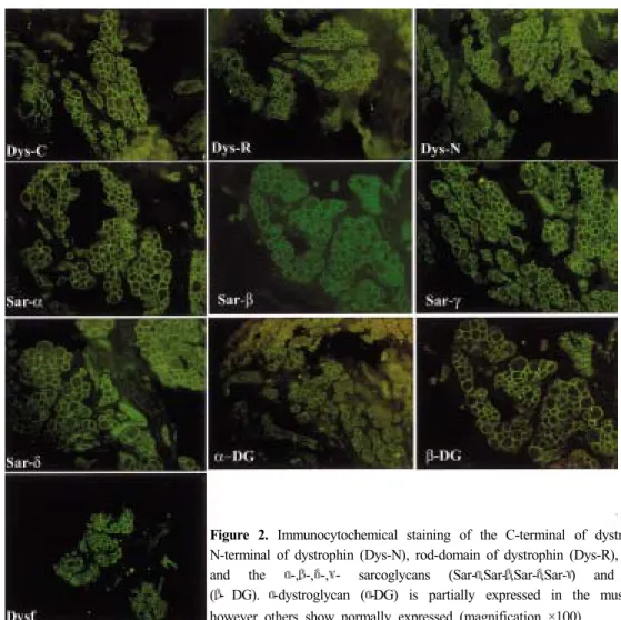
(152 beat/min). The motor and sensory nerve con-duction studies were normal. A needle electro-myography (EMG) revealed a small and polyphasic motor unit action potentials in the left adductor longus and vastus medialis muscles. A muscle biopsy was performed in the left vastus lateralis muscle. There were dystrophic myopathic changes with degenerating and regenerating fibers, increased fiber size variations, perimysial fibrosis and fatty infiltration (Fig. 1-A). Gomori trichrome staining showed no ragged red fibers or nemaline bodies (Fig. 1-B). On ATPase staining, there was no fiber type grouping (figure not shown). Immunohistochemistry using antibodies against C-terminal of dystrophin (NCL-DYS2, Novocastra), N-terminal of dystrophin (NCL-DYS1, Novocastra), rod-domain of dystrophin (NCL-DYS3, Novocastra), laminin α2 (NCL-MEROSIN, Novocastra), and α- sarcoglycan (NCL-a-SARC, Novocastra), β-sarcoglycan (NCL-b-SARC, Novocastra), γ-sarcoglycan (NCL- g-SARC, Novocastra), δ-sarcoglycan (NCL-d-SARC, Novocastra), α-dystrophin (VIA4-1, Upstate), β- dystrophin (NCL-b-DG, Novocastra) and dysferin (NCL-Hamlet, Novocastra) was perform. The immuno-reactivity against the laminin α2 chain was completely lost (Fig. 1-D) and α-dystroglycan was not expressed on some of the muscle membranes (Fig. 2). However,
dystrophin, α-,β-,γ-,δ- sarcoglycan, dysferin and β- dystroglycan were normally expressed (Fig. 2).
Discussion
Merosin (laminin-2 and -4), which contains the laminin α2 chains, is a major component of basal lamina (BL) of the skeletal muscle fiber. The BL surrounds each muscle fiber and is believed to have a critical role in maintaining the cytoarchitecture and homeostasis of the mature muscle fibers, along with the proper migration and proliferation of myogenic cells during myogenesis.4,5 Laminin α2 is expressed in numerous tissues including the skeletal muscle fibers, Schwann cells, the synaptic basal lamina of the peripheral nerves, the heart, trophoblast and skin. In skeletal muscle, the laminin (2 is located at the extracellular matrix and provides a link between the extracellular matrix and α-dystroglycan. Although laminin α2 deficiency is caused by merosin-deficient CMD, secondary reduction of laminin α2 had also been reported in other forms of CMD.6,7 In our case, the partial loss of α-dystroglycan is a secondary change caused by the selective loss of laminin (2, which is directly linked to α-dystroglycan.
The prevalence of CMD was estimated to be 0.7/100000 in a sample from north-east Italy.8 While merosin deficient CMD accounts for approximately 30% of the CMD cases in European countries, it occupies only 6% in Japan.3 Tome et al. first demon-strated merosin deficient CMD in 13 out of 20 patients with non-Fukuyama CMD.1
The human LAMA2 gene is located on 6q22-23. It spans over 260kb and consists of 64 exons.3 An analysis of the LAMA2 gene showed that nucleotide substitutions, small deletions, or insertions induce a complete merosin deficiency and a severe phenotype. Partial merosin deficient CMD is caused by a homo-zygous missense mutation, a missense mutation associated with a nonsense mutation, in-frame dele-tions in the LAMA2 gene, or nonsense mutadele-tions in the last exons of the G domain (exon58 to 64).9
The clinical features have been described as severe neonatal hypotonia, joint contracture, inability to walk, highly elevated serum creatine kinase, alterations in the somatosensory and visual evoked potentials, and abnormalities in the brain images. Most patients have a normal intelligence. To date, treatment is not available but the conditions of life can be improved by
Kyoung-Min Chae, Seong-Woong Kang, Tai-Seung Kim, Young-Chul Choi
대한신경과학회지 22권 6호 682
physiotherapy to reduce the contractures and arthro-desis in order to limit deformation.
We described a Korean patient with a merosin defi-cient CMD, who showed typical clinical features. On immunocytochemistry, there was complete absence of laminin α2 with partial loss of α-dystroglycan. How-ever, the other cytoskeletal proteins were present in the muscle membrane. This is the first case of Korean merosin-deficient CMD confirmed by immunocytoc-hemical staining.
REFERENCES
1. Tome FM, Evangelista T, Leclerc A, Sunada Y, Manole E, Estournet B, et al. Congenital muscular dystrophy with merosin deficiency. C R Acad SciIII 1994;317:351-357. 2. Voit T. Congenital muscular dystrophies: 1997 update. Brain
Dev 1998;20:65-74.
3. Hayashi YK, Tezak Z, Momoi T, Nonaka I, Garcia CA, Hoffman EP, et al. Massive muscle cell degeneration in the early stage of merosin-deficient congenital muscular dystrophy. Neuromuscul Disord 2001;11:350-359.
4. Gullberg D, Tiger CF, Velling T. Laminins during muscle development and in muscular dystrophies. Cell Mol Life Sci 1999;30:442-460.
5. Tachi N, Kamimura S, Ohya K, Chiba S, Sasaki K. Congenital muscular dystrophy with partial deficiency of merosin. J Neurol Sci 1997;151:25-27.
6. Auranen M, Rapola J, Pihko H, Haltia M, Leivo I, Soinila S, et. al. Muscle membrane-skeleton protein changes and histopathological characterization of muscle-eye-brain disease.
Neuromuscul Disord 2000;10:16-23.
7. Mostacciuolo ML, Miorin M, Martinello F, Angelini C, Perini P, Trevisan CP. Genetic epidemiology of congenital muscular dystrophy in a sample from north-east Italy. Hum
Genet 1996;97:277-279.
8. Allamand V, Guicheney P. Merosin-deficient congenital muscular dystrophy, autosomal recessive (MDC1A, MIM# 156225, LAMA2 gene coding for alpha2 chain of laminin).
Eur J Hum Genet 2002;10:91-94.
9. Guicheney P, Vignier N, Helbling-Leclerc A, Nissinen M, Zhang X, Cruaud C, et al. Genetics of laminin alpha 2 chain (or merosin) deficient congenital muscular dystrophy: from identification of mutations to prenatal diagnosis.
Neuro-muscul Disord 1997;7:180-186.
Figure 2. Immunocytochemical staining of the C-terminal of dystrophin (Dys-C),
N-terminal of dystrophin (Dys-N), rod-domain of dystrophin (Dys-R), dysferin (Dysf) and the α-,β-,δ-,γ- sarcoglycans (Sar-α,Sar-β,Sar-δ,Sar-γ) and β-dystroglycan (β- DG). α-dystroglycan (α-DG) is partially expressed in the muscle membrane, however others show normally expressed (magnification ×100).