Changwon Kang, Ji-Joon Song, Jaeok Lee, Mi Young Kim, Department of Biological Sciences, Korea Advanced Institute of Science and Technology, Daejeon 305-701, South Korea
Author contributions: Kang C and Kim MY contributed equally
to this work; Kang C, Song JJ and Kim MY designed and wrote the manuscript; and Lee J searched the literature and outlined the manuscript.
Supported by National Research Foundation of Korea, No.
2013056334
Correspondence to: Mi Young Kim, PhD, Department of
Bio-logical Sciences, Korea Advanced Institute of Science and Tech-nology, 291 Daehak-ro, Yuseong-gu, Daejeon 305-701,
South Korea. [email protected]
Telephone: +82-42-3502615 Fax: +82-42-3502610 Received: December 1, 2013 Revised: January 7, 2014 Accepted: February 17, 2014
Published online: June 7, 2014
Abstract
Cancers, like other diseases, arise from gene mutations
and/or altered gene expression, which eventually cause
dysregulation of numerous proteins and noncoding
RNAs. Changes in gene expression,
i.e.
, upregulation of
oncogenes and/or downregulation of tumor suppressor
genes, can be generated not only by genetic and
envi-ronmental factors but also by epigenetic factors, which
are inheritable but nongenetic modifications of cellular
chromosome components. Identification of the factors
that contribute to individual cancers is a prerequisite to
a full understanding of cancer mechanisms and the
de-velopment of customized cancer therapies. The search
for genetic and environmental factors has a long history
in cancer research, but epigenetic factors only recently
began to be associated with cancer formation,
progres-sion, and metastasis. Epigenetic alterations of
chroma-tin include DNA methylation and histone modifications,
which can affect gene-expression profiles. Recent
stud-ies have revealed diverse mechanisms by which
chro-matin modifiers, including writers, erasers and readers
of the aforementioned modifications, contribute to the
formation and progression of cancer. Furthermore,
functional RNAs, such as microRNAs and long
noncod-ing RNAs, have also been identified as key players in
these processes. This review highlights recent findings
concerning the epigenetic alterations associated with
cancers, especially gastric cancer.
© 2014 Baishideng Publishing Group Inc. All rights reserved.
Key words: Gastric cancer; Epigenetics; DNA
methyla-tion; Histone modificamethyla-tion; Gene expression
Core tip: The pathogenesis of gastric, or stomach
cancer has long been a topic of extensive research,
and these research efforts have resulted in
tremen-dous improvements in the diagnosis and treatment of
gastric cancer patients. However, research on gastric
cancer has been focused on the genetic and
environ-mental determinants of its formation and progression
while the role of regulators, another important set of
contributors to gastric cancer, has just begun to be
elucidated. In this review, we highlight our current
understanding of the epigenetic mechanisms by which
gastric cancer arises and progresses and discuss future
research directions.
Kang C, Song JJ, Lee J, Kim MY. Epigenetics: An emerg-ing player in gastric cancer. World J Gastroenterol 2014; 20(21): 6433-6447 Available from: URL: http://www.wjgnet. com/1007-9327/full/v20/i21/6433.htm DOI: http://dx.doi. org/10.3748/wjg.v20.i21.6433
INTRODUCTION
Gastric cancer is the fourth most frequently occurring
cancer worldwide and the second leading cause of
can-cer-related death
[1]. The occurrence of gastric cancer
var-ies with geographic area, with the highest incidence rate
of gastric cancer in East Asia, especially South Korea,
Mongolia, Japan and China. Although the mortality rate
TOPIC HIGHLIGHT
DOI: 10.3748/wjg.v20.i21.6433 © 2014 Baishideng Publishing Group Inc. All rights reserved.
Epigenetics: An emerging player in gastric cancer
WJG 20
thAnniversary Special Issues (8): Gastric cancer
has dramatically decreased due to improvements in
en-doscopy and surgical technology, the survival rate is still
less than 15% once gastric cancer metastasizes.
Gastric cancer can arise from precursor lesions or
de
novo and is commonly categorized into two main
sub-types, diffuse-type gastric cancer and intestinal-type
gas-tric cancer. Research in the past decades has provided us
with insights into the molecular mechanisms that drive
gastric cancer tumorigenesis and progression. As in other
types of cancer, genetic, epigenetic and
environmen-tal factors in combination contribute to gastric cancer
tumorigenesis and progression. Previous research has
mainly focused on genetic factors such as the inheritance
of gastric cancer susceptibility genes and environmental
factors including
Helicobacter pylori infection, salt
con-sumption, stress, smoking
etc. In recent years, however,
the epigenetic mechanisms governing gastric cancer have
been at the center of gastric cancer research.
EPIGENETIC REGULATION OF GENE
EXPRESSION
In multi-cellular organisms, different gene expression
patterns determine the fates of cells, causing them to
dif-ferentiate into various cell types. Therefore, it is critical to
precisely coordinate the gene expression pattern based on
cell types during developmental processes. Furthermore,
gene expression patterns need to be maintained
through-out the life span once established. Each cell appears to
‘‘memorize’’ the genetic information to be expressed and
precisely passes this memory on to its daughter cells after
cell division. This process is referred to as ‘‘epigenetic
cellular memory’’. Dysregulation of epigenetic memory
causes developmental defects, cancers and
neurodegen-erative diseases.
In the nucleus, DNA is packaged into a higher order
structure called chromatin, the physiological template for
transcription. Alteration of chromatin structure
via the
various modifications described below is the major factor
that controls gene expression in a temporal and spatial
manner, resulting in the establishment and maintenance
of epigenetic cellular memory.
Regulation by DNA methylation
Chromatin structure is modified and altered in several
lay-ers. First of all, DNA itself is methylated, and this event
mostly occurs at cytosines in CpG-rich regions. DNA
methylation at promoter regions generally occludes the
binding of transcription factors or recruits
methyl-DNA-binding proteins, leading to the inactivation of gene
ex-pression, with few exceptions in which DNA methylation
can be involved in preventing gene repression
[2]. DNA
methylation is a stable epigenetic mark that is inherited
by offspring or daughter cells once established.
Two different classes of DNA methyltransferases
(DNMTs) are responsible for establishing and
maintain-ing DNA methylation. DNMT1 maintains DNA
meth-ylation through its substrate preference for
hemimethyl-ated DNA at CpG regions. DNMT3 family members,
DNMT3A, DNMT3B and DNMT3L, are involved in
establishing
de novo DNA methylation patterns, although
DNMT3L is catalytically inactive and might cause gene
repression independent of DNA methylation
[3].
DNA methylation was once believed to be a
perma-nent epigenetic mark. So far, no enzyme has been
discov-ered that directly removes the methyl group from
meth-ylcytosine. However, TET family proteins were identified
to oxidize 5-methylcytosine to 5-hydroxymethylcytosine,
eventually leading to the removal of the methyl group
from methylcytosine
[4]. TET family proteins are involved
in regulating transcription during embryonic
develop-ment. The tight regulation of writing and erasing methyl
marks on DNA is required for proper gene expression,
and the imbalance between writing and erasing is
impli-cated in various cancers.
Regulation by histone modifications
The nucleosome, which is composed of 146 bp of
DNA and a histone octamer (dimers of H2A, H2B, H3
and H4) is the fundamental repeating unit of chromatin
structure and a major target of chromatin regulation
[5].
Histone proteins have long flexible N-terminal tails that
are subject to several covalent modifications including
acetylation, methylation, phosphorylation, ubiquitylation,
ADP-ribosylation, crotonylation and glutarylation
[6].
The combinations of different types and locations of
histone modifications, also known as histone codes, are
the main determinants of gene repression or activation.
Covalent modifications are regulated by a trio of writers,
erasers and readers. Writers and erasers add and remove
covalent modifications, respectively, while readers
rec-ognize specific modifications with specialized domains,
resulting in the recruitment of transcriptional machinery
or transcription-repression complexes. More detailed
de-scriptions are given in the sections below.
Regulation by histone lysine acetylation and
deacetylation
The first covalent modification identified was the
acetyla-tion of lysine (Lys or K) residues of histones by histone
acetyltransferases (HATs), more specifically called histone
lysine acetyltransferases (KATs). Many Lys residues of
histones are involved in interacting with DNA, and this
acetylation neutralizes the positive charge of Lys,
lead-ing to the weakenlead-ing of the DNA-histone interaction
and subsequent activation of transcription
[7]. In addition,
acetyllysine recruits other chromatin modifiers containing
a bromodomain that recognizes an acetyllysine to activate
transcription
[8].
Histone deacetylases (HDACs), the erasers of
acetyla-tion, have been shown to be directly involved in cancer
pathogenesis
via transcriptional repression of tumor
sup-pressor genes
[9]. Some HDAC inhibitors are currently in
Regulation by histone lysine methylation and
demethylation
Several Lys residues of histones can also be mono-, di-
or trimethylated. The different locations and levels of
histone methylation add another layer of complexity to
covalent modification of histones. Among histone
ly-sine methylations, those of histone H3 Lys4 (denoted as
H3K4) and histone H3 Lys27 (H3K27) are particularly
interesting because H3K4 and H3K27 methylations are
directly implicated in transcriptional activation and
re-pression, respectively
[10].
Methylation of H3K4 and H3K27 is catalyzed by
multi-subunit protein complexes. For example, KMT2A
(K-specific methyltransferase 2A, commonly called
MLL), which methylates H3K4 using its SET domain, is
complexed with WDR5, RBBP5 and ASH2L
[11]. H3K27
is methylated by PRC2 (polycomb repressive complex 2)
composed of EED, EZH2, SUZ12 and RBBP4
[12].
It is not clearly understood how H3K4 or H3K27
methylation regulates transcriptional activation or
repres-sion, respectively. However, it has been shown that H3K4
methylation recruits the BAF chromatin remodeling
com-plex
via its chromodomain to activate transcription
[13]. In
regard to H3K27 methylation, another polycomb
repres-sive complex, PRC1, recognizes trimethylated H3K27
(denoted as H3K27me3)
via the
chromodomain-contain-ing protein CBX1 (chromobox homolog 1) and induces
the compaction of chromatin, resulting in transcriptional
repression, although the requirement of H3K27me3 for
PRC1 function is controversial
[14-16].
Because histone methylation status is critical for gene
expression, the removal of histone methylation is highly
regulated by several histone Lys-specific demethylases
(KDMs). H3K4 is demethylated by KDM1 (commonly
known as LSD1) and KDM5B (JARID1), whereas
H3K27 is demethylated by KDM6A (UTX) and KDM6B
(JMJD3)
[10]. Because the balance between methylation
and demethylation of histones is critical for coordinating
gene expression, the disruption of this balance is found
in many cancers.
Regulation by histone arginine methylation
Arginine (Arg or R) residues in histones are also targets
for methylation. Arg methylation affects gene expression
by activating or repressing transcription depending on
the methylated sites
[17]. Arg can be monomethylated,
sym-metrically dimethylated, or asymsym-metrically dimethylated,
although the different biological consequences of
sym-metric
vs asymmetric Arg dimethylation are unclear.
The methylation of Arg functions in at least two
different ways. The methylation of Arg near a Lys in
histones can block the Lys methylation
[18]. Specifically,
methylation of histone H3 Arg2 (denoted as H3R2)
represses transcription by blocking H3K4 methylation,
which is critical for transcriptional activation
[19]. In
addi-tion, methylarginine may serve as a site-specific docking
stage for methylarginine-binding proteins, which recruit
other transcriptional regulators
[20].
Regulation by other histone modifications and
nucleosome variants
Other covalent modifications such as ubiquitylation,
cro-tonylation and glutarylation are also involved in
regulat-ing gene expression. However, the downstream pathways
of these modifications are not well studied.
A canonical nucleosome is composed of histones
H3, H4, H2A and H2B. There are also several histone
variants such as H3.3, H2A.Z, CENP-A and macroH2A,
which are incorporated into nucleosomes with other
histones to execute their specific functions. For example,
histone H3.3 is not only found in transcriptionally active
genes but is also involved in recruiting PRC2. H2A.Z
functions in the cell cycle, and the improper
incorpora-tion of H2A.Z is implicated in various cancers
[21].
Regulation by chromatin remodeling
The immediate consequence of forming a nucleosome is
to limit DNA accessibility by protein factors. Therefore,
cells have developed an elaborate system to remodel
nu-cleosomes using ATP as an energy source.
ATP-depen-dent chromatin remodeling complexes are classified into
five families depending on the type of ATPase subunit
of the complexes and are known as the SWI/SNF, ISWI,
CHD, INO80 and SWR1 families.
Each ATP-dependent chromatin remodeling family
is believed to remodel nucleosomes
via a distinct
mecha-nism and is involved in a distinct biological pathway, such
as gene repression, gene activation, histone exchange and
the DNA-damage response
[22]. Due to the importance
of the roles of ATP-dependent chromatin remodelers
in various physiological processes, mutations and
over-expression of the remodelers are often found in several
cancers.
Regulation by long noncoding RNAs
The most interesting but least studied epigenetic
regu-lator is long noncoding RNAs (lncRNAs), which are
defined as transcripts that are generally longer than 200
nucleotides and do not code for proteins. Most lncRNAs
are synthesized by RNA polymerase II, capped at the 5’
end and polyadenylated at the 3’ end
[23]. Although the
role of lncRNAs in chromatin regulation was first
identi-fied in X-chromosome inactivation several decades ago,
the significance of lncRNAs in chromatin regulation was
not fully recognized until the recent discovery that many
lncRNAs interact with chromatin modifiers and directly
control gene expression
[24].
Although exact mechanisms of lncRNAs are not well
understood, it is believed that lncRNAs function through
their binding partners in several different ways
[25]. They
function as a scaffold for multi-subunit protein complex
formation or recruit chromatin modifying complexes to
a specific locus leading to transcriptional activation or
repression. For example, HOTAIR (HOX antisense
in-tergenic RNA) transcribed from a
HOXC locus interacts
with PRC2 and LSD1
via its 5’ and 3’ ends, respectively,
to repress gene expression.
In addition to HATs and HDACs, proteins called
acetylation readers, which contain a bromodomain that
recognizes acetylated histones and recruits other
com-plexes, have been reported to undergo mutations or
translocations in certain tumor types, suggesting that
modification readers also contribute to tumorigenesis.
Histone lysine methylation writers in cancer
Histone methylation can take place at lysine, histidine
or arginine residues. However, we will mainly discuss
lysine and arginine methylation in this section. With
the discovery of histone lysine demethylases (KDMs),
the contribution of aberrant histone methylation status
to tumorigenesis and cancer progression has received
renewed attention in the field of cancer epigenetics in
recent years.
Alteration of histone methylation status can be a
con-sequence of translocation, amplification, deletion,
over-expression or repression of histone methyltransferase or
demethylase genes. The best-studied methyltransferase
that undergoes chromosomal translocation is KMT2A
(commonly known as MLL). This H3K4
methyltrans-ferase is often fused with another protein, such as AFF1
(AF4/FMR2 family, member 1), ELL (elongation factor
RNA polymerase
Ⅱ
, alternatively called ELL1), MLLT1
(myeloid/lymphoid or mixed-lineage leukemia;
translo-cated to, 1, or ENL) or MLLT3 (myeloid/lymphoid or
mixed-lineage leukemia; translocated to, 3, or AF9)
[36,39,40].
MLL-fusion proteins can cause aberrant H3K4
methyla-tion of target genes including
HOXA7 (homeobox A7)
and
HOXA9 (homeobox A9)
[41]. Intriguingly, several
MLL-fusion proteins have been reported to recruit other
histone methyltransferase such as DOT1L (DOT1-like
histone H3K79 methyltransferase) in leukemia
[42].
El-evated expression of another H3K4 methyltransferase,
SMYD, was found in breast cancer.
In contrast, overexpression of another H3K27
meth-yltransferase gene,
EZH2, has been observed in a wide
variety of solid tumors and exhibits a strong association
with tumor stage and aggressiveness. An inactivating
mutation of
EZH2 has also been found in lymphoid,
myeloid and T-cell acute lymphoblastic leukemia
(T-ALL)
[43]. Specifically, NOTCH1 antagonizes PRC2,
thus driving the formation of T-ALL. Another histone
methyltransferase that undergoes mutation, translocation
or repression is the H3K36-specific methyltransferase
NSD1
[44,45].
Histone lysine methylation erasers in cancer
In addition to histone methyltransferases, the role of
histone demethylases in cancer has been highlighted in
recent studies. The activity of histone demethylases can
be dysregulated by somatic mutations or changes in their
expression in cancer cells. So far, somatic mutations have
been found in
KDM5A (commonly known as JARID1A),
KDM5C (JARID1C) and KDM6A (UTX)
[28,46].
Specifi-cally, mutations in
UTX, a histone H3K27 demethylase
gene, have been found in 12 different types of cancer,
CANCER AND EPIGENETICS
Numerous studies have unraveled complex networks of
epigenetic regulation in several types of cancer. In this
section, we will highlight some of the epigenetic
mecha-nisms that contribute to tumorigenesis and tumor
pro-gression, mainly focusing on DNA methylation, histone
acetylation, histone methylation and lncRNAs.
DNA methylation in cancer
In general, cancer cells exhibit hypermethylation of the
CpG islands of some genes, including tumor suppressor
genes,
BRCA1 (breast cancer 1, early onset), CDKN2A
(cyclin-dependent kinase inhibitor 2A),
MLH1 (mutL
ho-molog 1) and
VHL (von Hippel-Lindau tumor
suppres-sor, E3 ubiquitin protein ligase)
[26-28]. In contrast, cancer
cells exhibit global hypomethylation at many genomic
sequences, which can result in chromosomal instability as
well as activation of proto-oncogenes
[29-31].
DNA methyltransferase genes have been shown to be
mutated in certain cancers. For example,
DNMT3A gene
is mutated in acute myelogenous leukemia,
myeloprolif-erative disease and myelodysplastic syndrome
[32]. In
adtion, the recently identified TET1 (tet methylcytosine
di-oxygenase 1) and TET2 (tet methylcytosine didi-oxygenase
2) proteins in the Tet (ten-eleven translocation) family of
DNA hydroxylases are involved in DNA demethylation
and found to be mutated in acute myelogenous leukemia,
myeloproliferative disease, myelodysplastic syndrome and
chronic myelomonocytic leukemia. Furthermore,
TET1
gene is also fused with the histone methyltransferase
MLL (myeloid/lymphoid or mixed-lineage leukemia)
gene in some cases of acute myelogenous leukemia
[33,34].
Histone modifications in cancer
As described above, histone modification status is finely
regulated by modification writers and erasers. Disruption
of this balance can cause aberrant histone modifications,
resulting in dysregulation of gene expression. In cancer
cells, one of the best-established changes in histone
mod-ifications is a global decrease in the acetylation of H4K16
and trimethylation of H4K20
[35]. Recent findings
regard-ing the roles of histone modifications in various types of
cancer are summarized in the sections below.
Histone acetylation modifiers in cancer
In cancer cells, mutations or changes in the expression
of HATs and HDACs are frequently observed. For
ex-ample, some genes that encode HATs such as CREBBP
(alternatively called CBP), EP300 (p300), KAT6A (MOZ),
KAT6B (MORF) and MOXD1 (MOX) have been shown
to be mutated, translocated, or overexpressed in solid and
hematological tumors
[35-38]. In addition, altered expression
of HDACs has been observed in a variety of cancers,
while somatic mutations are rarely found. Moreover, the
recruitment of HDACs to certain target genes
via
chime-ric fusion proteins, which can occur in leukemia, has been
shown to be another mechanism of gene repression
[39].
indicating a tumor-suppressive role of UTX in various
cancers. This concept was supported by a recent finding
showing that UTX controls the cell cycle by targeting the
RB1 (Rb) protein network
[47].
In contrast, the role of KDM6B (JMJD3), another
H3K27 demethylase, seems to vary depending on the
type of cancer. For example, JMJD3 has been shown
to function in oncogene-induced senescence,
suggest-ing a tumor-suppressive role of the protein
[48]. However,
upregulation of JMJD3 in metastatic prostate cancer
in-dicates a potential role of the protein in the progression
of prostate cancer
[48]. In addition, overexpression of the
histone H3K4 demethylase gene
KDM1A (LSD1) has
been associated with the recurrence of prostate cancer
[49].
Furthermore, LSD1 has been identified as a positive
regulator of neuroblastoma and breast tumors
[50,51].
Other enzymes with altered expression in cancer
include KDM2B (JHDM1B), KDM4C (JMJD2C),
KD-M5A (RBP2) and KDM5B (PLU1)
[52-57]. JMJD2C, a
mem-ber of the JMJD2 H3K9 demethylase family, has been
shown to be upregulated in various tumors including
breast cancer, prostate cancer, esophageal squamous cell
carcinoma and desmoplastic medulloblastoma
[54,55,58-60]. In
addition, its potential role as an oncogene has been
sug-gested by a recent study using immortalized mammary
epithelial cells
[61].
Histone lysine methylation readers in cancer
Like acetylation readers, methyllysine readers play a
pivot-al role in cancer. For example, ING (inhibitor of growth)
family proteins, which can bind di- and trimethylated
H3K4, have been found to be mutated or downregulated
by the loss of heterozygosity, supporting their role as
tu-mor suppressors in several types of cancer
[62-64]. Another
example of a methylated H3K4 binding protein, NUP98
(nucleoporin 98 kDa), is often fused with several subunits
of histone lysine methyltransferases, thereby contributing
to hematopoietic cancer
[65].
Histone arginine methylation in cancer
Although histone arginine methylation has not received
as much attention as lysine methylation, numerous studies
have implicated the function of protein arginine
methyl-transferases (PRMTs) in cancers. The best-studied PRMT1
has been reported to be overexpressed and/or aberrantly
spliced in various types of cancer, including breast,
pros-tate, lung and colon cancers
[66-69]. A recent study
demon-strated that H4R3 methylation has a strong positive
corre-lation with tumor stages in prostate cancer
[67].
In addition to PRMT1, other PRMTs, such as
PRMT2, PRMT5 and PRMT6, are overexpressed in
breast, gastric, colon and lung cancers, while elevated
PRMT3 activity without changes in its expression level
has been reported
[70-73]. Furthermore, somatic
muta-tions in PRMT8 were found in ovarian and skin cancers.
Finally, a non-PRMT family arginine methyltransferase
CARM1 (coactivator-associated arginine
methyltrans-ferase 1) has been shown to be overexpressed in breast,
prostate and colon cancers
[74-77].
The mechanisms by which the aforementioned
PRMTs contribute to tumorigenesis and metastasis have
been studied by several groups. For example, PRMT1 and
CARM1 are involved in the activation of WNT signaling,
a well-known tumor-promoting signaling pathway
[78,79].
In addition, elevated activity of PRMTs
via the various
mechanisms mentioned above can affect cell growth and
migration and the tumor microenvironment.
lncRNAs in cancer
It has become clear that lncRNAs have fundamental roles
in tumorigenesis and tumor progression. One of the
best-studied lncRNAs is HOTAIR. HOTAIR has been
shown to be overexpressed in breast and colon cancers
and esophageal squamous cell carcinoma and functions
via altering PRC2 target-gene occupancy
[25,80-83]. In
ad-dition, several lncRNAs have been implicated in cancer
with oncogenic functions. They include CDKN2B-AS1
(CDKN2B antisense RNA 1, or ANRIL), H19 (imprinted
maternally expressed transcript), MALAT1 (metastasis
associated lung adenocarcinoma transcript 1), PCAT1
(prostate cancer-associated transcript 1), PCBP2-OT1
(PCBP2 overlapping transcript 1, or TUC338), PCGEM1
(prostate-specific transcript), PRNCR1 (prostate cancer
associated non-coding RNA 1) and SPRY4-IT1 (SPRY4
intronic transcript 1). These lncRNAs are often found to
be upregulated in several types of cancer and exert their
oncogenic effects
via promoting cell proliferation or
in-hibiting apoptosis and senescence.
The mechanisms by which some of these lncRNAs
execute their oncogenic functions have been uncovered.
For instance, CDKN2B-AS1 functions by causing
aber-rant recruitment of the PRC2 complex to
CDKN2A
(cyclin-dependent kinase inhibitor 2A, or INK4A) or
CDKN2B (cyclin-dependent kinase inhibitor 2B, or
INK4B), thus suppressing their expression
[84,85], whereas
PCAT1 inhibits
BRCA2 (breast cancer 2, early onset)
expression
[86]. In contrast, other lncRNAs, such as GAS5
(growth arrest-specific 5), MEG3 (maternally expressed
3), PTENP1 (phosphatase and tensin homolog
pseu-dogene 1) and LincRNA-p21, have been suggested to
have tumor-suppressive effects
[87-92]. GAS5 induces the
expression of the proapoptotic protein BIRC3
(baculo-viral IAP repeat containing 3, or cIAP2) and has been
found to be downregulated in breast cancer
[87,88], while
LincRNA-p21 induces apoptosis by affecting the TP53
(p53) pathway
[92].
DNA METHYLATION IN GASTRIC
CANCER
As in other types of cancer, numerous studies have
shown that key players in gastric cancer are regulated by
changes in DNA methylation patterns at their promoter
CpG islands,
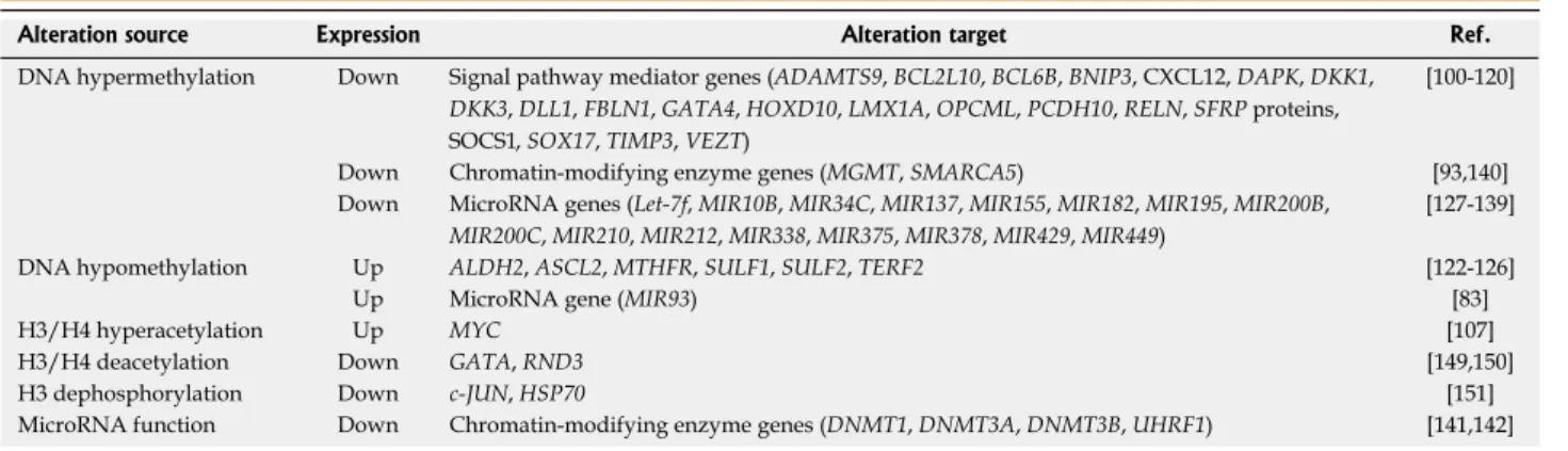
i.e., hyper- or hypomethylation (Table 1).
These genes include tumor-suppressor genes, oncogenes,
and genes that are involved in tumor progression and
metastasis. In addition, recent findings demonstrating
changes in the DNA methylation patterns of microRNA
genes in gastric cancer patient samples have revealed
more complexity in the epigenetic regulation of gastric
cancer.
DNA hypermethylation in gastric cancer
Hypermethylation of CpG islands results in the silencing
of neighboring genes, and promoters of
tumor-suppres-sor genes are often methylated in gastric cancer patient
samples. Widely studied genes with methylated
promot-ers include
CDKN2A, TP53 (tumor protein p53), MLH1,
CDH1 (cadherin 1, or E-cadherin), RUNX3 (runt-related
transcription factor 3),
APC (adenomatous polyposis
coli)
and RASSF1A (Ras association (RalGDS/AF-6)
domain family member 1)
[93-99]. In addition, recent studies
have identified numerous hypermethylated genes
encod-ing pro-apoptotic or anti-growth proteins (BCL2L10,
BCL6B, BNIP3, DAPK and FBLN1), transcription
fac-tors (GATA4, HOXD10, LMX1A and SOX17), enzymes
(KL), cell-cell interaction or migration-related proteins
(ADAMTS9, OPCML, PCDH10, RELN, TIMP3 and
VEZT), DNA-repair proteins (XRCC1), signaling
mol-ecules (CXCL12, DKK1, DKK3, DLL1, SFRP proteins
and SOCS1), an RNA binding-protein (QKI) and others
(NDRG2)
[100-121].
Hypermethylation of the aforementioned genes
generally promotes gastric cancer tumorigenesis and/or
metastasis
via several mechanisms. DNA methylation of
tumor-suppressor genes endows gastric cells with the
ability to overcome oncogene-induced senescence as well
as apoptosis. For example, downregulation of DKK1
(dickkopf WNT signaling pathway inhibitor 1) and
SOCS1 (suppressor of cytokine signaling 1) reactivates
the WNT and STAT3 pathways, respectively
[116,119,122].
DNA hypomethylation in gastric cancer
Hypomethylation causes derepression of target genes;
several genes involved in tumorigenesis, progression, and
metastasis of gastric cancers have been found to be
hy-pomethylated. For example, Kwon
et al
[122]demonstrated
that the promoter of
ASCL2 (achaete-scute family bHLH
transcription factor 2), which encodes a basic
helix-loop-helix transcription factor, shows hypomethylation in
gastric cancer samples compared to normal tissues, and
high expression levels of this gene are correlated with
poor survival of gastric cancer patients. In addition, the
promoter of the well-known oncogene
MYC has been
shown to undergo hypomethylation in gastric cancer
with lymph node metastasis
[123]. Yashiro
et al
[124]showed
that demethylation in
TERF2 (telomeric repeat binding
protein 2, or TRF2) and
ERAS (ES cell expressed Ras)
promoters causes reactivation of these genes in gastric
cancer
[124,125].
A recent study by Balassiano
et al
[126]reported that
gastric cancer patient samples contain hypomethylated
promoters of two cancer-associated genes,
ALDH2
(aldehyde dehydrogenase 2 family) and
MTHFR
(methy-lenetetrahydrofolate reductase). Furthermore,
overex-pression of
SULF1 (sulfatase 1) and SULF2 (sulfatase
1), members of the sulfatase family, caused by promoter
hypomethylation has been shown to be an independent
prognostic marker for lymph node metastasis. Finally, an
interesting study by Yuasa
et al
[127]showed an association
between hypomethylation of blood leukocyte DNA and
the risk of gastric cancer, indicating that changes in the
DNA methylation pattern in non-tumor cells in addition
to tumor cells themselves can be used as potential
prog-nostic markers in gastric cancer.
MicroRNA promoter methylation in gastric cancer
MicroRNAs (miRNAs) are small noncoding RNAs that
can regulate the expression of target genes at the
post-transcriptional level. Because a single miRNA can target
several messenger RNAs, dysregulation of miRNAs can
effectively affect multiple signaling pathways leading to
tumor formation and metastasis. As in other types of
cancer, recent studies have identified several miRNAs
as frequent targets of DNA methylation in gastric
can-cer (Table 1). For example, the suppression of several
miRNA genes, such as
MIR137, MIR210, MIR375 or
MIR449, via promoter methylation has been shown to
prevent apoptosis by alleviating the miRNA-induced
inhibition of pro-survival pathways such as MAPK1 (by
MIR137 and MIR210) and PDK1 (by MIR375) or by
in-hibiting pro-apoptotic pathways (by MIR449)
[128-130].
Table 1 Examples of epigenetic alterations found in gastric cancer
Alteration source Expression Alteration target Ref.
DNA hypermethylation Down Signal pathway mediator genes (ADAMTS9, BCL2L10, BCL6B, BNIP3, CXCL12, DAPK, DKK1,
DKK3, DLL1, FBLN1, GATA4, HOXD10, LMX1A, OPCML, PCDH10, RELN, SFRP proteins,
, SOX17, TIMP3, VEZT)
[100-120] Down Chromatin-modifying enzyme genes (MGMT, SMARCA5) [93,140] Down MicroRNA genes (Let-7f, MIR10B, MIR34C, MIR137, MIR155, MIR182, MIR195, MIR200B,
MIR200C, MIR210, MIR212, MIR338, MIR375, MIR378, MIR429, MIR449)
[127-139] DNA hypomethylation Up ALDH2, ASCL2, MTHFR, SULF1, SULF2, TERF2 [122-126]
Up MicroRNA gene (MIR93) [83]
H3/H4 hyperacetylation Up MYC [107]
H3/H4 deacetylation Down GATA, RND3 [149,150]
H3 dephosphorylation Down c-JUN, HSP70 [151]
In some cases, downregulation of miRNAs
via
meth-ylation activates tumor growth-promoting pathways such
as CDK6-VEGF (by MIR195 and MIR378), c-MYC
(by MIR212 and MIR429), cAMP response element (by
MIR182) and MAPRE1 (by MIR10B)
[28,131-133]. Thus,
methylation of the aforementioned miRNAs causes
over-all growth of gastric cancer.
In addition to regulating gastric cancer cell survival
and growth, DNA methylation of some miRNAs
pro-motes the ability of gastric cancer cells to invade and
mi-grate, thus increasing their metastatic potential. Examples
of these include Let-7f, MIR155 and MIR338, which
ex-ert their effects by altering the expression of
MYH9
(my-osin, heavy chain 9, non-muscle),
SMAD2 (SMAD family
member 2) and
SSX2IP (synovial sarcoma, X breakpoint
2 interacting protein), respectively
[134-136].
Downregulation of
MIR9 via hypermethylation in
gastric cancer has also been found to increase not only
proliferation but also cell migration and invasion, a
prerequisite for the formation of successful
metasta-sis, although their target genes have not been identified
yet
[137]. Finally, dysregulation of MIR34C can cause drug
resistance by affecting MAPT (microtubule-associated
protein tau)
[138], and dysregulation of the
MIR200BC/429
cluster can do so by altering the expression of BCL2
(B-cell CLL/lymphoma 2) and XIAP (X-linked inhibitor of
apoptosis)
[139].
Hypomethylation of miRNAs has also been studied.
For example, the loss of methylation at the promoter of
the
MIR196 gene and upregulation of this miRNA are
frequently found in primary gastric cancer, indicating the
tumor-suppressive role of MIR196
[83]. In addition, the
up-regulation of several oncogenic miRNAs such as MIR9,
MIR93, MIR106B and MIR222 in gastric cancer have
been reported, and their role in proliferation,
anti-apop-tosis and metastasis has been studied in gastric cancer
cell lines
[137,140,141]. However, the question of whether the
upregulation of the aforementioned miRNAs is a
conse-quence of DNA hypomethylation has yet to be answered.
Promoter methylation of chromatin-modifying enzyme
genes in gastric cancer
Chromatin-modifying enzymes (CMEs) can affect the
DNA methylation and histone modification status of
target genes, thus causing changes in chromatin
struc-ture. Alteration at the level of CMEs can
initiate several
epigenetic cascades that affect diverse pathways involved
in tumorigenesis and the progression and metastasis of
gastric cancer.
In gastric cancer, several CMEs are also the targets of
DNA methylation (Table 1). For example, Gigek
et al
[142]found that SMARCA5 (SWI/SNF related, matrix
associ-ated, actin dependent regulator of chromatin, subfamily
a, member 5), which has helicase and ATPase activity, was
often downregulated in gastric cancer patient samples
compared to normal tissue and as a consequence of its
promoter methylation. MGMT (O-6-methylguanine-DNA
methyltransferase) has also been frequently found to be
absent in gastric cancer due to promoter methylation
[93].
Moreover, the expression of several CMEs is
regu-lated by miRNAs. For example, upregulation of
UHRF1
(ubiquitin-like with PHD and ring finger domains 1)
ex-pression
via downregulation of MIR146A and MIR146B
causes aberrant DNA methylation in
CDH1, RUNX3
and
SLIT3 (slit homolog 3) genes
[143]. Furthermore,
DNMT1, DNMT3A and DNMT3B proteins are
down-regulated
via overexpression of MIR200B and MIR200C
in gastric cancer, and this may be a cause of global DNA
hypomethylation in gastric cancer cells
[144].
HISTONE MODIFICATIONS IN GASTRIC
CANCER
Histone modifications including acetylation, methylation,
phosphorylation and ubiquitylation can directly alter gene
expression. Several histone modifiers show aberrant
ex-pression patterns or mutations during tumorigenesis and
cancer progression as explained above. The mechanisms
by which alterations of histone modifications contribute to
tumorigenesis and metastasis have been intensively studied
in several types of cancer. In contrast, studies of histone
modifications in gastric cancer are lacking (Table 1).
Histone-modifying enzymes in gastric cancer
Most epigenetic studies of gastric cancer have been
fo-cused on DNA methylation. Thus, scientists only recently
started to investigate histone modifiers in gastric cancer.
Recent findings on the role of histone modifiers in
gas-tric cancer have shed light on the complex epigenetic
mechanisms governing the development and progression
of gastric cancer.
For example, histone H3K4 demethylase KDM1A
(LSD1) is upregulated in some gastric cancer cells, and
treatment of these cells with LSD1 inhibitors exerts
cyto-toxic effects as well as inhibitory effects on the migration
and invasion of these cells, suggesting an important role
for LSD1 in gastric cancer
[145]. In addition, it has been
shown that the histone deacetylase SIRT1 (sirtuin 1) plays
a tumor-suppressive role in gastric cancer development
via inhibition of NF-κB signaling and is downregulated
in gastric cancer
[146].
In contrast, the H3K9/K36 demethylase KDM4B
(commonly called JMJD2B) was recently discovered to
be a potent activator of cell proliferation as well as the
epithelial-mesenchymal transition (EMT) and correlated
with lymph node/distant metastasis
[147,148]. Another H3K9
demethylase, JMJD1C, is also upregulated in gastric
can-cer. In addition, the H3K27 methyltransferase EZH2 has
been shown to promote gastric cancer tumorigenesis in
various model systems and exhibits significant association
with patient survival as well as lymph node metastasis
[149].
Furthermore, the expression of the histone lysine
acetyl-transferase KAT5 (TIP60) has been shown to be reduced
in gastric cancer and to have a significant correlation with
lymph node metastasis
[150].
Genes deregulated by histone modifications in gastric
cancer
Several recent studies identified genes whose expression
is regulated by various histone modifications. These
in-clude the H3/H4 hyperacetylation of the
MYC promoter
via FOXO6/HNF4 axis, the repression of GATA by
deacetylation of histone H3/H4 at the promoter, and the
downregulation of
RND3 (RhoE)
[107,151,152]. In addition,
dephosphorylation of histone H3 serine 10 on c-
JUN
and
HSP70 genes has been shown to cause altered
ex-pression of these genes
[153].
Combinatorial modifications of DNA and histones in
gastric cancer
DNA methylation events are often accompanied by
histone modifications and
vice versa to tightly regulate
gene expression. Several studies also discovered that the
expression of several genes in gastric cancer can be
regu-lated by DNA methylation and histone modification
si-multaneously. For example, Meng
et al
[154]showed that the
promoter of the
CDKN2A gene undergoes both DNA
methylation and histone H3K9 dimethylation. Lee
et al
[97]showed that hypoxia silences
RUNX3, which is known to
be suppressed by DNA methylation,
via modification of
histones during the progression of gastric cancer.
Overexpression of
LAMB3 (laminin, beta 3) affects
several malignant phenotypes in gastric cancer cell lines,
and these genes not only undergo demethylation at CpG
islands but also exhibit an increase in H3K4
trimethyl-ation
[155].
MYO5B (myosin VB) gene is suppressed by
DNA methylation as well as histone deacetylation,
caus-ing persistent c-MET signalcaus-ing in gastric cancer
[156]. A
study by Ma
et al
[157]demonstrated that DNA
hypermeth-ylation and histone hypomethhypermeth-ylation of
PDX1 (pancreatic
and duodenal homeobox 1) causes downregulation of
this gene in gastric cancer. Finally, gene expression of
PRDM5 (PR domain containing 5), a member of the
kruppel-like zinc finger family, is downregulated
via DNA
methylation and H3K27 trimethylation, alleviating the
cell growth suppressive effect of PRDM5
[158].
LncRNAs in gastric cancer
LncRNAs, once thought to be junk in cells, have now
become a center of attention in various fields from
de-velopmental biology to the study of human diseases.
However, there are few studies of the role of lncRNAs
in gastric cancer. Arita
et al
[159]have examined several
ln-cRNAs previously shown to be involved in other cancers,
including H19, HOTAIR and MALAT1, and showed
that the plasma level of H19 was higher in gastric cancer
patients than in healthy controls, raising the possibility of
using lncRNA as a tumor marker in gastric cancer.
An-other study of H19 showed its role in the proliferation
of gastric cancer cells
[160].
Cao
et al
[161]compared the expression profiles of
almost 10000 lncRNAs in gastric cancer and normal
tis-sue samples and identified TUG1 (taurine upregulated
1), UCA1 (urothelial cancer associated 1), PVT1 (Pvt1
oncogene), SNHG1 (small nucleolar RNA host gene 1),
LINC00152 (long intergenic non-protein coding RNA
152) and LINC00261 (long intergenic non-protein
cod-ing RNA 1261) as differentially expressed lncRNAs in
gastric cancer. Studies by several groups revealed that the
expression of
HOTAIR is positively associated with
gas-tric cancer development and plays a role in invasion and
the epithelial-mesenchymal transition of gastric cancer
cells
[159,162,163].
DISCUSSION
Due to tremendous research efforts, it has become clear
that epigenetic modification is a major contributor to the
formation and metastasis of most, if not all, of cancers,
including gastric cancer. Epigenetic changes including
DNA methylation and histone modifications can be
caused by mutations and/or altered expression of
writ-ers, erasers and readers of these modifications. These
deregulated modifiers, in turn, facilitate uncontrolled
ex-pression of oncogenes and metastasis-promoting genes
while keeping that of tumor- and metastasis-suppressor
genes silenced.
The focus of epigenetic research in cancer has shifted
from mere identification of changes in chromatin
modi-fications to distinguishing epigenetic modimodi-fications that
truly drive cancer formation from bystanders. These types
of research are imperative to the design and development
of effective anti-cancer therapeutic drugs. In contrast to
the extensive studies on the epigenetic dysregulation of
other types of cancer such as breast cancer, similar
stud-ies on gastric cancer are still lagging behind, calling for
more vigorous research on this subject.
In particular, our understanding of the histone
modi-fications in gastric cancer is very limited compared to
that of other cancer types and to DNA methylation. In
contrast to the few types of DNA modifications, histone
modifications are more diverse, adding more layers of
complexity to the epigenetic mechanisms involved in
cancer. Thus, a better understanding of the network of
histone modifications in gastric cancer will provide not
only a complete picture of gastric cancer but also an
op-portunity to develop anti-gastric cancer therapeutics.
Another player whose importance in gastric cancer
has only recently been identified is noncoding RNA, such
as miRNAs and lncRNAs. Whereas miRNAs regulate
protein-coding RNAs
via direct binding, lncRNAs work
through guiding chromatin modifiers to the target genes.
Studies on the role of lncRNAs in gastric cancer have
only recently begun, and we are just starting to
under-stand their functions in gastric cancer. There is no doubt
that further studies on noncoding RNAs will reveal a new
paradigm in the field of gastric cancer research.
One of the remaining important needs in
understand-ing gastric cancer is to gain insights into the diversity of
epigenetic drivers in different types of gastric cancer. As
in genetic modifications, the types of epigenetic changes
that contribute to the formation of tumors vary
depend-ing on cancer types and subtypes even within tumors
originating from the same organ. For example, a given
modification contributing to intestinal-type gastric cancer
may not be the key factor for diffuse-type gastric cancer
development. Thus, it is crucial to understand cancer
type-specific epigenetic modifications in order to develop
personalized anti-gastric cancer therapeutics.
Finally, for cancer cells to grow and metastasize, they
must acquire abilities to exploit surrounding stroma,
em-phasizing the importance of distinction between
altera-tions in tumor cells and those in stromal cells. However,
most previous studies of gastric patient samples have
been performed on whole tumor tissues without
sepa-rating tumor cells and surrounding stromal cells,
mak-ing it hard to interpret the results. Very recently, several
research groups have utilized elegant methods including
fluorescence-activated cell sorting and laser capture
mi-crodissection to separate stromal cells from tumor cells.
They made the very intriguing discovery that stroma
cells also undergo alterations in gene expression profiles,
likely caused by epigenetic modifications. These
stroma-specific changes might have been masked by the tumor
cell gene expression profile if whole tumor tissue had
been used. Scientists argue that targeting the tumor
stroma might be a safer and more effective way to treat
cancer due to the relatively stable and homogeneous
fea-tures of stromal cells compared to heterogeneous and
rapidly evolving tumor cells. To this end, it is imperative
to accurately characterize stroma- and tumor cell-specific
epigenetic changes, particularly in the case of gastric
cancer.
As a final comment, the primary unmet needs for
gas-tric cancer are the development of an accurate way to
pre-dict patients at high risk for metastasis and the generation
of therapeutic drugs that effectively treat gastric cancer.
This will reduce unnecessary gastrectomy, thus improving
the quality of patients’ life and moving us one step closer
to conquering gastric cancer in the near future.
REFERENCES
1 Bou Kheir T, Futoma-Kazmierczak E, Jacobsen A, Krogh A, Bardram L, Hother C, Grønbæk K, Federspiel B, Lund AH, Friis-Hansen L. miR-449 inhibits cell proliferation and is down-regulated in gastric cancer. Mol Cancer 2011; 10: 29 [PMID: 21418558 DOI: 10.1186/1476-4598-10-29]
2 Smith ZD, Meissner A. DNA methylation: roles in mam-malian development. Nat Rev Genet 2013; 14: 204-220 [PMID: 23400093 DOI: 10.1038/nrg3354]
3 Denis H, Ndlovu MN, Fuks F. Regulation of mammalian DNA methyltransferases: a route to new mechanisms.
EMBO Rep 2011; 12: 647-656 [PMID: 21660058 DOI: 10.1038/
embor.2011.110]
4 Pastor WA, Aravind L, Rao A. TETonic shift: biological roles of TET proteins in DNA demethylation and transcription.
Nat Rev Mol Cell Biol 2013; 14: 341-356 [PMID: 23698584 DOI:
10.1038/nrm3589]
5 Luger K, Mäder AW, Richmond RK, Sargent DF, Richmond TJ. Crystal structure of the nucleosome core particle at 2.8 A resolution. Nature 1997; 389: 251-260 [PMID: 9305837] 6 Kim JH, Choi YK, Kwon HJ, Yang HK, Choi JH, Kim DY.
Downregulation of gelsolin and retinoic acid receptor
beta expression in gastric cancer tissues through histone deacetylase 1. J Gastroenterol Hepatol 2004; 19: 218-224 [PMID: 14731134]
7 Mizzen CA, Yang XJ, Kokubo T, Brownell JE, Bannister AJ, Owen-Hughes T, Workman J, Wang L, Berger SL, Kouza-rides T, Nakatani Y, Allis CD. The TAF(II)250 subunit of TFIID has histone acetyltransferase activity. Cell 1996; 87: 1261-1270 [PMID: 8980232]
8 Dhalluin C, Carlson JE, Zeng L, He C, Aggarwal AK, Zhou MM. Structure and ligand of a histone acetyltransferase bro-modomain. Nature 1999; 399: 491-496 [PMID: 10365964] 9 Marks P, Rifkind RA, Richon VM, Breslow R, Miller T, Kelly
WK. Histone deacetylases and cancer: causes and therapies.
Nat Rev Cancer 2001; 1: 194-202 [PMID: 11902574]
10 Black JC, Van Rechem C, Whetstine JR. Histone lysine meth-ylation dynamics: establishment, regulation, and biological impact. Mol Cell 2012; 48: 491-507 [PMID: 23200123 DOI: 10.1016/j.molcel.2012.11.006]
11 Yokoyama A, Wang Z, Wysocka J, Sanyal M, Aufiero DJ, Kitabayashi I, Herr W, Cleary ML. Leukemia proto-oncopro-tein MLL forms a SET1-like histone methyltransferase com-plex with menin to regulate Hox gene expression. Mol Cell
Biol 2004; 24: 5639-5649 [PMID: 15199122]
12 Cao R, Zhang Y. The functions of E(Z)/EZH2-mediated methylation of lysine 27 in histone H3. Curr Opin Genet Dev 2004; 14: 155-164 [PMID: 15196462]
13 Wysocka J, Swigut T, Xiao H, Milne TA, Kwon SY, Landry J, Kauer M, Tackett AJ, Chait BT, Badenhorst P, Wu C, Allis CD. A PHD finger of NURF couples histone H3 lysine 4 tri-methylation with chromatin remodelling. Nature 2006; 442: 86-90 [PMID: 16728976]
14 Francis NJ, Kingston RE, Woodcock CL. Chromatin compac-tion by a polycomb group protein complex. Science 2004; 306: 1574-1577 [PMID: 15567868]
15 Akiyama Y, Maesawa C, Ogasawara S, Terashima M, Ma-suda T. Cell-type-specific repression of the maspin gene is disrupted frequently by demethylation at the promoter region in gastric intestinal metaplasia and cancer cells. Am J
Pathol 2003; 163: 1911-1919 [PMID: 14578190]
16 Kaustov L, Ouyang H, Amaya M, Lemak A, Nady N, Duan S, Wasney GA, Li Z, Vedadi M, Schapira M, Min J, Arrowsmith CH. Recognition and specificity determinants of the human cbx chromodomains. J Biol Chem 2011; 286: 521-529 [PMID: 21047797 DOI: 10.1074/jbc.M110.191411]
17 Di Lorenzo A, Bedford MT. Histone arginine methyla-tion. FEBS Lett 2011; 585: 2024-2031 [PMID: 21074527 DOI: 10.1016/j.febslet.2010.11.010]
18 Hyllus D, Stein C, Schnabel K, Schiltz E, Imhof A, Dou Y, Hsieh J, Bauer UM. PRMT6-mediated methylation of R2 in histone H3 antagonizes H3 K4 trimethylation. Genes Dev 2007; 21: 3369-3380 [PMID: 18079182]
19 Migliori V, Müller J, Phalke S, Low D, Bezzi M, Mok WC, Sahu SK, Gunaratne J, Capasso P, Bassi C, Cecatiello V, De Marco A, Blackstock W, Kuznetsov V, Amati B, Mapelli M, Guccione E. Symmetric dimethylation of H3R2 is a newly identified histone mark that supports euchromatin mainte-nance. Nat Struct Mol Biol 2012; 19: 136-144 [PMID: 22231400 DOI: 10.1038/nsmb.2209]
20 Liu K, Guo Y, Liu H, Bian C, Lam R, Liu Y, Mackenzie F, Rojas LA, Reinberg D, Bedford MT, Xu RM, Min J. Crystal structure of TDRD3 and methyl-arginine binding character-ization of TDRD3, SMN and SPF30. PLoS One 2012; 7: e30375 [PMID: 22363433 DOI: 10.1371/journal.pone.0030375] 21 Kapoor A, Goldberg MS, Cumberland LK, Ratnakumar K,
Segura MF, Emanuel PO, Menendez S, Vardabasso C, Leroy G, Vidal CI, Polsky D, Osman I, Garcia BA, Hernando E, Ber-nstein E. The histone variant macroH2A suppresses mela-noma progression through regulation of CDK8. Nature 2010; 468: 1105-1109 [PMID: 21179167 DOI: 10.1038/nature09590] 22 Narlikar GJ, Sundaramoorthy R, Owen-Hughes T.
Mecha-nisms and functions of ATP-dependent chromatin-remodel-ing enzymes. Cell 2013; 154: 490-503 [PMID: 23911317 DOI: 10.1016/j.cell.2013.07.011]
23 Guttman M, Amit I, Garber M, French C, Lin MF, Feldser D, Huarte M, Zuk O, Carey BW, Cassady JP, Cabili MN, Jaenisch R, Mikkelsen TS, Jacks T, Hacohen N, Bernstein BE, Kellis M, Regev A, Rinn JL, Lander ES. Chromatin sig-nature reveals over a thousand highly conserved large non-coding RNAs in mammals. Nature 2009; 458: 223-227 [PMID: 19182780 DOI: 10.1038/nature07672]
24 Rinn JL, Kertesz M, Wang JK, Squazzo SL, Xu X, Brugmann SA, Goodnough LH, Helms JA, Farnham PJ, Segal E, Chang HY. Functional demarcation of active and silent chromatin domains in human HOX loci by noncoding RNAs. Cell 2007; 129: 1311-1323 [PMID: 17604720]
25 Gupta RA, Shah N, Wang KC, Kim J, Horlings HM, Wong DJ, Tsai MC, Hung T, Argani P, Rinn JL, Wang Y, Brzoska P, Kong B, Li R, West RB, van de Vijver MJ, Sukumar S, Chang HY. Long non-coding RNA HOTAIR reprograms chroma-tin state to promote cancer metastasis. Nature 2010; 464: 1071-1076 [PMID: 20393566 DOI: 10.1038/nature08975] 26 Esteller M. Epigenetics in cancer. N Engl J Med 2008; 358:
1148-1159 [PMID: 18337604 DOI: 10.1056/NEJMra072067] 27 Jones PA, Baylin SB. The epigenomics of cancer. Cell 2007;
128: 683-692 [PMID: 17320506]
28 Dalgliesh GL, Furge K, Greenman C, Chen L, Bignell G, Butler A, Davies H, Edkins S, Hardy C, Latimer C, Teague J, Andrews J, Barthorpe S, Beare D, Buck G, Campbell PJ, Forbes S, Jia M, Jones D, Knott H, Kok CY, Lau KW, Leroy C, Lin ML, McBride DJ, Maddison M, Maguire S, McLay K, Menzies A, Mironenko T, Mulderrig L, Mudie L, O’Meara S, Pleasance E, Rajasingham A, Shepherd R, Smith R, Steb-bings L, Stephens P, Tang G, Tarpey PS, Turrell K, Dykema KJ, Khoo SK, Petillo D, Wondergem B, Anema J, Kahnoski RJ, Teh BT, Stratton MR, Futreal PA. Systematic sequencing of renal carcinoma reveals inactivation of histone modify-ing genes. Nature 2010; 463: 360-363 [PMID: 20054297 DOI: 10.1038/nature08672]
29 Eden A, Gaudet F, Waghmare A, Jaenisch R. Chromosomal instability and tumors promoted by DNA hypomethylation.
Science 2003; 300: 455 [PMID: 12702868]
30 Howard G, Eiges R, Gaudet F, Jaenisch R, Eden A. Activa-tion and transposiActiva-tion of endogenous retroviral elements in hypomethylation induced tumors in mice. Oncogene 2008; 27: 404-408 [PMID: 17621273]
31 Rodriguez J, Frigola J, Vendrell E, Risques RA, Fraga MF, Morales C, Moreno V, Esteller M, Capellà G, Ribas M, Pei-nado MA. Chromosomal instability correlates with genome-wide DNA demethylation in human primary colorectal cancers. Cancer Res 2006; 66: 8462-9468 [PMID: 16951157] 32 Ley TJ, Ding L, Walter MJ, McLellan MD, Lamprecht T,
Lar-son DE, Kandoth C, Payton JE, Baty J, Welch J, Harris CC, Lichti CF, Townsend RR, Fulton RS, Dooling DJ, Koboldt DC, Schmidt H, Zhang Q, Osborne JR, Lin L, O’Laughlin M, McMichael JF, Delehaunty KD, McGrath SD, Fulton LA, Magrini VJ, Vickery TL, Hundal J, Cook LL, Conyers JJ, Swift GW, Reed JP, Alldredge PA, Wylie T, Walker J, Kalicki J, Watson MA, Heath S, Shannon WD, Varghese N, Nagara-jan R, Westervelt P, Tomasson MH, Link DC, Graubert TA, DiPersio JF, Mardis ER, Wilson RK. DNMT3A mutations in acute myeloid leukemia. N Engl J Med 2010; 363: 2424-2433 [PMID: 21067377 DOI: 10.1056/NEJMoa1005143]
33 Burmeister T, Meyer C, Schwartz S, Hofmann J, Molkentin M, Kowarz E, Schneider B, Raff T, Reinhardt R, Gökbuget N, Hoelzer D, Thiel E, Marschalek R. The MLL recombi-nome of adult CD10-negative B-cell precursor acute lym-phoblastic leukemia: results from the GMALL study group.
Blood 2009; 113: 4011-4015 [PMID: 19144982 DOI: 10.1182/
blood-2008-10-183483]
34 Meyer C, Kowarz E, Hofmann J, Renneville A, Zuna J, Trka J,
Ben Abdelali R, Macintyre E, De Braekeleer E, De Braekeleer M, Delabesse E, de Oliveira MP, Cavé H, Clappier E, van Dongen JJ, Balgobind BV, van den Heuvel-Eibrink MM, Bev-erloo HB, Panzer-Grümayer R, Teigler-Schlegel A, Harbott J, Kjeldsen E, Schnittger S, Koehl U, Gruhn B, Heidenreich O, Chan LC, Yip SF, Krzywinski M, Eckert C, Möricke A, Schrappe M, Alonso CN, Schäfer BW, Krauter J, Lee DA, Zur Stadt U, Te Kronnie G, Sutton R, Izraeli S, Trakhtenbrot L, Lo Nigro L, Tsaur G, Fechina L, Szczepanski T, Strehl S, Ilencikova D, Molkentin M, Burmeister T, Dingermann T, Klingebiel T, Marschalek R. New insights to the MLL re-combinome of acute leukemias. Leukemia 2009; 23: 1490-1499 [PMID: 19262598 DOI: 10.1038/leu.2009.33]
35 Ferrari R, Pellegrini M, Horwitz GA, Xie W, Berk AJ, Kurd-istani SK. Epigenetic reprogramming by adenovirus e1a.
Sci-ence 2008; 321: 1086-1088 [PMID: 18719284 DOI:
10.1126/sci-ence.1155546]
36 Chan EM, Chan RJ, Comer EM, Goulet RJ, Crean CD, Brown ZD, Fruehwald AM, Yang Z, Boswell HS, Nakshatri H, Gab-ig TG. MOZ and MOZ-CBP cooperate with NF-kappaB to activate transcription from NF-kappaB-dependent promot-ers. Exp Hematol 2007; 35: 1782-1792 [PMID: 17920756] 37 Yang XJ. The diverse superfamily of lysine acetyltransferases
and their roles in leukemia and other diseases. Nucleic Acids
Res 2004; 32: 959-976 [PMID: 14960713]
38 Rasti M, Grand RJ, Mymryk JS, Gallimore PH, Turnell AS. Recruitment of CBP/p300, TATA-binding protein, and S8 to distinct regions at the N terminus of adenovirus E1A. J Virol 2005; 79: 5594-5605 [PMID: 15827174]
39 Johnstone RW, Licht JD. Histone deacetylase inhibitors in cancer therapy: is transcription the primary target? Cancer
Cell 2003; 4: 13-18 [PMID: 12892709]
40 Krivtsov AV, Armstrong SA. MLL translocations, histone modifications and leukaemia stem-cell development. Nat Rev
Cancer 2007; 7: 823-833 [PMID: 17957188]
41 Ayton PM, Cleary ML. Transformation of myeloid pro-genitors by MLL oncoproteins is dependent on Hoxa7 and Hoxa9. Genes Dev 2003; 17: 2298-2307 [PMID: 12952893] 42 Chi P, Allis CD, Wang GG. Covalent histone
modifications--miswritten, misinterpreted and mis-erased in human can-cers. Nat Rev Cancer 2010; 10: 457-469 [PMID: 20574448 DOI: 10.1038/nrc2876]
43 Morin RD, Johnson NA, Severson TM, Mungall AJ, An J, Goya R, Paul JE, Boyle M, Woolcock BW, Kuchenbauer F, Yap D, Humphries RK, Griffith OL, Shah S, Zhu H, Kimbara M, Shashkin P, Charlot JF, Tcherpakov M, Corbett R, Tam A, Var-hol R, Smailus D, Moksa M, Zhao Y, Delaney A, Qian H, Birol I, Schein J, Moore R, Holt R, Horsman DE, Connors JM, Jones S, Aparicio S, Hirst M, Gascoyne RD, Marra MA. Somatic mu-tations altering EZH2 (Tyr641) in follicular and diffuse large B-cell lymphomas of germinal-center origin. Nat Genet 2010; 42: 181-185 [PMID: 20081860 DOI: 10.1038/ng.518]
44 Berdasco M, Ropero S, Setien F, Fraga MF, Lapunzina P, Los-son R, Alaminos M, Cheung NK, Rahman N, Esteller M. Epi-genetic inactivation of the Sotos overgrowth syndrome gene histone methyltransferase NSD1 in human neuroblastoma and glioma. Proc Natl Acad Sci USA 2009; 106: 21830-21835 [PMID: 20018718 DOI: 10.1073/pnas.0906831106]
45 Wang GG, Cai L, Pasillas MP, Kamps MP. NUP98-NSD1 links H3K36 methylation to Hox-A gene activation and leukaemogenesis. Nat Cell Biol 2007; 9: 804-812 [PMID: 17589499]
46 van Haaften G, Dalgliesh GL, Davies H, Chen L, Bignell G, Greenman C, Edkins S, Hardy C, O’Meara S, Teague J, But-ler A, Hinton J, Latimer C, Andrews J, Barthorpe S, Beare D, Buck G, Campbell PJ, Cole J, Forbes S, Jia M, Jones D, Kok CY, Leroy C, Lin ML, McBride DJ, Maddison M, Maquire S, McLay K, Menzies A, Mironenko T, Mulderrig L, Mudie L, Pleasance E, Shepherd R, Smith R, Stebbings L, Stephens P, Tang G, Tarpey PS, Turner R, Turrell K, Varian J, West S,