REVIEW
Clin Endosc 2020;53:142-166 https://doi.org/10.5946/ce.2020.032
Print ISSN 2234-2400 • On-line ISSN 2234-2443
Clinical Practice Guideline for Endoscopic Resection of Early
Gastrointestinal Cancer
Chan Hyuk Park1*, Dong-Hoon Yang2*, Jong Wook Kim3, Jie-Hyun Kim4, Ji Hyun Kim5, Yang Won Min6, Si Hyung Lee7, Jung Ho Bae8, Hyunsoo Chung9, Kee Don Choi2, Jun Chul Park10, Hyuk Lee6, Min-Seob Kwak11, Bun Kim12, Hyun Jung Lee9, Hye Seung Lee13, Miyoung Choi14, Dong-Ah Park14, Jong Yeul Lee15, Jeong-Sik Byeon2, Chan Guk Park16, Joo Young Cho17, Soo Teik Lee18 and Hoon Jai Chun19
1Department of Gastroenterology, Hanyang University Guri Hospital, Guri, 2Department of Gastroenterology, Asan Medical Center, Seoul, 3Department of Gastroenterology, Inje University Ilsan Paik Hospital, Goyang, 4Department of Gastroenterology, Yonsei University Gangnam Severance Hospital, Seoul, 5Department of Gastroenterology, Inje University Busan Paik Hospital, Busan, 6Department of Gastroenterology, Samsung Medical Center, Seoul, 7Department of Gastroenterology, Yeungnam University Medical Center, Daegu, 8Department of Gastroenterology, Seoul National University Hospital Healthcare System Gangnam Center, Seoul, 9Department of Gastroenterology, Seoul National University Hospital, Seoul, 10Department of Gastroenterology, Yonsei University Severance Hospital, Seoul, 11Department of Gastroenterology, Kyung Hee University Hospital at Gangdong, Seoul, 12Center for Colorectal Cancer, National Cancer Center, Goyang, 13Department of Pathology, Seoul National University Bundang Hospital, Seongnam, 14National Evidence-based Healthcare Collaborating Agency, Seoul, 15Center for Gastric Cancer, National Cancer Center, Goyang, 16Department of Gastroenterology, Chosun University Hospital, Gwangju, 17Department of Gastroenterology, Cha University Bundang Medical Center, Seongnam, 18Department of Gastroenterology, Jeonbuk National University Hospital, Jeonju, 19Department of Gastroenterology, Korea University Anam Hospital, Seoul, Korea
Although surgery was the standard treatment for early gastrointestinal cancers, endoscopic resection is now a standard treatment for early gastrointestinal cancers without regional lymph node metastasis. High-definition white light endoscopy, chromoendoscopy, and image-enhanced endoscopy such as narrow band imaging are performed to assess the edge and depth of early gastrointestinal cancers for delineation of resection boundaries and prediction of the possibility of lymph node metastasis before the decision of endoscopic resection. Endoscopic mucosal resection and/or endoscopic submucosal dissection can be performed to remove early gastrointestinal cancers completely by en bloc fashion. Histopathological evaluation should be carefully made to investigate the presence of risk factors for lymph node metastasis such as depth of cancer invasion and lymphovascular invasion. Additional treatment such as radical surgery with regional lymphadenectomy should be considered if the endoscopically resected specimen shows risk factors for lymph node metastasis. This is the first Korean clinical practice guideline for endoscopic resection of early gastrointestinal cancer. This guideline was developed by using mainly de novo methods and encompasses endoscopic management of superficial esophageal squamous cell carcinoma, early gastric cancer, and early colorectal cancer. This guideline will be revised as new data on early gastrointestinal cancer are collected. Clin Endosc 2020;53:142-166
Key Words: Early colorectal cancer; Early gastric cancer; Endoscopic resection; Guideline; Superficial esophageal squamous cell carcinoma
Open Access
Received: January 27, 2020 Revised: March 22, 2020 Accepted: March 23, 2020
Correspondence: Jong Yeul Lee
Center for Gastric Cancer, National Cancer Center, 323 Ilsan-ro, Ilsandong-gu, Goyang 10408, Korea
Tel: +82-31-920-1613, Fax: +82-31-920-2799, E-mail: [email protected] ORCID: https://orcid.org/0000-0001-8709-5097
Jeong-Sik Byeon
Department of Gastroenterology, Asan Medical Center, University of Ulsan Col-lege of Medicine, 88 Olympic-ro 43-gil, Songpa-gu, Seoul 05505, Korea Tel: +82-2-3010-3905, Fax: +82-2-3010-6157, E-mail: [email protected] ORCID: https://orcid.org/0000-0002-9793-6379
*These authors contributed equally to this study.
These guidelines are being co-published in Clinical Endoscopy, the Korean Journal of Gastroenterology, the Korean Journal of Helicobacter and Upper Gastrointestinal
Research, Intestinal Research, and Journal of Digestive Cancer Reports for the facilitated distribution.
cc This is an Open Access article distributed under the terms of the Creative Commons Attribution Non-Commercial License (http://creativecommons.org/licenses/by-nc/3.0) which permits unrestricted non-commercial use, distribution, and reproduction in any medium, provided the original work is properly cited.
InTRODuCTIOn
Endoscopic resection is a minimally invasive procedure for the treatment of early gastrointestinal cancers including esophageal, gastric, and colorectal cancers.1 In South Korea, the widespread use of upper gastrointestinal and colorectal endoscopies for screening purposes has increased the rate of early diagnosis of gastrointestinal cancers and, subsequently, the number of endoscopic resections performed for early gas-trointestinal cancers.2,3 The popularity of endoscopic submu-cosal dissection (ESD) has rapidly increased with over thou-sands of ESD procedures performed per year as the procedure allows the en bloc resection of a lesion regardless of its size and location.3,4 ESD was first introduced in South Korea in 1999 and has been widely accepted as a treatment method for early gastric cancer since 2003. It was performed in 45 tertiary medical institutions in 2014 and is being performed in 44% of the total 287 general hospitals in South Korea.5
Endoscopic resection does not require general anesthesia, has fast recovery time relative to the extent of resection, re-quires a short hospital stay, and is cost-friendly.6 However, since the procedure only resects primary local lesions and not the lymph nodes, it is important to screen patients for early gastrointestinal cancer without a possibility of lymph node metastasis before endoscopic resection.1,7,8 Additionally, even if endoscopic resection of a local lesion is successful, surgical resection must still be considered to minimize the possibility of cancer recurrence and metastasis when histopathological risk factors associated with cancer recurrence in the lymph nodes are detected in the endoscopic resection specimen. High-resolution endoscopy, image-enhanced endoscopy, chromoscopy, magnification endoscopy, endoscopic ultra-sound, and computed tomography (CT) are used in making the clinical decision of whether to perform endoscopic re-sections,9-15 and models have been developed that can predict patients with high likelihoods of lymph node metastasis.1,7,8,16,17 Evidence-based guidelines published from other countries help clinicians with decision-making regarding the examina-tion and treatment of gastrointestinal cancers.1 However, since
the incidence of gastrointestinal cancers and available medical resources vary greatly depending on the target organs (esoph-agus, stomach, and colon), countries, and regions, direct appli-cation of foreign guidelines to the medical situations of South Korea would be inadequate. South Korea still has no clinical practice guidelines for endoscopic resection of early gastro-intestinal cancers despite the high need for it, forcing physi-cians to refer to foreign clinical practice guidelines or review domestic literatures and apply their results in clinical practice. The present clinical practice guideline comprehensively re-views studies on endoscopic resection of early gastrointestinal cancers conducted in and outside Korea and proposes rec-ommendations for the examination and treatment of early gastrointestinal cancers after considering the epidemiological and clinical characteristics of early gastrointestinal cancers and medical environments in the country. This guideline con-sists of three sections, each discussing superficial esophageal squamous cell carcinoma (SESCC), early gastric cancer, and early colorectal cancer, and will be subject to revisions and modifications based on future research findings.
METhOD
Purpose and scope of developing clinical practice
guideline
We aimed to develop a treatment guideline for endoscopic resection of early gastrointestinal cancers that caters to the current medical situations in Korea and can be used in clini-cal settings. The target population for this guideline included male and female adults with SESCC, early gastric cancer, and early colorectal cancer requiring endoscopic resection. The users of this clinical guideline are gastroenterologists who per-form gastrointestinal endoscopy in primary, secondary, and tertiary medical institutions. To facilitate the understanding of gastroenterologists, the definitions of terms regarding endo-scopic resection were presented in Table 1. The purpose of the guideline is to help these physicians make decisions regarding patient diagnosis, preoperative evaluation, method of resec-Table 1. Definition of Terms Related to Endoscopic Resection
Term Definition
En bloc resection Resection of a tumor in one piece without visible residual tumor
Complete resection Resection of a tumor without histological evidence of tumor cell involvement on the lateral and vertical resection margins
Curative resection Resection of an early gastrointestinal cancer, which is considered curative based on complete resection and mini-mal to no risk of lymph node metastasis
The criteria for curative resection are different according to the type of cancers (early esophageal, gastric and col-orectal cancers)
tion, and postoperative management. It also aims to guide resident physicians and hospital employees in these aspects and provide patients and healthy persons with realistic and standard medical information.
Formation of the Clinical Practice Guideline
Com-mittee and development process
The Clinical Practice Guideline Committee consisted of the president (Hoon Jai Chun), congress chairman (Soo Teik Lee), and committee members of Korean Society of Gastrointesti-nal Endoscopy (KSGE) in November, 2017. The members of the committee established a development strategy, elected a director of clinical practice guideline project, and reviewed and approved budgets regarding the project. In addition, they reviewed proposed recommendations and ensured editorial independence and participation of all involved parties in the guideline editing process. The Clinical Practice Guideline Committee in January, 2020 (Joo Young Cho, the president and Chan Guk Park, the congress chairman) reviewed the final version of guideline and approved its publication.
The Clinical Practice Guideline Committee formed the KSGE Task Force on Clinical Practice Guideline, which di-rected the development of the clinical practice guideline for endoscopic resection of early gastrointestinal cancers. For multidisciplinary development of the clinical practice guide-line, Jeong-Sik Byeon, a gastroenterologist and member of KSGE, was appointed as the director of the KSGE Task Force on Clinical Practice Guideline along with the recommended medical experts of KSGE (Jong Wook Kim, Jie-Hyun Kim, Ji Hyun Kim, Yang Won Min, Chan Hyuk Park, Si Hyung Lee and Jong Yeul Lee), the Korean Society of Gastroenterology (Jung Ho Bae, Dong-Hoon Yang, Hyunsoo Chung and Kee Don Choi), the Korean College of Helicobacter and Upper Gastrointestinal Research (Jun Chul Park and Hyuk Lee), the Korean Association for the Study of Intestinal Diseases (Min-Seob Kwak), the Korean Society of Gastrointestinal Cancer (Bun Kim and Hyun Jung Lee), and the Korean Society of Pathologists (Hye Seung Lee) as members of the KSGE Task Force. Additionally, two experts (Dong-Ah Park and Miyoung Choi) of clinical practice guideline development from the Na-tional Evidence-based Healthcare Collaborating Agency par-ticipated in the guideline development. Three sub-committees were formed for each gastrointestinal cancer—SESCC (team leader: Kee Don Choi), early gastric cancer (team leader: Jong Yeul Lee), and early colorectal cancer (Dong-Hoon Yang)— to ensure systematic guideline development. The sub-committees selected key questions for the guideline, conducted a literature search, derived recommendations, and wrote and edited the first draft of the guideline.
To maintain consistency in guideline development among
the sub-committees, the KSGE Task Force on Clinical Practice Guideline held four meetings since December 22, 2017. The Task Force also held two workshops to establish a methodol-ogy for guideline development and review the development process (March 12, 2018 and November 10, 2018). These workshops were accompanied by an education session on the methods of guideline development, grading of scientific evi-dence and recommendations, and achievement of recommen-dation consensus. The Task Force chose the de novo guideline development approach. The sub-committees for guideline de-velopment developed the clinical practice guideline through online and in-person meetings.
Selection of key questions
Selection criteria were established, and a questionnaire was formed through the PICO process wherein key questions to be included in the clinical practice guideline were derived. P (population) represents patients with SESCC, early gastric cancer, and early colorectal cancer; I (intervention) represents interventions including diagnostic and treatment methods; C (comparison) includes patient groups for comparison between specific intervention methods; and O (outcome) represents the usefulness of diagnosis or treatment outcome. The PICO processes are presented in Supplementary material 1. The members of the sub-committees for guideline development gathered questionnaires containing key questions and rated the importance of each question to determine the questions to be included in the clinical practice guideline. Studies were excluded if any of the following was noted: (1) the studies did not involve human subjects or the target patients of the guideline’s key questions; (2) the studies did not conduct an intervention related to the key questions and an intervention for comparison; (3) the studies were review articles, case re-ports or abstracts only; (4) the studies were not published in English or Korean; and (5) the studies’ original copies could not be found. In case where ≥2 studies used the same groups of subjects, the smaller studies were excluded.
Literature search and selection
A literature search was conducted using the MEDLINE, EMBASE, Cochrane Library, KoreaMed, and the Guideline International Network in August 2018 by Miyoung Choi, a researcher from the National Evidence-based Healthcare Collaborating Agency. Keywords related to esophageal can-cer ((“esophageal” OR “esophagus” OR “oesophageal” OR “oesophagus” OR “gullet”) AND (“cancer$” OR “tumo?r” OR “carcinoma$” OR “adenocarcinoma$” OR “neoplas$”)), gastric cancer ((“stomach” OR “gastric”) AND (“cancer$” OR “tumo?r” OR “carcinoma$” OR “adenocarcinoma$” OR “neoplas$”)), colorectal cancer ((“colon$” OR “rectum” OR “colorectal” OR
“rectal”) AND (“polyp$” OR “cancer” OR “adenoma$” OR “adenocarcino$” OR “carcino$” OR “tumo?r”)), and endo-scopic resection ((“endoscop$”) AND (“dissection” OR “resec-tion” OR “treat$” OR “ESD”)) were used. Different keywords or different combinations of keywords were also used based on the key questions. Duplicate articles were removed. Two committee members were assigned to the key questions, and they independently selected articles according to the inclusion and exclusion criteria. They first removed articles irrelevant to the guideline development based on titles and abstracts and then reviewed the entire content of the remaining articles for further screening. Any disagreements between the two members were resolved through negotiations. In case they did not reach a consensus, the team leader of the corresponding sub-committee made the final decision. The flowchart show-ing the searchshow-ing process is shown in Supplementary material 2.
Bias assessment and summary of evidence and
rec-ommendation grade
The validity of the selected articles that would form the basis of the clinical practice guideline was assessed using systematic methods. The revised Cochrane Risk of Bias Tool was used to evaluate randomized comparative studies,18,19 and RoBANS 2.0 and Newcastle–Ottawa assessment scale were used to evaluate nonrandomized studies.20 The QUADAS-2 tool was used for diagnostic studies.21 The GRADE method was used to present summaries of evidence.22 Although, by default, randomized comparative studies have high levels of
evidence, and observational studies low levels of evidence, a study’s final level of evidence was determined as high, mod-erate, low, or very low based on various factors affecting the quality of research.
The grade of recommendation was determined as strong or weak based on the balance between positive and negative ef-fects of the recommendation, quality of evidence, values, and preferences. Strong recommendations are recommended to most patients since the recommendations have more positive than negative effects, are supported by high-quality evidence, are highly valuable, and are more strongly preferred than oth-er intoth-erventions. Weak recommendations are also beneficial for many patients although they have relatively small positive effects that are supported by weak evidence. Alternative inter-vention method may be chosen instead of the weakly recom-mended intervention depending on the health professionals’ values and preferences. Tables 2-4 summarize the recommen-dations with their grades of recommendation and levels of evidence, respectively.
Review and approval
An editorial committee consisting of 29 members of KSGE Steering Committee, 14 members from the Insurance Com-mittee, and 15 members from the Research Group for Endo-scopic Submucosal Dissection was formed in August, 2019. The members evaluated the first draft of the guideline using open-ended questions. The draft was revised by the KSGE Task Force on Clinical Practice Guideline and re-evaluated Table 2. Summary and Strength of Recommendations for Superficial Esophageal Squamous Cell Carcinoma
Statement E1: We recommend endoscopic resection for SESCC without distant or lymph node metastasis, excluding those with obvious submucosal invasion (Grade of recommendation: strong, Level of evidence: moderate).
Statement E2: We recommend Lugol chromoendoscopy and/or image-enhanced endoscopy to define the extent of lesion before endo-scopic treatment of SESCC (Grade of recommendation: strong, Level of evidence: moderate).
Statement E3: We recommend endoscopic ultrasound to define the stage of SESCC before endoscopic treatment (Grade of recommenda-tion: strong, Level of evidence: moderate).
Statement E4: We suggest magnifying endoscopy with narrow band imaging for SESCC to assess the depth of invasion before endoscopic treatment (Grade of recommendation: weak, Level of evidence: low).
Statement E5: We recommend endoscopic submucosal dissection rather than endoscopic mucosal resection for en bloc and curative re-section of SESCC confined to the mucosa (Grade of recommendation: strong, Level of evidence: moderate).
Statement E6: We recommend oral steroid or local steroid injection therapy for patients who develop mucosal defects in >75% of the esophageal circumference after endoscopic submucosal dissection to prevent esophageal stricture (Grade of recommendation: strong, Level of evidence: moderate).
Statement E7: No additional treatment is recommended after en bloc complete resection of SESCC invading no more than the lamina propria with no lymphovascular invasion because of a very low risk of lymph node metastasis (Grade of recommendation: strong, Level of evidence: moderate). As the risk of lymph node metastasis of a tumor invading into the muscularis mucosa without lymphovascular invasion is low, a close follow-up after en bloc complete endoscopic resection can be considered without additional treatment (Grade of recommendation: weak, Level of evidence: low). In case of a tumor with submucosal invasion, lymphovascular invasion, and/or positive vertical resection margin, additional treatment is recommended (Grade of recommendation: strong, Level of evidence: moderate). SESCC, superficial esophageal squamous cell carcinoma.
Table 3. Summary and Strength of Recommendations for Early Gastric Cancer
Statement G1: We recommend chromoendoscopy/image-enhanced endoscopy to determine the extent of lesion before endoscopic treat-ment of early gastric cancer (Grade of recommendation: strong, Level of evidence: moderate).
Statement G2: Endoscopic ultrasonography before endoscopic resection of early gastric cancer may be helpful in determining the depth of invasion in some patients with early gastric cancer (Grade of recommendation: weak, Level of evidence: moderate).
Statement G3: We recommend endoscopic resection for early gastric cancer of well or moderately differentiated tubular or papillary ad-enocarcinoma meeting endoscopically estimated tumor size ≤2 cm and endoscopically suspected mucosal cancer without ulcer (Grade of recommendation: strong, Level of evidence: moderate).
Statement G4: We suggest endoscopic resection for early gastric cancer of well or moderately differentiated tubular or papillary adenocar-cinoma meeting the following endoscopic findings: 1) mucosal cancer >2 cm without ulcer, or 2) mucosal cancer ≤3 cm with ulcer (Grade of recommendation: weak, Level of evidence: moderate).
Statement G5: We suggest endoscopic resection for poorly differentiated tubular adenocarcinoma, poorly cohesive carcinoma, and signet ring cell carcinoma meeting the following endoscopic findings: endoscopically estimated tumor size ≤2 cm, endoscopically mucosal cancer, and no ulcer in the tumor (Grade of recommendation: weak, Level of evidence: low).
Statement G6: We recommend prophylactic hemostasis of visible vessels on the post-resection ulcer caused by endoscopic resection of early gastric cancer to lower the risk of delayed bleeding (Grade of recommendation: strong, Level of evidence: low).
Statement G7: We recommend proton pump inhibitors to decrease the risk of symptoms and complications associated with iatrogenic ulcers caused by endoscopic resection of early gastric cancer (Grade of recommendation: strong, Level of evidence: high).
Statement G8: We recommend endoscopic closure as the first treatment option for perforation that occurred during endoscopic resection of early gastric cancer (Grade of recommendation: strong, Level of evidence: low).
Statement G9: We recommend surgical gastrectomy if histopathological evaluation after endoscopic resection of early gastric cancer meets the criteria for non-curative resection. An exception applies if cancer invasion is observed at the horizontal resection margin only (Grade of recommendation: strong, Level of evidence: moderate).
Statement G10: We recommend additional endoscopic management rather than surgical gastrectomy if histopathological evaluation of endoscopically resected early gastric cancer specimen shows positive involvement at the horizontal resection margin without any other findings compatible with non-curative resection (Grade of recommendation: strong, Level of evidence: moderate).
Statement G11: We recommend Helicobacter pylori eradication treatment after endoscopic resection of early gastric cancer in H.
pylo-ri-infected patients (Grade of recommendation: strong, Level of evidence: high).
Statement G12: We recommend regular surveillance endoscopy every 6–12 month for patients who have had curative endoscopic resec-tion of early gastric cancer based on absolute or expanded criteria for early detecresec-tion of metachronous gastric cancer (Grade of recom-mendation: strong, Level of evidence: low).
Statement G13: We suggest regular abdominopelvic computed tomography scan of 6–12 month interval for detection of extra-gastric re-currence after curative endoscopic resection of early gastric cancer based on absolute and expanded criteria (Grade of recommendation: weak, Level of evidence: low).
Table 4. Summary and Strength of Recommendations for Early Colorectal Cancer
Statement C1: Poor histologic types (poorly differentiated adenocarcinoma, signet ring cell carcinoma, and mucinous carcinoma), deep submucosal invasion, lymphovascular invasion, and intermediate-to-high–grade tumor budding at the site of deepest invasion are risk factors of lymph node metastasis in early colorectal cancer (Grade of recommendation: strong, Level of evidence: moderate).
Statement C2: Endoscopic resection of submucosal colorectal cancer with a high risk of lymph node metastasis has a higher recurrence rate than surgical resection. Therefore, we recommend additional surgery if histological signs after endoscopic resection suggest a high risk of lymph node metastasis (Grade of recommendation: strong, Level of evidence: high).
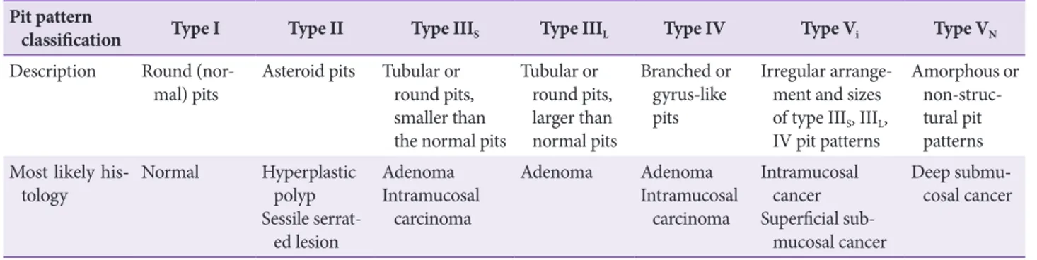
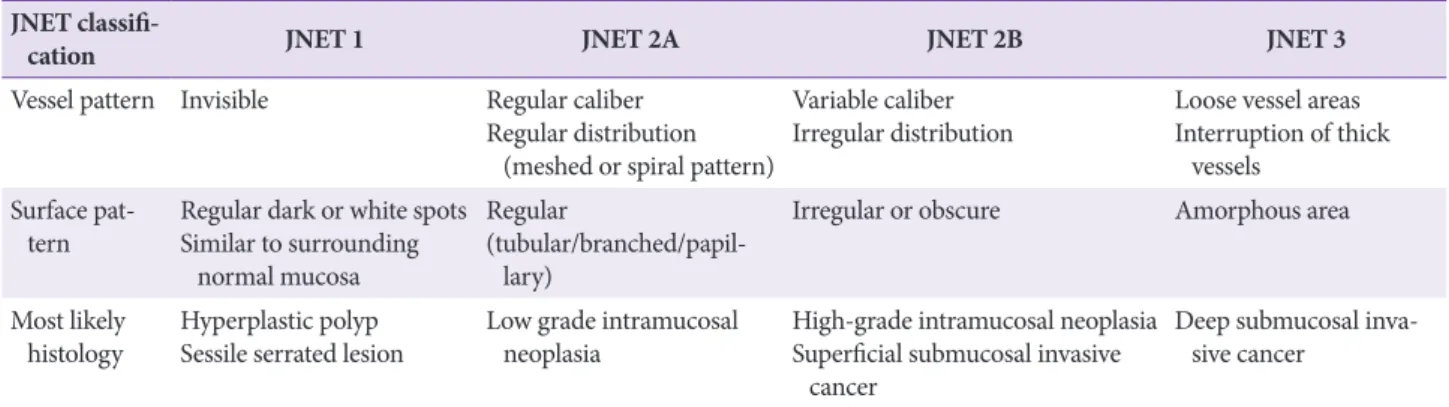
Statement C3: We recommend endoscopic assessment of pit patterns and vascular patterns to estimate the depth of submucosal invasion before endoscopic resection of early colorectal cancer (Grade of recommendation: strong, Level of evidence: high).
Statement C4: En bloc and histologically complete resection should be achieved for endoscopic treatment of a suspected or established early colorectal cancer. We recommend endoscopic submucosal dissection for the treatment of endoscopically resectable early colorectal cancer which cannot be resected en bloc using endoscopic mucosal resection technique (Grade of recommendation: strong, Level of evi-dence: moderate).
by the editorial committee to ensure information balance and guideline completion. For an external review of the guide-line, a public meeting in which 38 gastrointestinal endoscopy experts participated was held on August 18, 2019 during the 61st seminar of KSGE in which doctors and nurses from all over the country gathered. Opinions about the guideline were shared during the public meeting and the final draft of guide-line was made after its revision based on discussion during the public meeting.
Provision of clinical practice guideline and plans for
future updates
For wide provision and distribution of the clinical practice guideline, we plan to publish the guideline in Clinical Endos-copy, The Korean Journal of Gastroenterology, The Korean Journal of Helicobacter and Upper Gastrointestinal Research, Intestinal Research, and Journal of Digestive Cancer Reports. We will also upload the guideline on the website of KSGE and submit it to the Korean Medical Guideline Information Cen-ter. Because we expect slow distribution of guidelines among endoscopists through databases for clinical practice guidelines, KSGE, the main institution for developing the guideline, will send out the guideline for free via various routes including emails and will actively advertise the guideline in academic conferences, seminars, and workshops. Current recommen-dations in the clinical practice guideline are based on research conducted up to date and will be subject to revisions based on future findings.
Limitations
The most critical limitation of this clinical practice guide-line is the insufficiency of data pertaining to Koreans. Data from foreign countries cannot be directly used to develop a guideline for the Korean population since the epidemiological and clinical characteristics of early gastrointestinal cancers differ between Korean and foreign populations. In addition, this clinical practice guideline does not aim to provide an ab-solute treatment standard that physicians should use to man-age patients in real clinical settings but aims to help physicians make evidence-based clinical judgments with regard to the treatment of early gastrointestinal cancers. A physician must examine various clinical aspects of a patient before making any treatment decisions. This clinical practice guideline shall not be used to restrict medical practice of physicians, as health insurance criteria, or to make legal judgments regarding treat-ments performed on a particular patient.
Editorial independence
This clinical practice guideline is a project selected and funded by KSGE. KSGE did not influence the process of
guideline development in any manner. All parties involved in the guideline development had no conflict of interests regard-ing the guideline development.
SuPERFICIAL ESOPhAGEAL SquAMOuS
CELL CARCInOMA
Statement E1: We recommend endoscopic resection
for SESCC without distant or lymph node
metas-tasis, excluding those with obvious submucosal
in-vasion (Grade of recommendation: strong, Level of
evidence: moderate).
SESCC is squamous cell carcinoma of the esophagus that is localized to the mucosa or submucosa. SESCC without distant metastasis and with a low risk of lymph node metastasis is a good target of endoscopic resection. Although endoscopic resection of SESCC can conserve the esophagus, it is import-ant to carefully select patients who will receive the procedure since there is still a possibility of lymph node metastasis. The National Comprehensive Cancer Network guidelines rec-ommend endoscopic resection for SESCC localized to the mucosa and esophagectomy in the presence of submucosal invasion.23 The Japan Esophageal Society defines mucosal cancer invading only as deep as the lamina propria as an ab-solute indication for endoscopic resection as the cancer rarely metastasizes to the lymph nodes.17 The European Society of Gastrointestinal Endoscopy (ESGE) defines esophageal cancer invading the lamina propria without lymph node metastasis as an absolute indication for endoscopic resection.1 Thus, the depth of invasion of SESCC must be accurately determined using endoscopy, endoscopic ultrasound, and magnifying endoscopy with narrow band imaging (NBI) before perform-ing endoscopic resection of SESCC.24-28 However, the evalu-ation of invasion depth is not perfectly accurate. Therefore, it is advisable to perform endoscopic resection instead of esophagectomy to avoid unnecessary operations when there is no obvious submucosal invasion because esophagectomy has high morbidities and mortalities.29-31 Recent Korean studies report no significant difference in long-term survival between patients with SESCC without obvious submucosal invasion who underwent endoscopic resection as their first treatment and those who underwent esophagectomy, and the rate of postoperative complications was significantly higher in the operated patients.32 This may be because additional operations lowered the risk of recurrence following non-curative endo-scopic resection. A Chinese study also reported no significant difference in survival rates between patients with SESCC who underwent endoscopic resection and those who under-went surgical resection and reported a lower incidence of
treatment-related complications in the former group.33 These results show that performing endoscopic resection prior to a surgical resection of SESCC without obvious submucosal in-vasion may be an effective treatment strategy.
Statement E2: We recommend Lugol
chromoendos-copy and/or image-enhanced endoschromoendos-copy to define
the extent of lesion before endoscopic treatment of
SESCC (Grade of recommendation: strong, Level of
evidence: moderate).
SESCC manifests as erythematous lesions, subtle discol-oration, or nodules. In addition, synchronous SESCC lesions of various sizes are not rare, which are difficult to be dis-tinguished from noncancerous lesions only by white light endoscopy. Therefore, accurate measurement of the size and horizontal border of these lesions is challenging. Lugol chro-moendoscopy, which applies the fact that the keratin layer of the mucosa is destroyed by the cancer, is the most effective chromoendoscopy to identify the SESCC lesions. The normal esophageal mucosa turns dark brown when sprayed with Lugol’s solution, whereas the mucosa affected by SESCC ex-hibits a “pink-color sign”. That is, the mucosa remains light brown and turns pink 2–3 minutes after being sprayed with the solution.34 Studies evaluating the efficacy of Lugol chro-moendoscopy in diagnosing lesions suggestive of SESCC in white light endoscopy reported Lugol chromoendoscopy to be 73.8%–93.4% accurate in differentiating high-grade adeno-ma and SESCC from low-grade adenoadeno-ma and noncancerous lesions.34,35 Thus, Lugol chromoendoscopy effectively assesses the horizontal border of SESCC before endoscopic resection.
Image-enhanced endoscopy is a quick process that causes no inflammation around SESCC lesions unlike Lugol chromo-endoscopy. Image-enhanced endoscopy with NBI is the most widely studied image-enhanced endoscopy technique.36 In a study of 90 patients with high-grade adenoma and SESCC, the accuracy of image-enhanced endoscopy with NBI was significantly higher than that of white light endoscopy (92% vs. 67.8%), and was similar to that of Lugol chromoendoscopy (92% vs. 93.4%).37 In a prospective study that compared the diagnostic accuracy of detecting SESCC between Lugol chro-moendoscopy and image-enhanced endoscopy with NBI in 303 patients with high risk of SESCC, the accuracy of the im-age-enhanced endoscopy with NBI was 91.2%, which was not inferior to that of Lugol chromoendoscopy.38 Based on these results, we recommend Lugol chromoendoscopy or image-en-hanced endoscopy to determine the border of SESCC prior to endoscopic resection.
Statement E3: We recommend endoscopic
ultra-sound to define the stage of SESCC before
en-doscopic treatment (Grade of recommendation:
strong, Level of evidence: moderate).
Endoscopic treatments of SESCC have better prognoses when the depth of invasion is shallow, and unlike gastric cancer, SESCC invading the muscularis mucosa poses a risk of lymph node metastasis.39 Accurate staging of SESCC be-fore an endoscopic treatment is thus important. For SESCC, endoscopic ultrasound accurately determines the level of in-filtration by the primary tumor (T stage) and the presence or absence of lymph node metastasis (N stage). A study reported endoscopic ultrasound to be 81.6% sensitive and 99.4% specif-ic in staging esophageal cancer invading the mucosa and sub-mucosa.40 A meta-analysis of 19 studies reported endoscopic ultrasound as an excellent technique to differentiate mucosal invasion from submucosal invasion in SESCC (area under the summary ROC curve = 0.93).26 Additionally, in another meta-analysis that investigated whether endoscopic ultra-sounds can differentiate between esophageal cancer invasions in the lamina propria, muscularis mucosa, and submucosa, endoscopic ultrasounds showed an excellent diagnostic per-formance (area under the summary ROC curve = 0.98).11
Statement E4: We suggest magnifying endoscopy
with narrow band imaging for SESCC to assess
the depth of invasion before endoscopic treatment
(Grade of recommendation: weak, Level of
evi-dence: low).
A large-scale, multi-institutional prospective study that investigated whether magnifying endoscopy with NBI can accurately assess the depth of invasion of esophageal cancer reported magnifying endoscopy with NBI to be not superior to conventional endoscopy for squamous cell carcinoma (ac-curacy of magnifying endoscopy with NBI: 65.3%, ac(ac-curacy of conventional endoscopy: 71.4%, p=0.375).10 A prospective study conducted in Japan also reported that magnifying endoscopy with NBI is no better than high-resolution endos-copy and high-frequency endoscopic ultrasound.41 However, a recent large-scale retrospective study that investigated the performance of magnifying endoscopy with NBI in the as-sessment of the depth of SESCC reported a positive predictive value of 93% for epithelial/lamina proprial invasion, 65% for muscularis mucosal/superficial submucosal invasion, and 77% for deep submucosal invasion, demonstrating that magnifying endoscopy with NBI is useful for determining the depth of in-vasion of SESCC before an endoscopic treatment.42 Therefore, given that the operator is highly experienced, it may be useful to perform magnifying endoscopy with NBI to determine the depth of invasion of SESCC before endoscopic resection.
Statement E5: We recommend endoscopic
submu-cosal dissection rather than endoscopic musubmu-cosal
re-section for en bloc and curative rere-section of SESCC
confined to the mucosa (Grade of recommendation:
strong, Level of evidence: moderate).
Endoscopic mucosal resection (EMR) is popular as it is relatively easy to perform and is associated with low risks of complications. However, studies have reported high local recurrence rates of 2.8%–9.8% after EMR because en bloc resection is difficult by EMR, especially for large lesions.43-47 ESD is a technically demanding procedure with high risks of complications but is nonetheless considered appropriate for treating SESCC due to the high en bloc and curative resection rates and low risks of local recurrence. Resection techniques are usually chosen based on lesion size and auxiliary factors such as the patient’s conditions and the operator’s level of ex-perience.
There is no randomized study comparing EMR and ESD for SESCC. In a meta-analysis of retrospective studies, ESD had higher en bloc and curative resection rates than EMR re-gardless of the lesion size. ESD had a significantly lower rate of postoperative local recurrence than EMR (0.3% vs. 11.5%; odds ratio [OR], 0.08; 95% confidence interval [CI], 0.03–0.23; p<0.001). The rate of postoperative perforation was higher for ESD than for EMR (4.0% vs. 1.3%; OR, 2.19; 95% CI, 1.08–4.47; p=0.03). However, no significant difference was noted in the rate of stricture formation or bleeding between the two pro-cedures.48 Although studies report no difference in the rate of en bloc and curative resection rates between cap-assisted EMR and ESD for lesions measuring <10 to 15 mm,43,46,47 one meta-analysis reported a higher en bloc resection rate for ESD for lesions measuring <10 mm (OR, 3.58; 95% CI, 1.84–7.02; p<0.001).49
We recommend to consider ESD first regardless of the le-sion size since ESD has higher en bloc and curative resection rates than EMR for SESCC confined to the mucosa and has complication risks within an acceptable range. In case the op-erator lacks experience with ESD, performing EMR could be considered for only small lesions <10 mm in size.
Statement E6: We recommend oral steroid or local
steroid injection therapy for patients who develop
mucosal defects in >75% of the esophageal
circum-ference after endoscopic submucosal dissection to
prevent esophageal stricture (Grade of
recommen-dation: strong, Level of evidence: moderate).
ESD can lead to scar formation around the surgical site followed by esophageal strictures. Ono et al. reported that the risk of esophageal stricture increased in the presence of mu-cosal defects in >75% of the esophageal circumference.50
Fur-thermore, 66%–88% of patients with mucosal defects in >75% of the esophageal circumference that were left untreated after esophageal ESD developed an esophageal stricture.51-56 Patients who developed mucosal defects in 100% of the esophageal circumference required an average of 33.5 endoscopic balloon dilation (EBD) procedures to treat esophageal stricture.57
To prevent stricture after esophageal ESD, oral steroid ad-ministration or local steroid injection therapy is used. For oral steroid therapy, prednisolone is administered at 30 mg per day starting 1 or 2 days after the procedure, and the dose is decremented over 2–12 weeks.54,55,57 In six studies comparing patients who received oral steroids following ESD to those who did not, oral steroid administration significantly reduced the rate of esophageal stricture formation by 73% (69%–80% without oral steroids and 18%–23% with oral steroids) (OR, 0.27; 95% CI, 0.13–0.58).54,55 Local steroid injection therapy also effectively prevents esophageal stricture. Studies reported a 78% reduction in the rate of esophageal stricture formation following local injections of triamcinolone or dexamethasone at the site of ESD (OR, 0.32; 95% CI, 0.13–0.83).51-53,58
In a study that compared oral steroid therapy and preven-tive EBD, 32% of patients who underwent EBD twice per week for 8 weeks had an esophageal stricture, whereas only 5% of patients who were orally administered prednisolone at 30 mg per day starting 2 days after ESD and had the dose gradually decreased over 8 weeks had an esophageal stricture, demonstrating that oral steroid administration is superior to preventive EBD for the prevention of esophageal stricture.56
Studies on steroid administration for the prevention of esophageal stricture mostly involve patients who develop mu-cosal defects in >75% of the esophageal circumference and are at high risk of esophageal stricture. In a study that investigated the effect of local steroid injections in patients who under-went esophageal ESD regardless of the size of mucosal defect, local steroid injections had a 70% preventive effect on esoph-ageal strictures with the rate of stricture formation being 11% in patients who received local steroid injections and 36% in those who did not receive them (OR, 0.30; 95% CI, 0.13–0.83).58 However, considering the risk of esophageal perforation and adverse reactions associated with local steroid injections, it is advisable to consider oral steroid or local steroid injection therapy only for patients who develop mucosal defects in >75% of the esophageal circumference who are at high risk of esophageal stricture.
Statement E7: no additional treatment is
recom-mended after en bloc complete resection of SESCC
invading no more than the lamina propria with no
lymphovascular invasion because of a very low risk
of lymph node metastasis (Grade of
recommenda-tion: strong, Level of evidence: moderate). As the
risk of lymph node metastasis of a tumor invading
into the muscularis mucosa without lymphovascular
invasion is low, a close follow-up after en bloc complete
endoscopic resection can be considered without
additional treatment (Grade of recommendation:
weak, Level of evidence: low). In case of a tumor
with submucosal invasion, lymphovascular
inva-sion, and/or positive vertical resection margin,
ad-ditional treatment is recommended (Grade of
rec-ommendation: strong, Level of evidence: moderate).
Since the risk of lymph node metastasis associated with SESCC is closely related to a tumor’s depth of invasion, it is important to accurately evaluate the depth of invasion for deciding whether the endoscopic resection is curative or non-curative.59-61 Histopathological analyses of patients who underwent esophagectomy with dissection of the regional lymph nodes show that the risk of lymph node metastasis is 26.0%–53.8% in the presence of submucosal invasion of esophageal cancer.59-68 Moreover, 8.3%–53.1% of the patients had lymph node metastasis even when esophageal cancer invaded only the upper third of the submucosa. The Japan Esophageal Society and the ESGE guidelines define SESCC with a shallow submucosal invasion of ≤200 μm as a relative indication for endoscopic resection. However, data regard-ing the frequency of lymph node metastasis by SESCC with shallow submucosal invasion of ≤200 μm is limited.67 The rate of lymph node metastasis is lower but not negligible at 0.0%–15.4% for esophageal cancer confined to the mucosa.59-67 Among esophageal cancers confined to the mucosa, invasion of muscularis mucosa is at a higher risk of lymph node metas-tasis than invasion of lamina propria (8.0%–27.0% vs. 0.0%– 8.7%). The risk of lymph node metastasis is associated with vascular or lymphatic invasion.59,65,67,69,70 A large-scale Japanese study reported a five-year cumulative incidence of metastasis of primary esophageal cancer invading the muscularis mu-cosa following endoscopic resection of only 0.7%, suggesting that SESCC invading the muscularis mucosa can still be an indication for endoscopic resection if no lymphovascular invasion is observed.68 Two observational studies at Korean institutions supports the claim that endoscopic resection can be a safe and curative treatment option in SESCC invading to the mucosa. In those studies, no death due to esophageal cancer occurred during a long-term follow-up of patients who underwent endoscopic resection of SESCC with invasion up to the mucosa.71,72 There are conflicting research results regarding the association between undifferentiated esophageal cancer and the risk of lymph node metastasis, indicating that undifferentiated esophageal cancer cannot be yet used as an absolute contraindication of endoscopic resection for SESCC.
Additional data analyses are needed to derive more confirma-tive conclusions.59,61,65,68
Endoscopic resection is considered curative if histopatho-logical evaluation shows that SESCC does not invade be-yond the lamina propria and does not invade the vascular or lymphatic channels. In this case, a close follow-up may be conducted without additional operations. Following en bloc resection of SESCC with muscularis mucosal invasion and no lymphovascular invasion, a follow-up can be considered without additional operations after considering the patient’s age, accompanying diseases, conditions, and risk of operation since the risk of lymph node metastasis is low. In case of un-successful en bloc resection, follow-up strategies considering the possibility that the histopathological assessment may have been inaccurate are necessary.
Additional treatments are needed in case of noncurative resection such as positive vertical resection margin and sub-mucosal invasion and/or lymphovascular invasion suggesting the possibility of lymph node metastases. Esophagectomy with dissection of the regional lymph nodes is the standard treatment for noncurative endoscopic resection. However, since esophagectomy has high postoperative morbidities of 30%–40% and mortalities of 1%–2%, physicians must consider the patient’s conditions before deciding whether to perform the surgery or not.29-31 Studies have reported relatively satisfac-tory outcomes of chemoradiation therapy for noncurative en-doscopic resection of SESCC, suggesting that chemoradiation therapy might become a new treatment strategy for noncura-tive endoscopic resection.73-78
EARLy GASTRIC CAnCER
Statement G1: We recommend chromoendoscopy/
image-enhanced endoscopy to determine the
ex-tent of lesion before endoscopic treatment of early
gastric cancer (Grade of recommendation: strong,
Level of evidence: moderate).
Clearly identifying the horizontal border of the lesion before endoscopic resection reduces local recurrence and increases the likelihood of a complete resection. Chromoen-doscopy has been used widely to accurately measure lesion borders. Recent advances in endoscopy technology led to the advent of NBI and magnifying endoscopy now commonly used in clinical settings.12,79 A study reported that chromo-endoscopy using indigo carmine more accurately estimated lesion borders in early gastric cancer than white light endos-copy (75.9% vs. 50.0%), and chromoendosendos-copy using indigo carmine and acetic acid estimated lesion borders with 90.7% accuracy.80 A Korean study also reported that
chromoendos-copy using indigo carmine and acetic acid estimated lesion borders in early gastric cancer more accurately than white light endoscopy (84.1% vs. 66.9%).81 In a study that compared the accuracy of border prediction between magnifying endos-copy with NBI and chromoendosendos-copy using indigo carmine, the former technique estimated horizontal borders of lesions more accurately than the latter (81.1% vs. 72.6%).12 Magnifying endoscopy with NBI was also superior to white light endos-copy in terms of sensitivity and specificity for assessment of lesion borders (sensitivity, 92.9% vs. 42.9%; specificity, 94.7% vs. 61.0%).13 Based on these results, we recommend chromo-endoscopy and image-enhanced chromo-endoscopy to determine the extent of resection before endoscopic resection of early gastric cancer.
Statement G2: Endoscopic ultrasonography before
endoscopic resection of early gastric cancer may
be helpful in determining the depth of invasion in
some patients with early gastric cancer (Grade of
recommendation: weak, Level of evidence:
moder-ate).
Endoscopic ultrasonography is useful for assessing the depth of invasion in gastric cancer and determining the pres-ence or abspres-ence of lymph node metastasis. In a meta-analysis on 54 studies evaluating the efficacy of endoscopic ultraso-nography in predicting the depth of invasion of a primary tu-mor in 5,601 patients with gastric cancer, endoscopic ultraso-nography accurately differentiated between T3-T4 lesions and T1-T2 lesions with 86% sensitivity and 91% specificity.14 How-ever endoscopic ultrasonography before endoscopic resection of early gastric cancer has limited accuracy in predicting the depth of invasion.82-88 In a large-scale Korean prospective study, the accuracy of distinguishing mucosal and submucosal invasion in early gastric cancer was 67.4% with endoscopic ultrasonography, which was not superior to 73.7% of con-ventional endoscopy.84 Another Korean study also reported no significant difference in the accuracy of depth of invasion prediction in early gastric cancer between endoscopic ultraso-nography using a miniprobe and white light endoscopy (81.4% vs. 78.9%).83 However, some studies report that endoscopic ultrasonography may be useful for investigating the depth of invasion of early gastric cancer. According to a Japanese study, the diagnostic accuracy of endoscopic ultrasound was higher than that of white light endoscopy in predicting the depth of invasion of early gastric cancer (71% vs. 63%).85 A recent study reported that endoscopic ultrasonography had higher accura-cy than chromoendoscopy in predicting the depth of invasion of early gastric cancer (79.1% vs. 76.5%) and that the depth of invasion could be predicted with 88.3% accuracy using both chromoendoscopy and endoscopic ultrasonography.82
Based on these studies, there is still a role for endoscopic ul-trasonography to help differentiating mucosal or submucosal infiltration of early gastric cancer. Therefore, for patients who show signs of submucosal invasion in a white light endoscopic examination, endoscopic ultrasonography could be used to more accurately examine the depth of invasion of a tumor before endoscopic resection.89
Statement G3: We recommend endoscopic resection
for early gastric cancer of well or moderately
differ-entiated tubular or papillary adenocarcinoma
meet-ing endoscopically estimated tumor size ≤2 cm and
endoscopically suspected mucosal cancer without
ulcer (Grade of recommendation: strong, Level of
evidence: moderate).
Endoscopic resection is a local treatment for early gastric cancer with a negligible risk of lymph node metastasis. Before Gotoda et al.90 proposed expanded indications for endoscopic resection in 2000, well or moderately differentiated tubular or papillary gastric adenocarcinoma measuring ≤2 cm in diam-eter confined to the mucosa without ulcer and submucosal invasion was an indication for endoscopic resection. Thus, for these lesions, endoscopic resection must be considered as the first line of treatment. The risk of lymph node metastasis must be considered before performing endoscopic resection of early gastric cancer. Several studies have reported the risk of lymph node metastasis to be negligibly low in lesions that satisfy the aforementioned indications (0.0%–0.3%).90-92 In ad-dition, Korean studies reported no significant difference in the five-year survival rate between endoscopic resection and sur-gical resection (93.6%–96.4% vs. 94.2%–97.2%),93-95 They also reported no significant difference in the 10-year survival rate between endoscopic resection and surgical resection (81.9% vs. 84.9%).93 However, endoscopic resection had a higher five-year cumulative incidence of metachronous gastric cancer than surgical resection (5.8%–10.9% vs. 0.9%–1.1%).93-95 En-doscopic resection preserves the entire stomach, which can lead to metachronous tumor formation in the rest parts of the stomach. Therefore, even after curative endoscopic resection, regular follow-up endoscopy is necessary to look for meta-chronous gastric cancer. Endoscopic resection, which pre-serves the entire stomach, improves the quality of life, causes lesser complications, requires a shorter hospital stay, and is more cost-effective than surgical resection.93-98 In conclusion, we recommend endoscopic resection as the first line of treat-ment for well or moderately differentiated tubular or papillary gastric adenocarcinoma measuring ≤2 cm in diameter with-out endoscopic signs of ulcer and submucosal invasion since endoscopic resection is comparable to surgical resection in terms of survival, is associated with a satisfactory quality of
life, and is economical.93-96
Statement G4: We suggest endoscopic resection for
early gastric cancer of well or moderately
differenti-ated tubular or papillary adenocarcinoma with the
following endoscopic findings: (1) mucosal cancer
>2 cm without ulcer, or (2) mucosal cancer ≤3 cm
with ulcer (Grade of recommendation: weak, Level
of evidence: moderate).
The World Health Organization (WHO) histological classi-fication of gastric cancer published in 2000 is widely accepted as the standard classification system for gastric cancer, which defines undifferentiated carcinoma as carcinomas without glandular differentiation and squamous epithelial differen-tiation. However, most studies on endoscopic resection have classified well or moderately differentiated tubular or papil-lary adenocarcinoma as differentiated type adenocarcinoma and poorly differentiated tubular adenocarcinoma and poorly cohesive carcinoma as undifferentiated type adenocarcinoma. Expansion of existing indications for endoscopic resection should be considered only if there is no difference in the sur-vival rates between endoscopic resection and standard surgi-cal treatment. In addition, surgery-related mortalities must be compared and reviewed to determine the permissible range of risk of lymph node metastasis. The risk of lymph node metastasis is generally estimated based on the size of tumor, histologic type and grade, depth of invasion, and presence or absence of lymphovascular invasion. The risk of lymph node metastasis and distant metastasis has been reported to be 0.0%–0.21% for well or moderately differentiated tubular or papillary gastric mucosal adenocarcinoma measuring >2 cm without ulcers or mucosal cancer measuring ≤3 cm with ul-cers if the endoscopically resected tumor shows a negative re-section margin and no lymphovascular invasion. Considering that the risk of lymph node or distant metastasis (0.0%–0.21%) is similar to the mortality of gastrectomy (0.1%–0.3%), endo-scopic resection may be considered for the lesions described above.99-101 Additionally, studies reported no significant differ-ence between the five-year survival rate of surgical resection and endoscopic resection (92.0%–97.2% vs. 93.3%–96.4%), indicating that endoscopic resection is considered curative for the aforementioned lesions with no requirement of further treatment.95,100,102-109
Surgical resection is the standard treatment for clinically suspected submucosal invasive gastric cancer in preoperative evaluation. However, studies analyzing pathologic results of patients who underwent gastrectomy due to preoperatively suspected submucosal invasion reported that 28.8%–43.0% of these lesions could have been treated with endoscopic re-section.110,111 Therefore, further research is needed regarding
treatment methods for preoperatively suspected submucosal invasive early gastric cancer.
Statement G5: We suggest endoscopic resection
for poorly differentiated tubular
adenocarcino-ma, poorly cohesive carcinoadenocarcino-ma, or signet ring cell
carcinoma meeting the following endoscopic
find-ings: endoscopically estimated tumor size ≤2 cm,
endoscopically mucosal cancer, and no ulcer in the
tumor (Grade of recommendation: weak, Level of
evidence: low).
Undifferentiated type adenocarcinoma including poorly differentiated tubular adenocarcinoma, poorly cohesive car-cinoma and signet ring cell carcar-cinoma of the stomach has not been accepted as an indication for endoscopic resection due to reports that undifferentiated type adenocarcinoma is associ-ated with a high risk of lymph node metastasis.112,113 However, retrospective studies reported a low incidence of extragastric metastasis during follow-up and high five-year survival rates of 95.0%–98.6% among patients who underwent endoscopic resection for undifferentiated type adenocarcinoma with mucosal invasion, no ulcer and ≤2 cm in size.100,114-118 Studies also reported no significant difference in long-term outcomes between endoscopic resection and surgical resection for the aforementioned lesions.95,100,103,118 Thus, endoscopic resection may be recommended for undifferentiated type gastric adeno-carcinoma with mucosal invasion, no ulcer and ≤2 cm in size. However, since undifferentiated type adenocarcinoma tends to have unclear borders that contribute to low curative resec-tion rates (45.1%–70%),115,116 it is important to clearly identify borders and secure a sufficient resection margin during endo-scopic resection. In addition, even in undifferentiated type ad-enocarcinoma, poorly differentiated tubular adad-enocarcinoma, poorly cohesive carcinoma and signet ring cell carcinoma can have different biological behaviors, and further studies in this field are needed.
Statement G6: We recommend prophylactic
hemo-stasis of visible vessels on the post-resection ulcer
caused by endoscopic resection of early gastric
can-cer to lower the risk of delayed bleeding (Grade of
recommendation: strong, Level of evidence: low).
Statement G7: We recommend proton pump
inhibi-tors to decrease the risk of symptoms and
complica-tions associated with iatrogenic ulcers caused by
en-doscopic resection of early gastric cancer (Grade of
recommendation: strong, Level of evidence: high).
Reports on the incidence of bleeding associated with endo-scopic resection vary depending on the definition of bleeding.
Serious immediate bleeding that requires an intraprocedural blood transfusion or surgical treatment has been reported in <1% of patients.119 Delayed bleeding that occurs after endo-scopic resection is defined as bleeding from an iatrogenic ulcer that requires hemostasis, and its incidence has been reported to be 1.3%–11.9%. Delayed bleeding usually occurs within 24 hours after endoscopic resection but can occur up to 2 weeks after endoscopic resection.120,121 Prophylactic coagulation of visible vessels exposed on the base of an iatrogenic ulcer after endoscopic resection can effectively prevent delayed bleeding. One retrospective study reported a 2.47-fold increase in the risk of delayed bleeding when prophylactic coagulation was not performed.122 However, excessive prophylactic coagulation can increase the risk of post-coagulation syndrome or delayed perforation.123,124
Postoperative administration of proton pump inhibitors or histamine 2 (H2) receptor blockers can prevent delayed bleeding. Whether one drug is more effective than the other is unclear with some studies reporting proton pump inhibitors to be more effective in treating iatrogenic ulcers related to endoscopic resection,125,126 and others reporting the two drugs to be on a par with one another.127,128 One meta-analysis re-ported no difference between proton pump inhibitors and H2 receptor blockers in their ability to treat iatrogenic ulcers and relieve symptoms but reported a significantly lower incidence of delayed bleeding in patients who were administered proton pump inhibitors (OR, 0.49; 95% CI, 0.25–0.95).129 Various re-ports regarding the dose and administration period of proton pump inhibitors for iatrogenic ulcers and concomitant use of mucosal protective agents are available.130-142 Randomized trials reported that administration of proton pump inhibitors before endoscopic resection is not effective in preventing de-layed bleeding and that second look endoscopy also did not effectively prevent delayed bleeding.143-145
Statement G8: We recommend endoscopic closure
as the first treatment option for perforation that
oc-curred during endoscopic resection of early gastric
cancer (Grade of recommendation: strong, Level of
evidence: low).
The incidence of perforation resulting from excessive dam-age to the muscularis propria during endoscopic resection is 1.2%–5.2% and that of delayed perforation that occurs after endoscopic resection is reported at below 0.5%. The risk of in-traoperative perforation varies depending on the location and size of the lesion.146 Closure by endoscopic clipping effectively treats intraoperative perforation, and conservative treatments such as fasting and antibiotic administration after successful endoscopic closure usually lead to symptom relief without the need for additional surgical treatments.147-149 However, surgical
treatment must be considered in case of unsuccessful closure, signs of generalized peritonitis, or delayed perforation.150 The endoscopic closure must be performed by an experienced en-doscopist. If a patient becomes hemodynamically unstable or has respiratory problems due to tension pneumoperitoneum, rapid decompression of the intra-abdominal pressure using a percutaneous aspiration and/or drainage of intra-abdominal free air is required.151 It may also be useful to switch from oxygen to carbon dioxide infusion during endoscopy when perforation occurs.152
Statement G9: We recommend surgical gastrectomy
if histopathological evaluation after endoscopic
re-section of early gastric cancer meets the criteria for
non-curative resection. An exception applies if
can-cer invasion is observed at the horizontal resection
margin only (Grade of recommendation: strong,
Level of evidence: moderate).
Statement G10: We recommend additional
endo-scopic management rather than surgical
gastrecto-my if histopathological evaluation of endoscopically
resected early gastric cancer specimen shows
posi-tive involvement at the horizontal resection margin
without any other findings compatible with
non-cu-rative resection (Grade of recommendation: strong,
Level of evidence: moderate).
Curative resection of an absolute indication lesion is as-sumed when well or moderately differentiated tubular or papillary adenocarcinoma confined to the mucosa measuring <2 cm with no histopathological evidence of lymphovascular invasion and ulcers and a negative resection margin is ob-served following endoscopic en bloc resection of a lesion. Cu-rative resection of an expanded indication lesion is considered when any of the following conditions are observed after en bloc resection: (1) differentiated type mucosal adenocarcinoma such as well or moderately differentiated tubular or papillary adenocarcinoma with a negative resection margin and ulcers of ≥2 cm and without lymphovascular invasion; (2) mucosal adenocarcinoma of ≤3 cm accompanied by ulcers; (3) sub-mucosal invasive cancer of ≤3 cm with subsub-mucosal invasion depth ≤500 µm; or (4) undifferentiated type adenocarcinoma such as poorly differentiated tubular adenocarcinoma, poorly cohesive carcinoma, and signet ring cell carcinoma measur-ing ≤2 cm confined to the mucosa. Lesions that do not meet these criteria for curative resection are considered to have undergone non-curative resection.1,120,153 The risk of lymph node metastasis is reported to be 2.6%–3.0% for differentiated type adenocarcinoma that satisfy the criteria for non-curative resection, with the exception of differentiated type
adeno-carcinoma with a positive horizontal resection margin, and 5%–20% for undifferentiated type adenocarcinoma.90,154,155 In a large-scale cohort study on patients who received additional surgical treatments due to non-curative resection, lymphatic invasion was associated with the highest risk of lymph node metastasis. Large tumor size, positive vertical resection mar-gin, vascular invasion, and submucosal invasion depth >500 µm are reported to increase the risk of lymph node metastasis to a similar extent.156 Many studies have demonstrated that lymphovascular invasion is an important risk factor of lymph node metastasis.157-159 Considering the risk of lymph node metastasis, patients may require additional surgical gastrecto-my including regional lymphadenectogastrecto-my when they satisfy the criteria for non-curative resection, with the exception of having a positive horizontal resection margin only. Differen-tiated type mucosal adenocarcinomas less than 2 cm in size that show lymphovascular invasion are reported to have low risk of lymph node metastasis, in which case the need for additional surgical procedures is not clear.160 Although some studies comparing surgical resection following non-curative resection vs. no surgery reported no additional benefits of ad-ditional surgery,161-163 most retrospective studies have reported an increase in overall survival and disease-specific survival in patients who underwent surgical resection158,159,164-167 Further-more, additional surgical resection following non-curative en-doscopic resection has also been reported to increase survival among patients of advanced age.168-170
Differentiated type adenocarcinoma with a positive hor-izontal resection margin that meet all the other criteria for curative resection following en bloc resection is associated with a low risk of lymph node metastasis. A cohort study that followed up patients who had this type of cancer and did not receive additional treatments reported low five-year recur-rence rates among these patients and that recurrent tumors could be curatively treated without any mortality associated with gastric cancer.171 Therefore, additional endoscopic re-section or argon plasma coagulation treatment may be used instead of surgical resection for tumors with a positive hori-zontal resection margin. Retrospective cohort studies reported favorable prognoses following these endoscopic treatments and suggested that additional endoscopic treatments within 3 months after the initial endoscopic resection are associated with low recurrence rates.172-174 However, it is impossible to de-termine whether resection is curative following argon plasma coagulation since histological evaluation is impossible; thus, a close follow-up is required after argon plasma coagulation.
Statement G11: We recommend Helicobacter pylori
eradication treatment after endoscopic resection of
early gastric cancer in H. pylori-infected patients
(Grade of recommendation: strong, Level of
evi-dence: high).
H. pylori eradication may be considered for patients who test positive for H. pylori following an endoscopic treatment of early gastric cancer to reduce the risk of metachronous re-currence. Fukase et al. reported that H. pylori eradication sig-nificantly reduced the risk of metachronous gastric cancer in patients with early gastric cancer after endoscopic resection in their multicenter randomized controlled study in 2008 (hazard ratio [HR], 0.339; 95% CI, 0.157–0.729).175 Based on this study, most clinical practice guidelines recommend H. pylori erad-ication after endoscopic resection of early gastric cancer.176-180 Four meta-analyses investigating whether H. pylori eradica-tion after endoscopic reseceradica-tion of early gastric cancer prevents metachronous gastric cancer reported that H. pylori eradi-cation significantly reduces the risk of metachronous gastric cancer to 0.42–0.51.181-184 A Korean prospective randomized double-blind, placebo-controlled trial published in 2018 also showed that 50% and 68% of metachronous gastric cancer was reduced in patients who received eradication therapy and for those in whom eradication was successful after endoscopic treatment of early gastric cancer, respectively.185 Another Kore-an prospective rKore-andomized study published in 2018 reported a 2.02-fold increase in the risk of metachronous gastric cancer in the control group compared with the eradication group.186 Therefore, we recommend H. pylori eradication following endoscopic resection of early gastric cancer for prevention of metachronous gastric cancer.
Statement G12: We recommend regular surveillance
endoscopy every 6–12 months for patients who have
had curative endoscopic resection of early gastric
cancer based on absolute or expanded criteria for
early detection of metachronous gastric cancer
(Grade of recommendation: strong, Level of
evi-dence: low).
Surveillance strategy for the patients after endoscopic re-section has similar follow-up plans compared with those after surgical gastrectomy. However, more careful follow-up should be performed after endoscopic resection of early gastric cancer as it is associated with high incidence of synchronous multiple gastric cancers and metachronous gastric cancers. The Korean Clinical Practical Guideline for Gastric Cancer recommends patients who undergo endoscopic treatment for early gastric cancer to have a follow-up endoscopic examina-tion on a yearly basis.187 The Japanese guideline for endoscopic resection for early gastric cancer recommend an endoscopy examination at a 6–12 month interval following curative en-doscopic resection to detect metachronous gastric cancer and additionally for those with expanded curative criteria