저작자표시-비영리-변경금지 2.0 대한민국 이용자는 아래의 조건을 따르는 경우에 한하여 자유롭게 l 이 저작물을 복제, 배포, 전송, 전시, 공연 및 방송할 수 있습니다. 다음과 같은 조건을 따라야 합니다: l 귀하는, 이 저작물의 재이용이나 배포의 경우, 이 저작물에 적용된 이용허락조건 을 명확하게 나타내어야 합니다. l 저작권자로부터 별도의 허가를 받으면 이러한 조건들은 적용되지 않습니다. 저작권법에 따른 이용자의 권리는 위의 내용에 의하여 영향을 받지 않습니다. 이것은 이용허락규약(Legal Code)을 이해하기 쉽게 요약한 것입니다. Disclaimer 저작자표시. 귀하는 원저작자를 표시하여야 합니다. 비영리. 귀하는 이 저작물을 영리 목적으로 이용할 수 없습니다. 변경금지. 귀하는 이 저작물을 개작, 변형 또는 가공할 수 없습니다.
Master’s Thesis
The Role of Angiotensin Type 1
Receptor and Src Kinase in Stretch
Induced Cardiac Fibroblast Injury
Model
Dany Chi
Department of Biomedicine and Drug Development
Graduate School
Jeju National University
신전손상으로 유도된 심장 섬유모세포에서의
안지오텐신 1형 수용체와 Src kinase의 역할
지도교수 김 기 석
치 데 니
이 논문을 의학 석사학위 논문으로 제출함
2016 년 2 월
치데니의 의학 석사학위 논문을 인준함
심사위원장 _______________________
위 원 _______________________
위 원 _______________________
제주대학교 대학원
2016 년 2 월
The Role of Angiotensin Type 1 Receptor and
Src Kinase in Stretch Induced Cardiac
Fibroblast Injury Model
Dany Chi
(Supervised by Professor Ki-Seok Kim)
A thesis submitted in partial fulfillment of the requirement for
degree of Master of Science in Biomedicine and Drug Development
2016.02
This thesis has been examined and approved.
...
...
...
Department of Biomedicine and Drug Development
Graduate School
The Role of Angiotensin Type 1 Receptor and
Src Kinase using Stretch-Induced Cardiac
Fibroblast Injury Model
Dany Chi
Department of Biomedicine and Drug Development
Graduate School
Jeju National University
CONTENTS
LIST OF ABBREVIATIONS ... i
LIST OF FIGURES... ii
1. ABSTRACT ... 1
2. INTRODUCTION ... 2
3. MATERIALS AND METHODS ... 4
3.1. Cells culture ... 4
3.2. Incubation of agonist and antagonists ... 4
3.3. Mechanical stretch ... 4
3.4. Western blotting ... 5
3.5. Statistical analysis ... 6
4. RESULTS ... 7
4.1. Effect of Angiotesin II on human cardiac fibroblasts. ... 7
4.2. Effect of Losartan dose-dependent on ERK1/2 phosphorylation ... 12
4.3. Effect of Losartan and AZD0530 on Ang II-induced ERK1/2, Src and Akt phosphorylation... 14
4.4. Effect of mechanical stretch on ERK1/2, Src and Akt phosphorylation 17 4.5. Effect of Losartan dose-dependent on mechanical stretch inducedERK1/2, Src and Akt phosphorylation. ... 19
4.6. Effect of Losartan and AZD0530 on mechanical stretch induced ERK1/2, Src and Akt phosphorylation. ... 21
4.7. Effect of starvation time on ERK1/2, Src and Akt phosphorylation ... 23
5. DISCUSSION... 25
i
LIST OF ABBREVIATIONS
Ang II Angiotensin IIAT1R Angiotensin II type 1 receptor
ARB Angiotensin II type 1 receptor blocker ERK Extracellular-signal-regulated kinase EGFR Epidermal growth factor receptor
Src Proto-oncogene tyrosine-protein kinase Src Akt Protein kinase B (PKB)
p38 p38 mitogen-activated protein kinase MAPK Mitogen-activated protein kinase JNK c-Jun N terminal kinase
PI3K Phosphoinositide 3-kinase Lo Losartan
AZD AZD0530
ECM Extracellular matrix
μM μmol/L
min minute
ii
LIST OF FIGURES
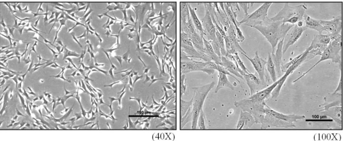
Figure 1. Photography of Human cardiac fibroblast (HCF). ... 8
Figure 2. Effect of Angiotensin II dose-dependent on ERK1/2 activation. ... 9
Figure 3. Time course of ERK1/2 and Src activated by Ang II. ... 10
Figure 4. Effect of Angiotensin II on ERK1/2, Src and AKt activation. ... 11
Figure 5. Effect of Losartan dose-dependent on ERK1/2 activation. ... 13
Figure 6. Effect of Losartan on Ang II induced ERK1/2, Src and Akt activation ... 15
Figure 7. Effect of AZD0530 on Ang II induced ERK1/2, Src and Akt activation ... 16
Figure 8. Effect of mechanical stretch on ERK1/2, Src and Akt activation. ... 18
Figure 9. Effect of Losartan dose-dependent on mechanical stretch induced ERK1/2, Src and Akt activation. ... 20
Figure 10. Effect of Losartan and AZD0530 on mechanical stretch induced ERK1/2, Src and Akt activation. ... 22
1. ABSTRACT
Cardiac hypertrophy is a central risk factor of cardiac morbidity and mortality. Mechanical stretch activated Angiotensin II type I (AT1) receptor without the involvement of Angiotensin II (AngII) during the development of cardiac hypertrophy and causes secretion of Ang II, which plays a central role in mechanical stretch-induced hypertrophy. Here, we attempted to show the role of AT1 receptor and Src kinase in mechanical stretch-induced cardiac fibroblasts injury. Western blot analysis showed the differentiation of stimulation by Ang II and mechanical stretch triggered ERK1/2 phosphorylation was suppressed by Losartan, Src phosphorylation was suppressed by AZD0530 or both inhibitors on Akt phosphorylation. The phosphorylation of ERK and Src were increased by Ang II (100nM) and by mechanical stretch in 1hour. The effect of Ang II and stretch on ERK was blocked by Losartan and Src phosphorylation was suppressed by AZD0530. However, both Losartan and AZD0530 did not effect on Akt phosphorylation induced by Ang II, but they are could inhibit in stretch-induced Akt phosphorylation. We investigated that Losartan and AZD0530 can be took part in effect of mechanical stretch and Ang II induced cardiac fibroblast dysfunction. Moreover, mechanical stretch and Ang II induced all of three kinases phosphorylation that involves AT1 receptor. But they are might be located in different pathways.
Key words: Angiotensin II, Mechanical stretch, AT1 receptor, Losartan,
2. INTRODUCTION
Cardiac fibroblasts are the most abundant cell type in the myocardium which plays a pivotal role in the maintenance of cardiac structure and functions (Qang et
al., 2013; Wang et al., 2003; Fan et al., 2012). They are the essential cell type
responsible for secreting components of the extracellular matrix (ECM) in the heart (Atance et al., 2004; Cheng et al., 2003; Tian et al., 2003; Wang et al., 2013). The activation of cardiac fibroblasts and their differentiation contribute to cardiac fibrosis and hypertrophy (Wang et al., 2003). Cardiac hypertrophy is one of the independent risk factor responsible for myocardial injury and remodeling (Wang
et al., 2014). Mechanical stretch can induce cardiac hypertrophy by activating
angiotensin II (Ang II) type 1 (AT1) receptor through an Ang II-independent pathway (Balakumar and Jagadeesh, 2010; Dostal et al., 2014; Malhotra et al., 1999; S. Wang et al., 2014; Zablocki and Sadoshima, 2013) and by secretion of Ang II, which plays a central role in cardiovascular diseases including stimulates cardiac fibroblast proliferation, collagen synthesis and ECM production(Chen et
al., 2004; Heandeler and Ber, 2000; Malhotra et al., 1999; Wang et al., 2013; Zou
et al., 1998). AT1 receptor is a well-known receptor, which significant
contribution for the development of cardiac hypertrophy (Dostal et al., 2014). On binding to AT1 receptor, Ang II activates an array of protein kinases which is important for its growth-promoting effects such as Src family kinase, epidermal growth factor receptor (EGFR), ERK1/2, p38 MAPK, and Akt/protein kinase B (Ruwhof and Laarse, 2000; Schiffrin and Touyz, 2004; Taniyama et al., 2004; Yamazaki et al., 1998). The ERK1/2 plays a key role in cardiac hypertrophy and failure (Balakuma and Jagadeesh, 2010). However, it is still unclear whether
ERK1/2 is a critical mediator of hypertrophic responses (Bernardo et al., 2010). In cardiac fibroblasts, Ang II activates ERK1/2 through a pathway including the Gβγ subunit of Gi protein and Src family tyrosine kinases (Yamazaki et al., 1998). Blockade of ERK1/2 signaling pathways prevented the progression of cardiac hypertrophy (Balakuma and Jagadeesh, 2010). Although, the pathological and physiological mechanisms were associated with pressure overload-inducted cardiac hypertrophy that focus about intense scientific investigation for over 3 decades, the detail molecular mechanisms of how mechanical stress-induced AT1 receptor activation and its inhibition by angiotensin II receptor blockers (ARBs) still remain elucidation (Dostal et al., 2014).
To elucidate the role of AT1 receptor and Src kinase in stretch induced human cardiac fibroblast injury, we examined the inhibition effect of mechanical stretch and Ang II by using Losartan (AT1 receptor antagonist) including AZD0530 (Src inhibitor). Using mechanical stretch machine, Flexcell Tension System (FX-5000T), as a stress from disease such as high blood pressure. The human cardiac fibroblasts were stretched with/without inhibitors, which block the pathology signaling making hypertrophy.
3. MATERIALS AND METHODS
3.1. Cells culture
Human cardiac fibroblasts were purchased from Cell Applications Inc. Cells were culture in Fibroblast Growth Medium in a humidified incubator with 5% CO2 at 37oC. For all experiments, five passages cells were cultured to 80% - 90% confluence were used for treatment and mechanical stretch experiment.
3.2. Incubation of agonist and antagonists
Cells were seeded in 6-wells plate until the cells were cultured to 70% - 80% confluence. The medium was changed to serum starvation for 4-12 hour or without serum starvation prior to experiment treatment. The serum-free media was changed before the cells were treated with Ang II (#A9525) from sigma-Aldrich. Some cells were pretreated with/without the selective antagonists Losartan (#61188) from sigma-aldrich or/and AZD0530 (#A2133) from Apexbio before stimulated with Ang II or take mechanical stretch, as indicated.
3.3. Mechanical stretch
Bio Flex culture plates-collagen are 6-weels cultures plate consisting of Flexible rubber membrane percolated with collagen I (BF-3001C, Flexcell international corp., US). After plated cells are confluence of 70-80%, the media were changed and transferred to serum starvation for 4-12 hours (or no serum starvation). In some experiment, cells were also stimulated with Ang II 100nM. Both mechanical stretch and Ang II were applied to cells in the absence and presence inhibitors, including Losartan (20μM), AZD0530 (20μM). These
concentrations were used base on previous studies that demonstrated selectivity and sensitivity. For all inhibitor studies, cells were treated with the inhibitors 10-30 minutes before stimulation with Ang II or stretch. Mechanical stretch was applied to cells using the Flexcell FX-5000 tension system (Flexcell international corp., US). For the experiment in this study, cyclic sinusoidal stretch of 20% elongation and 0.4 frequencies was used for 5 minutes to 4 hours for our investigation.
3.4. Western Blotting
After incubated with Ang II or/and applied for mechanical strain, the cells were washed with PBS (Biosesang, KR), scrapped with lysis buffer (RIPA, Biosesang, KR) contained protease inhibitor (protease inhibitor cocktail, Sigma, US) and collected into micro tubes (1.5ml) in ice. The collected cells were kept in ice for 30minutes with vortex very often. Lysate was centrifuged at 15 000 rpm at 4oC for 15 minutes and transferred only the supernatant into new micro tube. Protein was measured using BCA protein assay kit (thermo scientific, Rockford, US). Following stimulation, proteins (5-10μg) of total proteins were extracted from cardiac fibroblasts, separated by 10% polyacrylamide gel electrophoresis and transferred onto a nitrocellulose (NC) or PVDF transfer membrane(Whatman TM, a part of GE Healthcare Life Science Ltd, UK) in transfer buffer. Membranes were incubated with 3-5% non-fat dry milk or 3-5% albumin traction V (BSA) (biobasac canada inc, CA) in Tris-buffered saline with Tween (TBS-T) for 1-2 hours was used to block nonspecific binding sites. The membranes were then incubated for overnight at 4 oC with phosphor-specific or total antibodies diluted
in TBS-T. The primary antibodies were used as follows: Anti- Phospho-44/42 MAPK (ERK 1/2) (#4377), 44/42 MAPK (ERK 1/2), Akt (#9272), Anti-Phospho-Akt (Ser473) (#9271), Src (#2108), Anti-Phospho-Src (Tyr416) (#2101) were purchased from Cell Signaling Technology, US. Then the membranes are fully washed with TBS-T buffer, and incubated with secondary antibody. The secondary antibody; goat anti-rabbit IgG HRP (SC-2030) from Santa Cruz Biotechnology, US. After washed with TBS-T, western lightning Plus-ECL reagents (NEN Life Science Products Inc, Boston, US) were used to detection immunoreactive protein bands of membrane and exposed on to an X-ray film blue.
3.5. Statistical analysis
Image J software were used to transform images into numerical values. Student's t-test was used to determine the statistical significance of differences compare to control. Data represent as the mean of relative to control ± standard deviation from at least three replications.
4. RESULTS
4.1. Effect of Angiotensin II on human cardiac fibroblasts
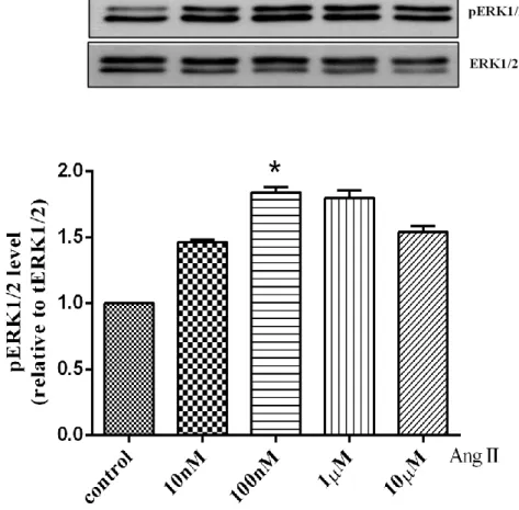
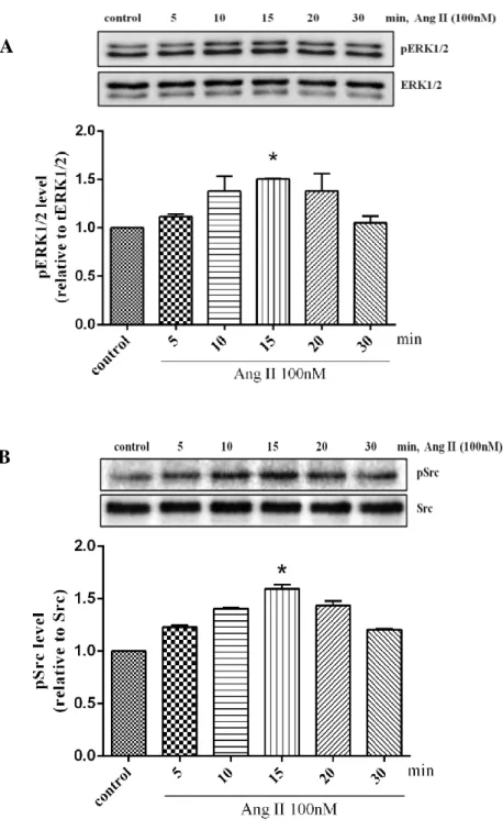
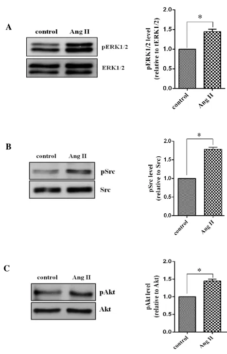
Ang II is kwon as activator which have multiple effects in cardiac fibroblast, which induces cardiac fibroblast proliferation, fibrosis and hypertrophy (Janet et al. 2000). In previous study showed that Ang II activates an array of protein kinases important for its promoting effects such as Src, ERK1/2, and Akt/protein kinase B via binding to AT1 receptor (Taniyama et al., 2004). To examine that Ang II stimulates cardiac fibroblast activation via AT1 receptor, we treated Ang II in proportion of its concentration for 10 minutes. According to western blot analysis, we investigated that phosphorylation of ERK1/2 was started increase by Ang II 10nM and the significantly increased by 100nM to 1µM before started decrease by 10µM (Fig. 2).Furthermore,the effect of time courses of Ang II was also determined. Cells were stimulated with Ang II 100nM in different time course. Ang II induced a rapid phosphorylation of ERK1/2 at 5 min and peaked at 10 or 15 min. The activity was returned to the basal level after 30min (Fig. 3A). These results showed the same in phosphorylation of Src. The activation of Src phosphorylation was started increase at 5 min until 15min and it was decreased at 20min (Fig. 3B). Similarly, we also found that ERK1/2, Src and Akt phospho-rylation significantly increased by Ang II 100nM at 10min (Fig. 4A-C). These results suggested that Ang II stimulated ERK1/2, Src and Akt phosphorylation depended on dose and time dependent maybe involved to AT1 receptor.
Figure 1. Photography of Human cardiac fibroblast (HCF) derived from normal human heart at second passage
Figure 2. Effect of Angiotensin II dose-dependent on ERK1/2 activation.
Human cardiac fibroblasts were stimulated with Ang II (10nM -10μM) for 10min and lysed. Proteins were separated by SDS-PAGE. Western analysis was performed by use of phospho-specific antibody to detect ERK1/2 phosphorylated. Values are mean ± SD compared to control. *P <0.005.
Figure 3. Time course of ERK1/2 and Src activated by Ang II.
Human cardiac fibroblasts were stimulated with Ang II 100nM for indicated times and lysed. Proteins were separated by SDS-PAGE. Western analysis was performed by use of phospho-specific antibodies to detect ERK1/2 and Src phosphorylated. Values are mean ± SD compared to control. *P <0.005.
B A
A
B
C
Figure 4. Effect of Angiotensin II on ERK1/2, Src and Akt activation.
Human cardiac fibroblasts were stimulated with Ang II 100nM for 10min and lysed. Proteins were separated by SDS-PAGE. Western analysis was performed by use of phospho-specific antibodies to detect ERK1/2, Src and Akt phosphorylated. Values are mean ± SD compared to control. *P <0.05.
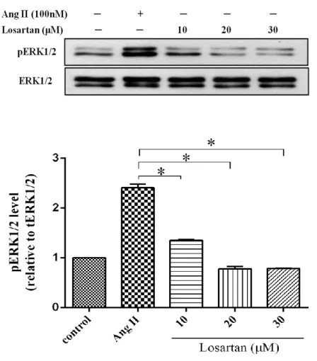
4.2. Effect of Losartan dose-dependent on ERK1/2 phosphorylation To examine the best concentration of Losartan for use to inhibit AT1 receptor, cells were treated with Losartan (10µM to 30µM), whereas some cells were stimulated with Ang II 100nM for 10min at the same time with Losartan. In this result indicated that Losartan 20µM significantly blocked the ERK1/2 phosphorylation compared to 10µM. So, we assumed that Losartan 20µM is the best concentration for used to block AT1 receptor-stimulated by Ang II compared to 10µM (Fig.5).
Figure 5. Effect of Losartan dose-dependent on ERK1/2 activation.
Human cardiac fibroblasts were stimulated with Ang II 100nM and Losartan (10μM-30μM) for 10min and lysed. Proteins were separated by SDS-PAGE. Western analysis was performed by use of phospho-specific antibody to detect ERK1/2 phosphorylated. Values are mean ± SD compared to Ang II. *P <0.005.
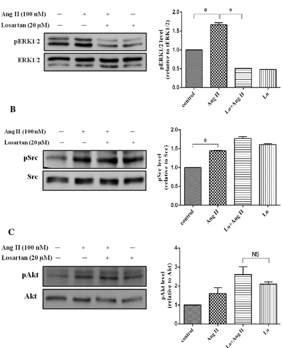
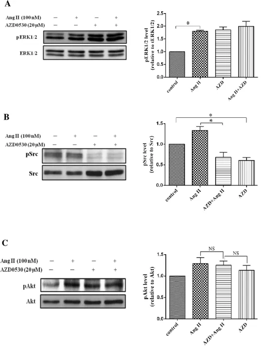
4.3. Effect of Losartan and AZD0530 on Ang II-induced ERK1/2, Src, and Akt phosphorylation
In previous study, we have demonstrated that Ang II stimulates ERK1/2, Src and Akt activation in cardiac fibroblasts maybe via AT1 receptor. To conform that Losartan and AZD0530 can block AT1 receptor and inhibit Src pathway induced by Ang II, we treated Losartan 20µM, AZD0530 20µM. Losartan 20µM or AZD0530 20µM and Losartan 20µM or AZD0530 20µM plus Ang II 100nM were treated in the same time for 10minutes. Other cells we stimulated with Ang II 100nM for 10 minutes same. From these experiments, we found that Ang II still induced ERK1/2, Src including Akt phosphorylation. The phosphorylation of ERK1/2 was decreased in Losartan plus Ang II and more attenuated when we treated with Losartan alone (Fig. 6A). However, Losartan had no effect on Src and Akt phosphorylation. (Fig. 6B, C). Conversely, AZD0530 has inhibited Ang II induce phosphorylation of Src (Fig. 7B), but it could not inhibit ERK1/2 and Akt phosphorylation (Fig.7A, C). These results illustrated that Ang II induced ERK1/2 phosphorylation via AT1 receptor, whereas Src and Akt phosphorylation induced by Ang II were not involved AT1 receptor.
A
B
C
Figure 6. Effect of Losartan on Ang II induced ERK1/2, Src and Akt activation.
Human cardiac fibroblasts were treated with Losartan 20μM then stimulated with Ang II 100nM for 10min in the same time and lysed. Proteins were separated by SDS-PAGE. Western analysis was performed by use of phospho-specific antibodies to detect phosphorylated of ERK1/2, Src and Akt phosphorylated. Values are mean ± SD compared to Ang II. *P <0.005
Figure 7. Effect of AZD0530 on Ang II induced ERK1/2, Src and Akt activation.
Human cardiac fibroblasts were pretreated with AZD0530 20μM then stimulated with Ang II 100nM for 10min in the same time and lysed. Proteins were separated by SDS-PAGE. Western analysis was performed by use of phospho-specific antibodies to detect ERK1/2, Src and Akt phosphorylated. Values are mean ± SD compared to Ang II. *P <0.05.
A
B
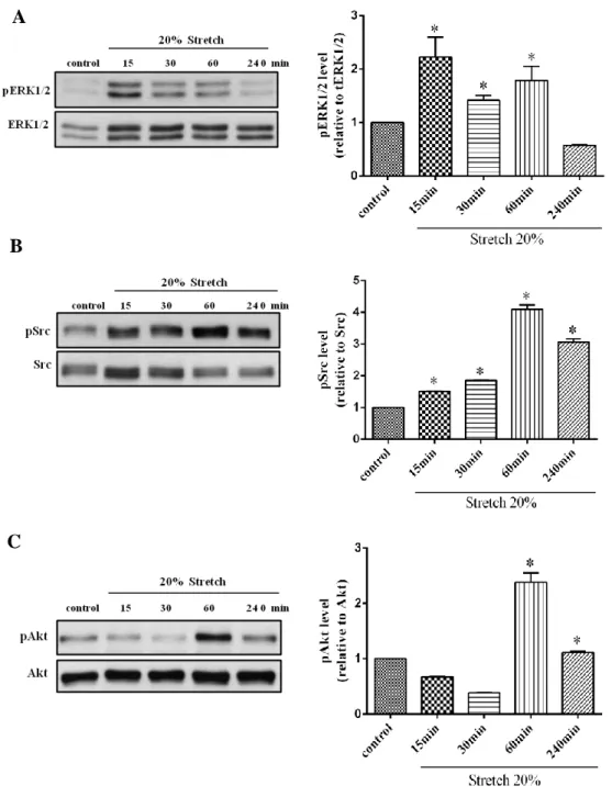
4.4. Effect of mechanical stretch on ERK1/2, Src and Akt phosphorylation
To conform whether mechanical stretch effect on ERK1/2, Src and Akt phosphorylation in human cardiac fibroblasts. Cells were stretched without present of Ang II at 20% elongation in different times (15min, 30min, 1h, and 4h). The acute cyclic stretch in human cardiac fibroblast induced phosphorylation of ERK1/2, Src and Akt were started increased at 15 minutes. Then it decreased at 30 minutes. But the activity was increased again in 60 minutes and decrease again at 4hr (Fig.9A-C). These results suggested that without involvement of Ang II, mechanical stretch also effect on ERK1/2, Src and Akt phosphorylation. However, the effect of mechanical stretch was depending on time dependent.
A
B
C
Figure 8. Effect of mechanical stretch on ERK1/2, Src and Akt activation.
Human cardiac fibroblasts were taken mechanical stretch in different times and lysed. Proteins were separated by SDS-PAGE. Western analysis was performed by use of phospho-specific antibodies to detect ERK1/2, Src and Akt phosphorylated. Values are mean ± SD compared to Ang II alone. * P<0.05.
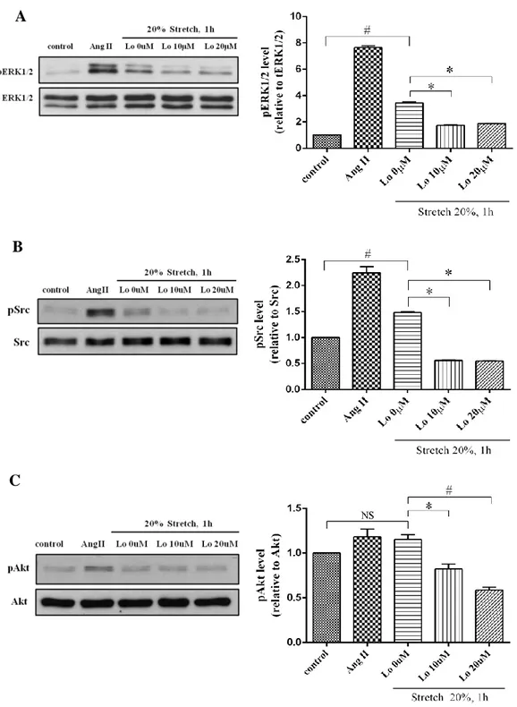
4.5. Effect of Losartan dose-dependent on mechanical stretch induced ERK1/2, Src and Akt phosphorylation
Dostal et al reported that pretreatment with AT1 receptor blockers can attenuate mechanical stretch induced hypertrophic responses (Dostal et al., 2014). To verify whether AT1 receptor was involved in mechanical stretch induced ERK1/2, Src and Akt phosphorylation in cardiac fibroblasts, we treated the cells with Losartan in order concentration (10μM and 20μM) for 10minutes before took mechanical stretch 20% elongation for 1hour. We investigated that Losartan 20μM is the best concentration for use to block AT1 receptor in mechanical stretch induced ERK1/2, Src and Akt phosphorylation (Fig. 10A-C). This result demonstrated that mechanical stretch induced ERK1/2, Src and Akt phospho-rylation was involved to AT1 receptor, which could be block by Losartan.
A
B
C
Figure 9. Effect of Losartan dose-dependent on mechanical stretch induced ERK1/2, Src and Akt activation.
Some cells were stimulated with Ang II 100nM for 10min and some cells were treated with Losartan (10 and 20 μM) for 10min before stretch for 1h and lysed. Proteins were separated by SDS-PAGE. Western analysis was performed by use of phospho-specific antibodies to detect ERK1/2, Src and Akt phosphorylated. Values are mean ± SD. # P <0.001 compared to control. * P <0.005 compare to Lo 0μM.
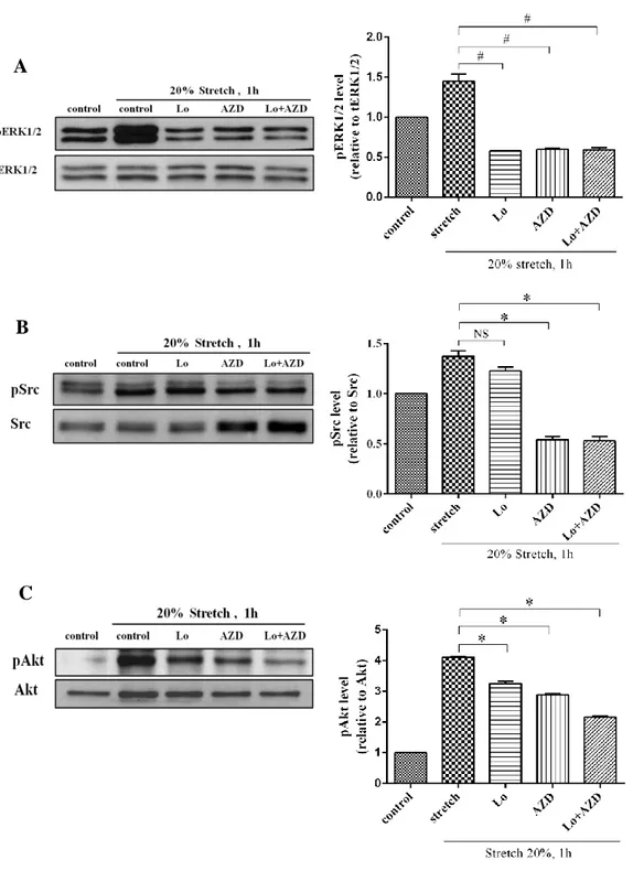
4.6.Effect of Losartan and AZD0530 on mechanical stretch induced ERK1/2, Src and Akt phosphorylation
To make sure of inhibitory effect of mechanical stretch on AT1 receptor induced ERK1/2, Src and Akt phosphorylation, we treated the cardiac fibroblasts with Losartan 20μM, AZD0530 20μM and Losartan plus AZD0530 20μM for 15min before took mechanical stretch 20% elongation for 1h. We found that Losartan and AZD0530 20μM compressed activation of ERK1/2 phosphorylation by stretch (Fig.11A). However, Losartan 20μM could little block the activation of Src phosphorylation by stretch, but the Src phosphorylation could significantly blocked by AZD0530 and Losartan plus AZD0530 20μM (Fig.11B). In contrast, both inhibitors could block the effect of mechanical stretch on Akt phosphorylation (Fig. 11C). From these results, we can assume that effect of acute mechanical stretch on AT1 receptor activated ERK1/2, Src, and Akt phosphorylation could be decrease by Losartan and AZD0530 20μM. So, Both AT1R and Src play role in stretch induced cardiac fibroblast dysfunction
A
B
C
Figure 10. Effect of Losartan and AZD0530 on mechanical stretch induced ERK, Src and Akt activation.
Cells were treated with Losartan 20μM and AZD0530 (20μM) for 10min before stretch for 1h and lysed. Proteins were separated by SDS-PAGE. Western analysis was performed by use of phospho-specific antibodies to detect ERK1/2, Src and Akt phosphorylated. Values are mean ± SD compared to stretch. #P<0.05, *P<0.005.
4.7. Effect of starvation time on ERK1/2, Src and Akt phosphorylation
To know the baseline of ERK1/2, Src and Akt phosphorylation, we investigated starvation time dependent on phosphorylation of the three kinases. We demonstrated that ERK1/2 and Akt phosphorylation were increased at 4hrs until 12hrs and decreased after 12hrs until 24hrs (Fig.12A, C). Src phospho-rylation was decreased and kept the expression same until 24hrs (Fig. 10B). These result showed starvation time has effect on the expression of ERK1/2, Src and Akt phosphorylation.
Figure 11. Effect of starvation time on ERK1/2, Src and Akt activation.
Cells were changed to serum starvation in different times and lysed. Proteins were separated by SDS-PAGE. Western analysis was performed by use of phospho-specific antibody to detect ERK1/2, Src and Akt. Values are mean ± SD compared to stretch. *P<0.005.
A
B
5. DISCUSSION
In this study, we demonstrated the signaling pathways that mediate ERK1/2, Src and Akt activation by Ang II and mechanical stretch in human cardiac fibroblast. Finding from the present study demonstrated that Ang II 100nM induced the activation of ERK1/2, Src and Akt phosphorylation in 10-15minutes. Mechanical stretch induced the activation of ERK1/2, Src and Akt phospho-rylation at 1hour. Both Ang II and mechanical stretch induced phosphophospho-rylation of ERK1/2 was significantly blocked by Losartan 20μM in 10minutes, but it had no effected on Src phosphorylation. However, phosphorylation of Src was blocked by AZD0530 20μM in 10minutes. In contrast, AZD0530 was increased the phosphorylation of ERK1/2. Although the both inhibitors can inhibit ERK1/2 and Src phosphorylation, but it had no effect on Akt phosphorylation.
In human cardiac fibroblasts, Ang II stimulates cells proliferation and collagen synthesis (Chen et al., 2004). Cardiac fibroblasts increased the production of ECM proteins when the heart is exposed to a different of injuries. The increasing number of cardiac fibroblasts and the content of proteins in ECM while cardiac remodeling is one of the major cause of cardiac dysfunction (Cheng
et al., 2003; Zou et al., 1998). Ang II inducted of both cardiomyocyte hypertrophy
and fibroblast proliferation via AT1 receptor and increased activation of ERKs, JNK, and p38MAPK pathways (Cheng et al., 2003; Wang et al., 2013). Ang II activates ERKs through a pathway including the Gβγ subunit of Gi protein, tyrosine kinases including Src family tyrosine kinases (Yamazaki, Komuro, and Yazaki, 1998). Src and Src-family protein tyrosine kinases are regulatory proteins that play major roles in cell differentiation, motility, proliferation, and survival
(Roskoski, 2005). Src was induced upregulation by Ang II (Yamazaki, Komuro, and Yazaki, 1998). In knockout mice, Src was recruited to the cell membrane and interact with AT1 receptor during stretching. The major role of Src in regulating AT1 receptor induced intracellular signaling transduction was stimulated by mechanical stretch (Wang et al., 2014; Takeishi et al., 2001).
Mechanical stretch activates AT1 receptor and integrins. The activation of these receptors can initiate several downstream signaling pathways, such as MAPK and Akt. In the heart, the Akt is implicated in the control of physiological cardiac hypertrophy, contractile function and Ca2+ handling (Dostal et al., 2014). One of the principal targets of PI3K signaling is the serine/threonine kinase Akt, PI3K-Akt signaling pathway is essential in the induction of physiological cardiac hypertrophy (Rohini et al., 2010). Mechanical stretch also activates AT1 receptor without the involvement of Ang II and caused an increase in the phosphorylation levels of ERKs through the Ang II independent pathway (Dostal et al., 2014; Wang et al., 2014; Yatabe et al., 2009). Pretreatment of cardiomyocytes with AT1 receptor blockers such as candesartan can suppresses AT1 receptor activation by both Ang II and mechanical stretch-induced hypertrophic responses (Dostal et al., 2014; Paravicini et al., 2012). Moreover, stretch of cardiomyocytes caused activation of ERKs, indicating that ERKs and mechanical stress-induced hypertrophy may be linked. ERKs may partly participate in the mechanisms of mechanical stress-induced hypertrophy (Cheng et al., 2003). Wang et al also reported that the Ang II-induced upregulation Src phosphorylation was inhibited by both candasartan and valsartan (AT1 receptor blockers) (Wang et al., 2014), whereas our study indicated that Losartan could not block the activation of Src
phosphorylation. On the other hand, the report of Yamazaki et al dimonstrated that inhibitors of tyrosine kinases abolish Ang II-induced ERKs activation in cardiac fibroblasts, while Rohini et al showed that inhibition the function of Src family tyrosine kinases, also has no effect on Ang II induced activation of ERKs in cardiac myocytes, which similar to the results in recent study (Rohini et al., 2010; Yamazaki, Komuro, and Yazaki, 1998).
Taken together, the results suggest that Ang II and mechanical stretch effect on AT1 receptor and Src kinase induced cardiac fibroblast dysfunction such as fibrosis and hypertrophy. Ang II and mechanical stretch induced ERK1/2 phosphorylation was compressed by AT1 receptor blocker, Losartan, whereas Src phosphorylation increased by Ang II and mechanical stretch was inhibited by Src inhibitor, AZD0530. So, AT1 receptor and Src kinase play an important role involve in mechanical stretch induced cardiac fibroblast injury, which can be attenuated by Losartan and AZD0530.
6. REFERENCES
Agrawal, R., Agrawal, N., Koyani, C. N., and Singh, R. (2010). “Molecular Targets and Regulators of Cardiac Hypertrophy.” Pharmacological Research 61(4): 269-280.
Atance, J., Michael, J. Y., and Wayne, C. (2004). “Influence of the Extracellular Matrix on the Regulation of Cardiac Fibroblast Behavior by Mechanical Stretch.” Journal of Cellular Physiology 200(3): 377-386.
Balakumar, P., and Gowraganahalli, J. (2010). “Multifarious Molecular Signaling Cascades of Cardiac Hypertrophy: Can the Muddy Waters Be Cleared?.”
Pharmacological Research 62(5): 365-383.
Bernardo, B. C., Kate, L. W., Pretorius, L., and Julie, R. M. (2010). “Molecular Distinction between Physiological and Pathological Cardiac Hypertrophy: Experimental Findings and Therapeutic Strategies.” Pharmacology and
Therapeutics 128(1): 191-227.
Chen, K., Chen, J., Li, D., Zhang, X., and Metha, J. J. (2004). “Angiotensin II Regulation of Collagen Type I Expression in Cardiac Fibroblasts: Modulation by PPAR- Ligand Pioglitazone.” Hypertension44(5):655-661.
Cheng, T. H., Chen, P. Y., Shin, N. L., Chen I. B., Wang, D. L., Chen, J. J. (2003). “Involvement of Reactive Oxygen Species in Angiotensin II-Induced Endothelin-1 Gene Expression in Rat Cardiac Fibroblasts.” Journal of the
American College of Cardiology 42(10):1845-1854.
Dostal, D. E., Feng, H., Nizamutdinov, D., Golden, H. B., Afroze, S. H., dostal, J. D., Jacob, J. C., Foster, D. F., Tong, C., Glaser, S. and FNU, G. (2014). “Mechanosensing and Regulation of Cardiac Function.” Journal of clinical
experimental cardiology 5(5): 314.
Fan, D., Takawale, A., Lee, L., and Kassiri, Z. (2012). “Cardiac Fibroblasts, Fibrosis and Extracellular Matrix Remodeling in Heart Disease.”
Fibrogenesis Tissue Repair 5(1):15.
Haendeler, J., and Berk, B. C. (2000). “Angiotensin II Mediated Signal Transduction. Important Role of Tyrosine Kinases.” Regulatory peptides 95(1-3): 1-7.
Janet, M. S. and Willa, A. H. (2000). “Angiotensin II, adhesion, and cardiac fibrosis.” Cardiovascular Research 46 (2000): 264–268
Ricky, M., Sadoshima, J., Brosius III, F. C., and Izumo, S. (1999). “Mechanical Stretch and Angiotensin II Differentially Upregulate the Renin-Angiotensin System in Cardiac Myocytes In Vitro.” Circulation Researchculation 02215(85): 137-146.
Paravicini, T. M., Montezano A. C., Yusuf, H. and Touyz, R. M. (2012). “Activation of Vascular p38MAPK by Mechanical Stretch Is Independent of c-Src and NADPH Oxidase: Influence of Hypertension and Angiotensiin II.”
Journal of the American Society pg Hypertension 6(3): 169-178.
Roskoski, R. J. (2005). “Src Kinase Regulation by Phosphorylation and Dephoshorylation.” Biochemical and Biophysical Research Communications 331(1): 11-14.
Ruwhof, C. and Laarse, A. D. (2000). “Mechanical Stress-Induced Cardiac Hypertrophy: Mechanisms and Signal Transduction Pathways.”
Cardiovascular Reseach 47(1): 23-37.
Schiffrin, E. L. and Touyz, R. M. (2004). “From bedside to bench to bedside: role of renin-angiotensin-aldosterone system in remodeling of resistance arteries in hypertension.” Am J Physiol Heart Circ Physiol 287: H435–H446.
Takeishi, Y., Huang, Q., Abe, J., Glassman, M., Che, W., Lee, J. D., Kawakatsu, H., Lawrence, E. G., Hoit, B. D., Berk, B. C., and Walsh, R. A. (2001). “Src and Multiple MAP Kinase Activation in Cardiac Hypertrophy and Congestive
Mechanical Stretch.” Journal of Molecular and Cellular Cardiology 33(9): 1637-1648.
Taniyama,Y., Ushio, F. M., Hitomi, H., Rocic, P., Kingsly, J. M., Pfahnl, C., Weber, D. S., Alexander, R. W., and Griendling, K. K. (2004). “Role of p38 MAPK and MAPKAPK-2 in Angiotesin II-Induced Akt Activation in Vascular Smooth Muscle Cells.” American journal of physiology. Cell physiology 2287(2): C494-499.
Tian, B., Liu, J., Bitterman, P., and Bache, J. R. (2003). “Angiotensin II Modulates Nitric Oxide-Induced Cardiac Fibroblast Apoptosis by Activation of AKT/PKB.” American journal of physiology. Heart and circulatory physiology 285(3): H1105-1112.
Yamazaki, T., Komuro, I., Kudoh, S., Zou, Y., Shiojima, I., Mizuno, T., Takano, H., Hiroi, Y., Ueki, K., Tobe, K., Kadowaki, T., Nagai, R., and Yazaki, Y. (1995). “Angiotensin II Partly Mediates Mechanical Stress-Induced Caediac Hypertrophy.” Circulation Res 77(2): 258-265.
Wang, J., Chen, H., Seth, A., and McCulloch, C. A. (2003). “ Mechanical Force Regulation of Myofibroblast Differentiation in Cardiac Fibroblasts.”
American journal of physiology. Heart and circulatory physiology 285(5):
H1871-1881.
Wang, L. P., Wang, Y., Zhao, L. M., Li, G. R., Deng, X. L. (2013). “Angiotensin II Upregulates KCa3.1 Channels and Stimulates Cell Proliferation in Rat Cardiac Fibroblast.” Biochemical Pharmacology 85(10): 1486-94.
Wang, S., Gong, H., Jiang, G., Ye, Y., Wu, J., You, J., Zhang, G., Sun, A., Komuro, I., Ge, J., and Zou, Y. (2014). “Src Is Required for Mechanical Stretch-Induced Cardiomyocyte Hypertrophy through Angiotensin II Type 1 Receptor-Dependent β-Arrestin2 Pathways.” PLoS ONE 9(4): e92926. Yamazaki, T., Komuro, I., and Yazaki, Y. (1998). “Sighnalling Pathways for
Cardiac Hypertrophy STRESS DIRECTLY INDUCES.” 10(10): 693-98. Yatabe, J., Sanada, H., Yatabe, M. S., Hashimoto, S., Yoneda, M., Felder, R. A.,
Jose, P. A., and Watanabe, T. (2009). “Angiotensin II Type 1 Receptor Blocker Attenuates the Activation of ERK and NADPH Oxidase by Mechanical Strain in Mesangial Cells in the Absence of Angiotensin II.”
American jouenal of physiology. Renal physilogy 296(5): F1052-1060.
Zablocki D., and Sadoshima J. (2013). “Solving the Cardiac Hypertrophy Riddle the Angiotensin II–Mechanical Stress Connection.” Circulation Research (113): 1192-1195
Zou, Y. et al 1998. “Cell Type Specific Angiotensin II Evoked Signal Transduction Pathways: Critical Roles of G Subunit, Src Family, and Ras in Cardiac Fibroblasts.” Circulation Research 82(3): 337-345.
ACKNOWLEDGEMENT
These two years, I finish my thesis under professor Ki-Seok Kim guidance. He give me chances to do experiments and provided me everything for support my study and living during I stayed in Korea. I am also grateful to professor Dong-Sun Lee for introducing me to the laboratory. I am thankful to Dortor Young-mee Kim for helping, Doctor Hack Sun Choi for giving advices and supported in my experiment. Thank to my laboratory researchers, Sae-byeok Hwang, Sophors Phol, Eun-Jung Kwon, Ga-young Seo and Eun-Jin Yang.