Respiratory tract infections (RTIs) are major causes of morbidity among children worldwide. RTIs are caused by various pathogens. Of several respiratory pathogens, respiratory viruses have emerged as the major ones that even affect hospitalized children1). However, no clinical or radiological characteristics were helpful to differentiate pathogens2).
Recently, multiplex reverse transcription polymerase chain reaction (RT-PCR) has been used to detect causative pathogens of RTIs in many hospitals. Before RT-PCR was used, many RTIs by respiratory virus were treated by antibiotics. Using the RT-PCR, the unnecessary usage of antibiotics could be reduced. Furthermore, we could better understand the etiology of pediatric respiratory infections.
Characteristics of respiratory viruses affecting children were reported worldwide, however the etiology of respiratory viruses in Jeju Island has not been reported. This study was conducted to determine the viral etiology of RTIs in hospitalized children.
Study population and respiratory specimens
We recruited children (age ≤15 years) who were admitted
to the Jeju National University Hospital (JNUH) and who underwent nasopharyngeal aspiration for the diagnosis of respiratory viruses. Respiratory specimens were collected by performing gentle nasopharyngeal aspiration during routine patient care. Respiratory samples were collected from patients who were hospitalized because of febrile episodes, respiratory symptoms, or radiologic abnormalities. The study was conducted from April 2014 to March 2016, and a consistent method was used to detect respiratory viruses since 2014 in JUNH.
Viral diagnosis
By using RT-PCR, all nasopharyngeal aspirates were tested for 12 respiratory viruses that included human rhinovirus (hRV); human adenovirus (ADV); respiratory syncytial virus (RSV); parainfluenza virus (PIV) types 1, 2, and 3; influenza virus types A and B; human coronavirus (hCoV) 229E and OC43; human metapneumovirus (hMPV); and human bocavirus (hBoV).We extracted viral nucleic acid by using the QiaAmp MinElute Virus Spin kit (Qiagen, Valencia, CA, USA). The extracted nucleic acid was amplified by One-step RV Detection kit (Biosewoom, Seoul, Korea) according to the manufacturers’ instructions. PCR was performed by using ABI 7500 Real-Time PCR Instrument System (Applied Biosystems, Foster City, CA, USA).
Clinical analysis
Medical records were retrospectively reviewed to determine accurate timings of the diagnosis of respiratory viral infections and patient characteristics. In this study, other
The Etiology of Respiratory Viral Infection in Children in Jeju Island
Yoon Joo Kim
1, Young Ree Kim
2, Jae Hong Choi
11Department of Pediatrics, Jeju National University Hospital, Jeju-si, Korea 2Department of Laboratory Medicine, Jeju National University, Jeju-si, Korea (Received May 16, 2016; Revised May 23, 2016; Accepted May 30, 2016)
Viral etiology of respiratory tract infections in hospitalized children was investigated from April 2014 to March 2016 in Jeju Island. The etiology of respiratory viral infection in Jeju Island wasnot different from that of mainland Korea. (J Med Life Sci 2016;6(1):25-29)
Key Words
: Etiology of Respiratory Viral Infection, Jeju IslandIntroduction
Correspondence to : Jae Hong Choi
Department of Pediatrics, Jeju National University School of Medicine, 15, Aran 13gil, Jeju-si, Jeju Special self–governing province, 63241, Republic of Korea
E-mail : [email protected]
Abstract
bacterial pathogens and clinical symptoms of patients were not considered because we focused only on the prevalence of respiratory viruses.
Statistical analysis
Differences between categorical variables were tested by using the chi-squared test and Fisher’s exact test. All data were analyzed by using SPSS version 18.0, and results were considered statistically significant when the Pvalue was < 0.05.
Ethics statement
This study was approved by the institutional review board of JNUH (No. 2016-04-007).
Patients and respiratory specimen characteristics In total, 2331 nasopharyngeal aspirates from 1834 hospitalized children were reviewed during the study period. There were 1021 (55.7%) boys and 813 (44.3%) girls. The mean age was 2.54 years; furthermore 61.4%, 35.0%, and 3.7% were < 24 months, 2-9 years, and ≥ 10 years in age, respectively.
Respiratory viruses were identified in 1819 (78.0% of the total aspirates) nasopharyngeal aspirates from 1367 children. The mean age of the group of children in which respiratory viruses were identified was 2.15 years.
Prevalence of respiratory viruses
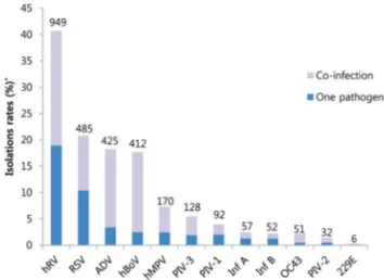
The most commonly detected virus was hRV in 949 (40.7%) of total aspirates, RSV in 485 (20.8%), ADV in 425 (18.2%), hBoV in 412 (17.7%), hMPV in 170 (7.3%), PIV-3 in 128 (5.5%), PIV-1 in 92 (3.9%), influenza A in 57 (2.4%), influenza B in 52 (2.2%), hCoV-OC43 in 51 (2.2%), PIV-2 in 32 (1.4%), and hCoV-229E in 6 (0.3%) (Fig. 1). Influenza B virus was identified in the oldest group of children (age, 4.62 years), and hCoV-OC43 was identified in the youngest group of children (age, 1.36 years).
Of 1819 nasopharyngeal aspirates that tested positive for viruses, only one respiratory virus was identified in were 1046 (57.5%) samples and more than two respiratory viruses (coinfections) were identified were in 773 (42.5%) samples. The most commonlydetected respiratory virus with other viruses simultaneously was hBoV (355/412, 86.2%); followed
by ADV in 81.4% (346/425), and hRV in 53.5% (508/949).
Seasonal pattern and age distribution
The monthly distributions of detected viruses are shown in Fig. 2. ADV was detected throughout the year, and RSV was mainly detected between November and February (82.1% of the total RSV). The prevalence of hMPV peaked between February and May (80.6% of the total hMPV), and PIV-3 was prevalent from April to June (75.8% of the total PIV-3). Influenza A and B weremainly distributed between December and March (91%, data not shown).
The age proportions of the detected respiratory virus are shown in Fig. 3. The proportions of hRV, influenza A, and influenza B infections increased with age. In contrast, the proportions of RSV and hBoV infections decreased with age. Compared to influenza B, RSV was predominantly detected among younger children (P<0.05).
Figure 1. Respiratory viruses detected in children. *The percentages of all nasopharyngeal aspirates in which a specific respiratory virus was detected
The numbers above the bar represent the cases of detected respiratory viruses.
hRV, human rhinovirus; RSV, respiratory syncytialvirus; ADV, adenovirus; hBoV, human bocavirus; hMPV, human metapneumovirus; PIV, parainfluenza virus; Inf, influenza; OC43, human coronavirus OC43; 229E, human coronavirus 229E
Figure 2. Monthly distribution of major 5 respiratory viruses in children
Figure 3. Detection proportion of respiratory virus according to age group
hRV, human rhinovirus; RSV, respiratory syncytialvirus; ADV, adenovirus; hBoV, human bocavirus; hMPV, human metapneumovirus; PIV, parainfluenza virus; Inf, influenza; OC43, human coronavirus OC43; 229E, human coronavirus 229E
In this study, we described the prevalence of 12respiratory viruses that were identified by using multiplex RT-PCR for the nasopharyngeal aspirated of the children who was admitted to the JNUH. Particularly, we focused on the age-dependent distribution, seasonal patterns of respiratory virus, and viral coinfections. In JNUH, multiplex RT-PCR for nasopharyngeal aspirates was generally used when children had fever and respiratory symptoms. We are not confident that all the identified respiratory viruses were related with respiratory tract infections because we did not review the respiratory symptoms related with the respiratory viruses. Therefore, we focused on the etiology of respiratory viral infections in children and their age.
Respiratory viruses were identified in 78.0% of the nasopharyngeal aspirates. The most common virus was hRV (40.7%); followed by RSV (20.8%), and ADV (18.2%). This predominance was not different from that of the mainland Korea (3). Influenza A and B were detected in 2.5% and 2.2% cases,respectively. This proportion could possibly be underestimated because of our hospitalization policy for children with influenza. In the 2014-2015 season, rapid antigen tests for influenza were conducted when the children were suspected to have influenza-like illness. If the results were positive, single rooms were allotted to the children suspected with influenza. In that season, many children suspected with influenza could not be admitted to the hospital. However, in 2015-2016 season, we modified the nosocomial influenza control policy by providing a room for five children who were suspected with influenza-like illness. Owing to this change of influenza cohort, most influenza viruses in this study were identified in 2015-2016 season (70.6%, 77/109).
Knowledge aboutthe seasonal prevalence of each respiratory virus could be useful for prescribing antibiotics and controlling infection. Similar to its prevalence in other temperate climate countries, RSV was mainly distributed in winter season (from November to February); influenza A prevalent from winter to early spring (from December to March), and hMPV was prevalent in spring (from February to May). Despite of the warmer climate in Jeju island, seasonal distribution of respiratory viruses was similar with that of the mainland Korea (3,4).
We found that Influenza (A/B), RSV, and hBoV exhibited specific age distribution patterns. RSV was typically found in
younger children. Of 568 virus children ≤1 year old who were identified with viruses, RSV was identified in 207 children (36.4%). The proportion of influenza increased with age of children (P < 0.05).
hBoV was discovered in 2005 in Sweden by Allander and colleagues (5). Several studies have shown the prevalence of hBoV infection worldwide ranging from 1.5% to 18% (6). In this study, hBoV was identified in 17.7% of the total nasopharyngeal aspirates which is larger than the prevalence (9.4-12.2%) observed in mainland Korea (7,8). Asymptomatic cases of hBoV infection and high frequency of coinfections have led to a doubt for its role for RTIs. Only 13.8% of all the hBoV infections were identified without other respiratory viruses in this study. Seasonal distribution was similar with that of hMPV and PIV-3 ranging from May to April (Fig 2). The age of children detected with hBoV was mainly < 5 years, and this distribution was similar with other results (9).
Rates of respiratory viral coinfections vary from 1% to 40% (10,11). In this study, coinfections were observed at a rate of 42.5%, and the threemajor commonly detected viruses co-occurring with the other respiratory viruses were hBoV, ADV, and hRV. We considered that hBoV and hRV were less likely to be true pathogens causing RTIs because of the previously mentioned characteristics of hBoV and longer shedding period of hRV. At first, ADV was suspected to manifest more severe symptoms when coinfection was observed. However, there is no consensus on the relationship between viral coinfection and clinical severity. Many recent studies showed viral coinfection is not associated with increased clinical severity (12,13).
This study included a relatively large number of children in an isolated region. However, there were several limitations. First, the identified respiratory virus was not correlated with its clinical symptoms which could have revealed more specific pathogenic roles of the virus besides the etiology. Second, RT-PCR method is a highly sensitive technique, and pathogen detection might have been overestimated. Despite these limitations, this study is meaningful because it is the first report on respiratory viral infections in children in Jeju Island.
In conclusion, the etiology of respiratory viral infection in Jeju Island isnot different from that of mainland Korea. Further investigations including the clinical status could provide more accurate etiology of the infection and its clinical characteristics in children.
1) Jain S, Williams DJ, Arnold SR, Ampofo K, Bramley AM, Reed C, et al. Community-acquired pneumonia requiring hospitalization among U.S. children. N Engl J Med. 2015;372(9):835–45.
2) Korppi M, Don M, Valent F, Canciani M. The value of clinical features in differentiating between viral, pneumococcal and atypical bacterial pneumonia in children. Acta Paediatr. 2008;97(7):943–7.
3) Seo Y Bin, Song JY, Choi MJ, Kim IS, Yang TU, Hong K-W, et al. Etiology and clinical outcomes of acute respiratory virus infection in hospitalized adults. Infect Chemother. 2014;46(2):67–76.
4) Choi EH, Lee HJ, Kim SJ, Eun BW, Kim NH, Lee JA, et al. The association of newly identified respiratory viruses with lower respiratory tract infections in Korean children, 2000-2005. Clin Infect Dis. 2006;43(5):585–92. 5) Allander T, Tammi MT, Eriksson M, Bjerkner A,
Tiveljung-Lindell A, Andersson B. Cloning of a human parvovirus by molecular screening of respiratory tract samples. Proc Natl Acad Sci. 2005;102(36):12891–6. 6) McIntosh K. Human bocavirus: developing evidence for
pathogenicity. J Infect Dis. 2006;194(9):1197–9. 7) Ahn JG, Choi SY, Kim DS, Kim KH. Human bocavirus
isolated from children with acute respiratory tract
infections in Korea, 2010-2011. J Med Virol. 2014;86(12):2011–8.
8) Choi JH, Paik JY, Choi EH, Lee HJ. Epidemiologic characteristics of human bocavirus-associated respiratory infection in children. Korean J Pediatr Infect Dis. 2011;18(1):61–7.
9) Weissbrich B, Neske F, Schubert J, Tollmann F, Blath K, Blessing K, et al. Frequent detection of bocavirus DNA in German children with respiratory tract infections. BMC Infect Dis. 2006;6(1):109.
10) Nascimento MS, Souza AV de, Ferreira AV de S, Rodrigues JC, Abramovici S, Silva Filho LVF da. High rate of viral identification and coinfections in infants with acute bronchiolitis. Clinics (São Paulo). 2010;65(11):1133–7.
11) Sly PD, Jones CM. Viral co-detection in infants hospitalized with respiratory disease: is it important to detect? J Pediatr (Rio J). 2011;87(4):277–80.
12) Harada Y, Kinoshita F, Yoshida LM, Minh LN, Suzuki M, Morimoto K, et al. Does respiratory virus coinfection increases the clinical severity of acute respiratory infection among children infected with respiratory syncytial virus? Pediatr Infect Dis J. 2013;32(5):441–5. 13) Lim FJ, de Klerk N, Blyth CC, Fathima P, Moore HC.
Systematic review and meta-analysis of respiratory viral coinfections in children. Respirology. 2016;21(4):648-55.