I TheJourn강김Medicineand L맨 Science Vol. 7,No,1,2010 ‘
Mechanism of ethanol-induced purkinje cell death in developing rat cerebellum: Its implication in apoptosis and oxidative damage
Ji.Hoon Song
,
Youngki LeeDepartment01Hislology‘Jeju NationalUniversilySch。이01Medicine,Jeju,Korea
Abstract
Ethanol trealment during the brain growth spurl period has been known to induce the death 01 Pur에nje cells. The underlying molecular mechanisms and the role 01 reaclive oxygen species (ROS) in triggering ethanoHnduced Purki미e cell death are, however,largely unresolved. We underlook TUNEL slaining,Eeslern blotting assay and immunohistochemistry for the cleaved 10rms 01 caspase-3 and -9,with calbindin D28K double immunostaining to identily apoptotic Purkinje celts. The possibility 01 ROS-induced Purkinje cell death was immunohistochemically determined by using anti-8-hydroxy-2'--cleoxyguanosine (S-QHdG), a specilic cellular marker fα
。
xidative damage. The results show thal Purkinje cell death of PD 5 rat cerebellum foll。에
n9 elhanol administration is mediated by the activalion of caspase-3 and -9. However,unexpecledly,TUNEL staining did nol reveal any posilive Purkinje cells while there were some TUNEL-posilive cells in lhe internal and external granular layer. 8-OHdG was delecled in the Purkinje cell layers at 8 h,peaked al 12-24 h,bul nol at 30 h posl-ethan이Irealmenl. No 8-0HdG immunoreactive cells were detected in the internal and external granular layer. The lobule speclfic S-QHdG staining pat1erns 1011。에
ng ethanol exposure are consislenl wilh thal 01 elhanoHnduced Purkinje cell loss. Thus. we suggest that elhanol-induced Purkinje cell death may nol occur by lhe classical apoptolic pathway and oxidalive damage is involved in ethanol-induced Purkinje cell death in lhe developing cerebellum. (J Med니fe SCi 2010:7:98-104)Key Words : Elhanol,Purkinje cell‘Apoptosis,Oxidalive damage
Introduction
Ethanol exposure dlll;l1g br상in development. prodllces a wide aITay of abnonnalities. I'esulting from disnlption of nonnal development. In humans such early ethanol exposure rnay callse a neurological sy:ndrome called fetal alcohol syndrome (FAS)‘a condilion characterized by a variely of neul'opathologies accornpanying behavioral and functional disturbancesil. 1꺼e characteristic features of FAS include facial dysrnorphology. prenalal and postnalal gl'owth retardation. and central nervous systern dysfunction. Brain is particularly sensitive
ω
lhe neurotoxic effecl of elhan이dllring the period of synaplogenesis,also know:n as the brain growth spurt period. which occurs postnatally in rat bul prenatally during the last trirnestel' of gesta디on m humans2,3). Microencephaly‘or a reduced brain:bα1y wei양lt ratio is one of the mosl featured symptoms of FAS4) Reduction of bl'ain:body wei영lt ratio may be attributed 1.0 loss of neurons. shrinkage of neuronal cell bodies 01 reduction in the number and exæ:nt of dendrites5)
Addressfor correspondence: YoungkiLee
Department01Hislology.Je씨NalionalUniversilySchoot01 Medicine.66 Jejudaehakno,690-756,Jeju. Korea E-mail:yklee38@jejunu.ac.kr
98
Of various regions of bl밍n sensitive to early ethanol exposme. cerebellum 1Sone of the most vulnerable region헌 7). Etha:nol has been show:n to produce a significant changes in neurogenesis. neuronal morphology‘and enhanced cell death of differenl1ated neurons in cerebellurn8). Therefore the I'at cerebel1urn is an excellent rnodel for evalualing 어latomical deficits. as well as cellular and moleculal' evenl..c; I'esulting from developrnental alcohol exposure. In anirnal models. a single heavy eηosure of ethanol on postnatal day 4-6‘a period of bl'ain growth spwt. reslllts in severe loss
。
f Purkinie cel1s and granule cells in cerebellum while similar expo:sure slight1y later in the postnatal period produces little if any loss9. 10). Purkinie cells wiU1in lhe different lobules of the cerebellurn are differentially vulnerable to the ethanol exposure. Lobules 1. II‘IlI,IX andX are the most vulnerable and lobules VI and VII al'e the least vulnerable1Jl. In addition to timing and lobllle specificity lo alcohol-indllced PurJμnje cell loss. many celllllar and molecular events have been identified that occur in c10seproximily to the 10ssof cells. and these cellular 비ld molec비ar events have been assmned to drive the 1085of Purkirue cellsl2l. Howevel‘how the Pul'kinie cells exposed to ethanol are led to death is largely undetermined. although ethanol-induced Purkinje ceU loss has been know:n to occur
Mechanismof ethan이 induced purkinje cell death in developingrat cerebeJlurn:Its implicationin apoptosis and oxidativedamage
due to clirect cell death riot to defects of neuronal migration
。
r neurogenesis dwing developmentThe 띠잉chan념ms of cell death induced by ethanol administration. in the developing rodent brain has been lllves디gated morphologically and biochemically,킹1d revealed to occur via apoptosis13 ,14) Neurons begin showing apoptotic morphological changes 2 to 4 h after ethanol treatment ,외ld by 10 to 12 h,abnost all of the affected neurons are manifesting very advanced apoptotic changes, including condensation and disintegration of the cell body and dendritic tree into numerous small independent bodies Biochemical studies also support the ethanol-induced
',apoptotic neurodegenration in developing rodent brain
Caspase-3 ,executioner of cell death in apoptosis ,is activated and cytochrome c is released in various regions of brairi‘i.t:lresponse to a single heavy exposure of ethanoP5) ln hÓ1ni‘pzygous Bax-deficient mice, neither caspase-3 activation nor cytochrome c re1ease are induced and neurodegeneration does not occur16J,the result implying that ethanol-induced neurodegenration is Bax-dependent and thus involves the intrinsic pathway of apoptotic cell death
。
ne of various factors which can trigger apoptosîs is oxidative stress,an excessive accumulation of free radicals in the cell. Free radicals are highly reac디ve molecules that can be formed dwing many biochemical reactions in the cell Many of these free radicals contain oxygen 킹1d are calledreactive oxygen species (ROS). To overcome 。잉da디ve stress,
mammalian cells have a complex antioxidant defense sysæm that includes non-enzymatic antloxidants (e.g. glutathione, thioredoxine) and enzymatic activities (e,g. superoxide dismutase,catalse). Thus the survival of a cell depends on the balance between ROS and antioxidants17l. lf ROS levels exceed the cell’S 외)ility to eliminate them. or if the normal antioxidnat levels are reduced due to a toxic insult such as alcohol,난len oxidative stress c잉1 occur. The cellular damage
caused by oxidative stress is a consequence of lipid peroxidation 1에,alteration of nucleic acid19l,and proteins20l These non-specific oxidative damages to cellular components have been known to provoke necrotic cell death However, it was also suggested that ROS may induce cytochrome c release,and subsequent activation of caspase 9 and -3,which lead to apoptotic cell death21l. lnteractions between DNA and ROS produce DNA strand breaks and base modification22),which are frequently
The present study was designed to investigate the mechanism of ethanol-induced Purkinje cel! death by using developing PD 5 rat. lmmunohistochemistrγ and wesæm blotting assay for the cleaved caspase-9 and -3 were carried out to show whether the degeneration of Purkinje cells in response to ethanol exposure occurs via apoptotic pathway. Furthermore ,to confirm the possible role of oxidative stress in ethanol-mediated Purkinje cell death the production of 8-0HdG was assessed by îmmunohistochemical analysis
i Materials and Methods
1. Animals and ethanol administration
Postnatal day (PD) 5 Sprague Dawley rats were used in this study,They were assigned to one of two groups ethanol (n=8) or control (n=7). On PD5,ethan이(6g!kg) in normal saline was administered subcutaneously in tw
。
separate treatments. 2 h apart,each treatment qeliverîng3g/kg,and control rats were treated saline 。띠y
2. Measurement 01 blood ethanol concèntration
Blood samples were collected from heárts of PD5 rats to determîne the blood ethanol concentration (BEC),BEG was measured in all pups on 2,4,6,8,12,J8 and 24 h following ethanol treatment by using alcohol kit (Sigma,MO, USA). Brief1y,the blood samples were centrifuged at 12,000 rpm for 5 min. Deionîzed waær ,ethanol sændard solution (0.08%) and samples were added to alcohol reagent and then mixed. All mixtures were incubated for 10 min at room temperature. Absorbance of samples was measured at 340 rnn wavelength
3. Tissue preparation
η1e tissue samples were prepared at 2‘4,6‘8‘12,18 and 24 h after ethanol or saline treatment by an overdosE of pentobarbital and subsequently perfused with 0.9% saline followed by 4% paraformaldehyde in O.lM phosphate buffer The cerebellum was removed from the brain and stored in the same fIxative. F'ixed cerebelli were immersed in 20% sucrose solution and sections of 40.μm thickness were
。
btained by cryostat sectioning. For 난1e western blotting assay the cerebelli were rapidly removed after decapitation, and stored at -70"C until used4. Western blot analysis
Ji-HoonSong.Youn양i Lce
(Upslate. USA)‘and UlChomogenates were centrifuged at 12.000 rprn for 15 rnin at 4t. The sl.lpernatanl was collected and protein concentTationwas detennined with a BCA protein a잃ay kit (Pierce. USA). The protein samples were separated by SDS-PAGE and transfen:ed to PVDF membrane. Non-specific immunoreactivity was blocked
。
vemight with 5% non-fat dry mi1k mixed in a solution of 0.1% 1Ween-20 in Tris buffered saline. Prirnary antibody for c1eaved caspase-3. and -9 (1:3000. Cell Signalling. USA) were incubated ovemight at 4'C. Mernbranes were washedand incubated with anti-rabbit IgG conjugated horseradish peroxidase (I:4()()().Sanl.a Cruz. USA) at roorn temperat따e for an hour. The signa1 was delected with enhanced chemiJuminescence(ECL)detection system
5. Immunohistochemistry
Vecl.ashield Hard-set mounting medium and analysed by confocal laser scanning microscope (Olympus)
6. TUNEL staining
For the analysis of DNA fragmentation. ApopTag kits Ontergen. NY. USA) were used according to the manufacturel ’s manual. BrieOy. the paraffin-embedded
tissue sections were deparaffinized with xylene and ethanol and Ihen apply proteinase K (2아LglmI) ω the specimen fOl 15 min at room temperatl.lre.The sections were washed and incubated with the TdT eI17..yme solution containing the digox.igenin-dNTP at 37t for 1 h. After several rinses. anti-digoxigen peroxidase conjugate was app1ied to the slides for 30 minutes at room temperature and then develop color in DAB solution for 3 to 6 minl.lte.Negative controls were obtained by exclusion of the TdT enzyme
600
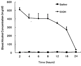
1. Blood ethanol concentration
24 18 12 -+-E10H .••← Salinu 8 Time (hours) 6 4 2
o
200 400 500 100 300..
,
E g m : 용 g‘
J 5。
〈§
。
1Blood ethanol concentrations (BECs) were measured in PD5 rats after ethanol and sa1ine administration 꺼1e mean
BECs at variOl.lStirne points were shown in Figure 1. The peak BEC was 509 mg/dl and achieved 2 h arter the ethanol 따atment in the pups. with near t이.al c1e81밍1ce of ethanol at the end of 24 h post-treatment. Saline-u'eated pups showed basal 1evelsof BECs
Figure 1. ßlood ethanol concentrations in PD5 rats after ethanol and saline treatrnenl. Ethanol W3Ssl.lbcul.aneously administered in two iniections(3g/kg x 2) spaced 2h apart
끼1e peak blood ethanol concentrations were achieved 2 h after ethanol treabnent and then decreased gradually to the control value at 24h post-treatment. n=6-7 each treatmcnt group
L
Res 비성 __,__.JImmunohistochemistryfor c1eavedcaspase-3 and -9 was carried out by free floating rnethod. The sections were rinsed in O.OIMPBS and blocked in 1% l-hOJto inactivate endogenous peroxidase π1e sections were incubated in PBS cont.aining3% nonnaI horse serum at room ternperature for 30 min. and then incubaled with rabbit anti-c1eaved caspase-3 and -9 (J:1oo. Cell Signalling. USA)ovemight at 4'C. Sections were reacted for 90 min with biotinylaled anti-rabbit IgG (1:200. Vector. USA) and Ihen incubated wilh ABC reageniß (Vector elite kit). For detection of 8-0HdG. sections were mOLmted00 2% ge1atin coated slide. dried‘
rinsed in PßS. and b10ckedendogenous peroxidase in 0.3% I-ù02. Subsequently sections were treated with 150 μg/mI
RNase A for an hour at 37'C (to exclude interference effecl
。
f oxidative RNA products) and 50 nM s。이
um hydroxide in 40% ethanol for 10 min (for denaturation of DNA)and then incubated for 30 min in 3% nonnal goat serum. The sections were incubated with mouse anti-8-0HdG (1:100. Chemicon‘USA)ovemight‘biotinylated anti-rnouse IgG for 90 rnin and
then ABC reagents. π1e sections were washed in PBS tlu'ee tirnes and incllbated for 6 rnin in DABsollltion and rnounted by po1ymount
1"01'double nuorescent immunostaining with cleaved caspase-3,-9 and ca1bindinD28K. sections were incubated ovemighl with first primalγ antibody,cleaved caspase-3 01' 9 at 4t and thell with 까TC-c。이ugated goat anti-rabbil
IgG at room ternperaωre for 90 min. After washing. the sections were incllbat.ed with second primary antibody of mouse anti-calbindin D28K(1:1αXl.Sigma. USA)for 90 min The sections were incubated with 1'exas-Red conjugated anti-mouse IgG (Vector. USA) ror 90 min. and mounted gelatin coated slide. Finally the sections were rnounted by
Mechanismof ethanoHnducedpurkiniecelldeathin developingral cercbellum:lts implicationin apoptosisand oxidativedamage
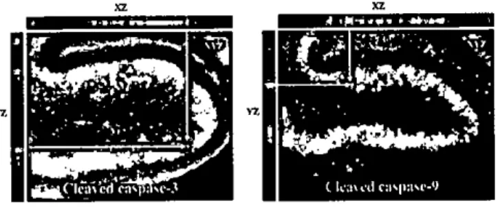
Figure 3. Confocal microscopic images for the colocalization of caspase-3 or -9 (FITC). and calbindin D28K (Texas red) in lobule X of cerebellum fol1owing ethanol exposure. M어lY calbindin 0 28K-positive Purkinie cells (red color) contain the caspase-3 or -9
immunoreaclivity (green color). XZ and YZ cross sections of arrows-indicated cells further prove the true colocalization events. Scale bar=60,um
Figure 4. Lobule-specific vulnerability foll
。、
.ving ethanol treatrnent in the midsagitt.al section of cerebellar vermis as revealed by caspase-3 immunohistochemisUy,Caspase-3 immunoreactive cells are mainly confined to the Purkinje cell layer in all 10 lobules. However. lobule 1. 11. lIJ. JX‘
and X of ear1y maturing lobules show more numerous immunoreactive cells than the lat.e maturing lobules. V. VJI.VlI and VlII. Scale bar=IOOpm
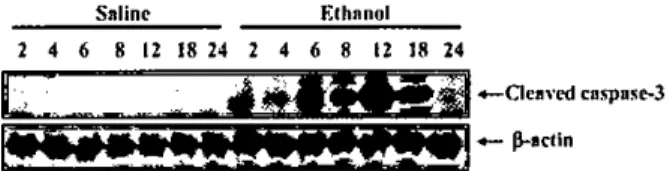
we examined the cerebellar vennis e셔racts from control and ethano!-exposed pups at the same time points of imrnunohistochemicalanalysis by using westem blots and antibodies for c1eaved caspase-3. Caspase-3 was not activated in saline σ'eated control extracts,consistent with the low levels of naturally 0
∞
urring cell death observed immunohistochemically (FIg. 5). However the ethanol administration dramatical1y induced the activation of caspase-3 and the appearance of cleaved caspase-3 immunoreactivitywas transient: lt was increas밍d between 6and 8 h postethanol exposure,pe와,ed at 12 b and then
dropped abrup디y to the saline control levels at 24 h 와'!er ethanol exposure (Fig. 5)
Next. we conducted TUNEL staining at the same time points of caspases immunostaining to obtain the furlher
g
vz.1
‘
';'.
vz2. Äctivation 01 caspas
‘←
3/9 and TUNEL stainingWe previously showed iliat ethanol induces p1디rkinie cell degeneration in PD5 rat pups by Fluoro-Jade B stainin링5l In the present study,we undertook immunohistochemistIy for the c1eavedfOlms of caspase-3 없ld -9,two important specific makers for apoptosis‘to rcveal that eilianol-induced Purkinje cell death occurs via apoptotic pathway,Cleaved
caspase-3 잉ld -9 immunoreactive cells be탱n to appear in
Purkinje cell layer of cerebellar vennis at 6-8 h,maximally detected betwèen 12-18 h and almost absent 24 h 외'!er ethanol administration (Fig. 2A밍ld 28),To confirm these
immunoreactive cells in the Purkinje cell layer are Purkinie cell per se. double f1uorescent immunostaining was conducted with anti-calbindin D28K. a marker for Purlånie cell. Confocal microscopic images with O.5.um optical section showed that c1eavedcaspase-3 or -9 immunoreactive cells (green color) were colocalizedwith calbidin D28K (red color) (I.'ig. 3),Thus most of active caspase-3 and -9
immunoreactivecells in the Purkinie cell layer proved to be Purkinie cells. As an attempt to evaluate any lobu!e-specific activation of caspases in the cerebell와 vennis of ethan이 treated rat pups,we canied out immunohistochemist1ywith the whole vennis at 12 h after ethanol treatment. Lobules L n.
m.
IX. and X showed the most numerous active caspase-3 in the Purki~e cell layer and lobuies VJ and VJI did the least (Fig. 4)To confirm the results of immunohistochemicalana1ysis,
Figure 2. lmmunohistα;hemisσy for the c1eaved caspase-3 (A). caspase-9 (8). and 8-0HdG (C) in the lohule J and X at various time points following ethanol exposure. A11the positive cells were mostly confined
ω
the Purkinie cell layer in the cerebellwn of PD 5 rat. Cleaved caspase-3 and -9 immunoreactive cells began to appear at 6-8 h. were maximally detected at 12-18 h and were almost absent 24 h aft.er etho.nol treatment. The temporal and spatial pattern of 8-0HdG immunoreactivity were similarω
those of the c1eaved forms of caspases but 8-0HdG immunoreactive cells in the Purl‘’
nje cell layer were persisted at 24 h post-treatment. Scale bar=50.urn앓펴짧쩔
훌평텔쩔뚫짧홉룹혔
뚫필탤펼
짧룹평짧펴표:쩔
쩔쩔폼嚴變뚫꿇품홉
Jì-HoonSong. Youn명이낭e
evidence of apoptotic .Purkinje cell death by ethanol. Figure 6D shows that TUNEL-positive cells in the .Purkinje cell layer were very rare although some posi니ve cells were present in U1e interna1 granular layer
3. Immunohhistochemical detection 01 S-DHdG To elucidate that ethanol-induced Purkinje cell death may be associated
ω
ROS generation. immunohistochemistry for 8-0HdG was peñonned with the vennal section of PD5 rat cerebellum following ethanol administration. 8-0HdG immunoreactive cells began to be detected in the Purkinie cel1 layer of cerebellwn at 8 h‘pe빼ed at 12-24 h. and disappeared almost completely at 30 h after ethanol Figure 5. Western blotting analysis for the temporal pattern of c1eaved caspase-3 immunoreactivity. Protein extracts of saline-tre:lted controJ :lnd eth:lnol-exposed vermis of cerebellum (2. 4. 6. 8 12. 18‘and 24 h posUreatment) were prepared and analyzed for c1eaved caspase-3 immunoreactivity. Cleaved caspase-3 immunoreactivity were readiJy detected by 6-8 h and pe따ed 12 h :lfter ethanol exposure and then returnedω
the b:lsal levels :lt 24 h. ,8-actin was used as a protein loading control Salinc Elhnnol 2 4 6 8 12 18 24 2 4 6 8 12 J8 24E
~~lií.ü
톨.를를빼
← 0"、
cdcnspns허'-훌‘
.:;;:ia‘_.-‘-‘_~
0=._ ß.•llctlnl
‘~~~~:::!~~~I::: τ;'.~.&."""-1
Figure 6. Immunohistochemistry for 8-0HdG (A). the c1eaved forms of caspase-3 (B). -9 (C)' and TUNEL siaining (0) 10 reveal the death mechanism of Purki이e cell
following ethanol exposure. Immunoreactive cells for the 8-0HdG. cleaved caspase-3 and -9 were readily dete야ed
in the Purkinje ceJl layer. while 1'UNEL staining did nol show any positive cells in the Purl。이e layer. Note the numerous TUNEL positive cells in theint.ernal granul31 layer. Seale bar=50Jßl1
102
σea미lent. Granular and molec비ar layer of cerebellum did not show any 8-0HdG immunoreactivity (Fig 2C) Cerebellum of saline treaied 밍um외s did not show any
8-。
iHdG immunoreacti씨tyDiscussion
This study was designed to investigate the possibJe mechanisrn of ethanol-induced Purkinje cell death in developing cerebellum 까1e cerebellar Purkinje ceJls are most vuJnerable 10 ethan이 during the early neonataJ period
in rat. Experimental s!;udies suggested that eU1anol activates lhe proapop1otic moJecules24l and leads to neurodegeneration by activation of caspase-3 in developing br.밍n3. 15}.However‘
lhe mechanism leading to Purkinje ceJl degene1'ation following ethanoJ administration is not clearly understood
Our previous study25l showed that FJB-positive ceJJs in the Purkinie cell layer afler ethanol exposure on PD5 1'at were more numerous in the early maturing lobuJes Oobules J. 11. m. lX and x) than those fOW1din late maturing lobules Oobules VJ and VII). This pattern of neurodegeneration reveaJed by FJB staining was welJ consistenl with that of lobule-specific Purkinje cell loss and confirmed that ethanol-induced Purkinie cell loss is due
ω
direcl Pu1'kinje cell death nol to the failure of their division 01' migration from the deep cerebellar nucleus. 1'0 further expJore the underJying mechanism of ethanoJ-induced Purkinje cell death. we undertook the immunohis1ochemical staining for active caspase-3 and -9. the key regulator in the processes of apop1otic cell death. Jt revealed that ethanol-induced Purl이nie cell death occurs by activation of easpase-3 and9 꺼1e spatial and temporal patterns of caspase-3 and -9 immunoreactive Purkinje cell were very simila1' to those seen in FJB staining 끼1ese 1'esul잉 well match the previotls 1'eports by other groupSl4l. Next. we introduced TUNEL staining to delermine whether Purkinje cell death is associated wiU1 DNA fragmentation. However. unexpectedly TUNEL-positive Purkinje cell was very rare and there was no lobule-specific pattern of TUNEL staining which was seen in ilie FJB s어ining. caspase-3 and -9 immunostaining
These results were contrary to the previous report by other groupl4) 끼1e TUNEL staining seems 10 be properly canied out since many πJNEL-positive cells could be seen in the granular layer and deep cerebelJa1' nucleus of the same tissue section which showed negative staining for Purkinie cell. In the present. we have no idea why such different 1'esults were obtained between two studies. One possible explanation fo1' ou1' result is tl1at ethanol rnay act as a
Mechanismof ethan이 induced purki미e cell death in dev허oping rat cerebellum:Its implicationin apoptosis and oxidativedamage
NMDA receptor antagonist3 ,26) 1n such circumstance ,
ethanol may block the entry of Ca2’into the cell and 야1e resulting low level of intracellu1ar Ca2’concentration cou1d inhibit the Ca'ν1Mξ dependent endonuclease activity,which 1ead to no DNA fragmentation27). In support of this idea‘
some apoptotic cell death do not show DNA fragmentation although cytochrome c release and caspase activation are
。
ccurred28). Thus, we tentative1y suggest that Purkinje cell death in PD 5 rat cerebellum following ethanol administration may not occur via the classical apoptotic pathwayln the present study ,8-0HdG immunoreactivity in response to ethanol treatment was obseπed in cells of Purkinje cell layer of PD5 rat cerebellum but was not detected at PD7 and PD14. Considering that vl니lnerability of Purkinje cells to ethanol treatment confines 0띠ly to narrow developmental period (PD4-6) and is little îf any at slightly 1ater ethanol-resistant period (PD7-9 or later)lO,24),it implies that oxidative stress may p1ay a role in the ethanol mediated p1니rkinje cell death. 1n support of this suggestion, lobu1e-spec퍼c pattem of 8-0HdG irnmunoreactive cells in the Purkinje cell 1ayer was very sin띠ar to those of active caspase-3!9 iImmmohistochemistry and FJB staining in the present and previous study,respecti
、
relyThe ability of oxidative stress to provoke necrotic cell death as a résu1ï of massive cellular damages associated t
。
1ipid peroxidation18) and a1teration of proteins20) and nucleic acids19) have been well docwnented for a long time. ROS has been a1so know끼 to act as signalling molecu1es in apoptotic pathway. For example,some anti-oxidants can inhibit activation of caspase and subsequent steps leading to apoptotic cell death17l. 1n the present study ,8-0HdG imnnmoreactivity in cells of Purkinje cell layer was observed for a longer time unti1 24 h 따'ter ethanol administration ,at which time point the active caspase-3 and -9 immunoreactivities were gone by. This longer duration of 8 OHdG immunoreactivity may reflect the persistent production of ROS leve1s in response to ethanol exposure,since it has been demonstrated 낭lat ROS levels in the extract of whole cerebellum remains elevated by 24 h after ethano1 administration24l. Therefore ,it is plausible that the 1ater increase of ROS levels may convert the apoptotic pathway of p1미r매매e cell death into necrotic pathway,which may also reflect the absence of DNA fragmentation in TUNEL stainingTaken together ,this study has shown that ethanol induc
DNA damage. However,the differential temporal pattem of cleaved caspases and 8-0HdG immunoreactivity Ìn the present study suggest that the Purkinje cells committed t
。
apoptosis may shift toward necrotic death due to a later burst in cellular ROS levelsReferences
1) West JR Chen WA,P밍]떠zis NJ. Fetal alcohol syndrome the
、
ru1nerability of the developing brain and possible mechanisms of damage. Metab Erain Dis 1994;9:291-322 2) West JR Goodlett GR,Bonthius DJ,Hamre KM,Marcussen BL. Cell population dep1etion associated with fetal alcohol brain .damage: mechanisms of BAC dependent cell 10ss. Alcohol Clin Exp Res 1990;14:813-8 3) 1konomidou C,Bittigau P,lshimaru MJ,Wozniak DF,
Koch C,Genz K,et a1. .Ethano1-induced apoptotic neurodegeneration and feta1 alcohol syndrome. Science 2000;287;1056-60
4) Bellinger FP,Davidson MS,Bedi KS,Wilce PA. Neonatal ethano1 exposure reduces AI\.1PAbut not NMDA receptol levels in the rat neocortex. Dev Brain Res 2002;136:77 84
5) Brooks PJ. Br없1 atrophy and neuronal loss in alcoholism a ro1e for DNA damage? Neurochem 1nt 2000;37:403-12 6) Good1ett CR,Marcussen BL,West JR. A single day of
alcoho1 exposure dming the brain groψ야1 spurt induces brain weight restriction and cerebellar Purkînje cell 10ss Alcohol 1990;7(2):107-14
7) Thomas JD. Good1ett CR,West JR. Alcoho1-induced Purkinje cell 10ss depends on developmental timing of alcohol exposure and corre1aæs with motor -perronnance Dev Brain Res 1998;105;159-66
8) Mou1der KL,Tao Fu,Melbostad H,Connier RJ,Isenberg KE,Zorumski CF,et a1. Ethanol-induced death of postnatal hippocampal neurons. Neurobiol Dis 2002;10;396-409
9) Goodlett CR, Lundahl IιR. Temporal determinants of neonatal -alcohol-induced cerebellar damage and motor performance deficits. Pharmacol Biochem Behav
1996;55;531→40
10) Pierce DR,Williams DK,Light DK. Purkinje cell vulnerability to developmental ethanol expos니re in the
rat cerehellum. Alcohol Clin Exp Res 1999;23;1650-9 11) Hamre KM,West JR. The effects of the timing of
ethanol exposure dwing the brain growth spurt on the number of cerebellar Purkinje cell nuclear profiles Alcohol Clin Exp Res 1999;17;610-22
Ji-Hoon Song. Youn밍ι Lee
12) Heaton. MB,Paiva M,Madorsky 1,Siler-Marsiglio KI, Shaw G. Effect of bax deletion on ethanol sensitivity in the neonatal rat cerebelllUll. J Neurobiol 2006;66:95-101 13) Dikranian K,Ishimaru MJ,Tenkva T,Labruyere J,Qin
YQ,Ikonomidou C,et a1. Apoptosis in the in VÌv
。
marnmalian forebrain. NeW'obiol Dis 2001;8:359-7914) Light KE,Beleher 3M. Pieree DR Time eourse and manner of Purkπ1,je neW'on death following a single ethanol exposW'e on posmatal day 4 in the developing rat. Neurosci 2002;114:327-37
15) Olney JW. Tenkova T,Dikranian K,Qin YQ,Labruyere J, Ikonomidou C. Ethanol-indueed apoptotic neurodegeneration in developing C57BLl6 mouse brain. Dev Brain Res 2002;133:115-26
16) YC때g C,K10eke BJ,Tenkova T,Choi J,Labruyere J
Ethanol-indueed neuronal apoptosis in VÌvo requires BAX in the developing mouse brain. Cell Death DiffeI 2003:10:1148-55
17) Fleurγ C,Mì밍10tte B,Vaysiere JL. Mitoehondrial reaetive
。
xygen species in cell death signalling. Biochimie 2002:84:131-4118) Sun AY. Chen YM,Kracke MH,Wixom P,Cheng Y Ethanol←indueed eell death by lipid peroxidation in PC12
cells,Neurochem Res 1997:22:1187-92
19) Navasumrit P,Ward TH,Dodd NJ,0’Conner PF Ethanol-indueed free radicals and hepatic DNA strand breaks are prevented in vivo by antioxidants: effect.5 of aeute and chronic ethanol exposure. Carcinogenesis 2000:21 :93-9
20) Sun AY,Mayhan WG. 3uperoxide dismutase ameliorates impaired nitric oxide synthetase-dependent dilatation of
the basilar aπerγ during chronic alcohol conslUllption Brain Res 2001:892:116-22
21) Yuan J,Murrell GA. Triekett A. Wang :MX. Involvement of cytochrome c release and caspase-3 activation in the oxidative stress-induced apoptosis in human tendon fibroblasts ,Biochim Biopbys Acta 2003:1641:35-41 22) Won MH,Kang T,Jeon G,10e J‘Kìm D,Choi E,el al
Immunohistochemieal detection of oxidative DNA damage induced by ischemia-reperfusion insult in gerb 니
hippoeampus in vitro. Erain Res 1999;836:70-8
23) Kasai H. Analysis of a form of oxidative DNA damage, 8-bydroxy-2 ↑ deoxyguanosine ,as a marker of cellular
。
xidative stress during careinogenesis. Mutation Res 1997:387:147-6324) Heaton MB,Paiva M,Mayer J,Mil1er R. Ethanol-mediated generation of reactive oxygen species in developing rat cerebellum. Neurosei 1ett 2002;334:83-6 25) Lee Y,Rowe J,Eskue K,West JR Maier SE. A1eohol
exposure on postnatal day 5 induces Purkinje cell loss and evidence of Purkinje eell degradation in lobule 1 of rat cerebellum. Alcohol 2008:42:295-302
26) Hoffman PL,Rabe CS,Moses F,Tabakoff B N-methyl-D-aspartate receptors and ethanol: inhibition of calcium flux and cyclic GMP production. J Neorochem 1989:52:1937-40
27) Cohen JJ. Duke RC. Glucorcorticoid activation of a calcium-dependent endonuclease in thymocyte nuclei lead 10 cell deatb,J lmmnnol 1984:132:38-42
28) Schwartz LM,Smith SW,Jones 11E,Osborne BA. Do all programmed eell deaths occur via apoptosis? Proc Natl Acad Sc! USA 1993:90:980-4