INTRODUCTION
Polycyclic aromatic hydrocarbons (PAH) are formed due to incomplete combustion of fossil fuels and are ubiquitously present in the environment. Benzo[a]pyrene (B[a]P) is one of a large number of PAH and is frequently used as a represen-tative indicator of total PAH exposure (1). B[a]P is usually inhaled and deposited in the lower lung, where it is readily absorbed. It is then metabolically activated by cytochrome P450 to highly reactive electrophilic forms, including phenol, trans-dihydrodiols, arene oxides, and diol-epoxides, which may be responsible for most of its observed cytotoxic, muta-genic, and carcinogenic effects (1, 2). Biotransformation of chemicals involves metabolic activation and detoxification pathways, which are known to play a primary role in chemi-cal carcinogenesis (3).
Polymorphisms in the genes for metabolism are largely re-sponsible for the different abilities of individuals to activate and detoxify genotoxic agents. Glutathione S-transferase (GS T), which forms a multigene family of phase II detoxification enzyme, is involved in the metabolic detoxification of PAH (4-6). This enzyme plays an important role in protecting DNA against damage and adduct formation, by glutathione conju-gation to electrophilic substances, particularly those with lipo-philic compounds (7). The polymorphism of GSTM1 and GSTT1 gene loci is caused by a gene deletion, which causes a virtual absence of enzyme activity in individuals with the
GSTM1- and GSTT1-null genotypes. The GSTM1-null geno-type has been known to be associated with an increased risk for various environmentally-induced cancers (8, 9) and simi-larly, the GSTT1-null genotype with an increased risk for primary brain tumors and myelodysplastic syndromes (10). However, the genotoxic effect of GSTT1 polymorphism is not well understood compared to that of the GSTM1 polymor-phism (4, 11). The coordinated activities of polymorpolymor-phisms of GST may influence cytotoxic, mutagenic, and carcinoge-nic effects (5, 12). Epidemiologic studies indicate that the coordinated activities of polymorphisms in the GSTM1 and GSTT1 genes are associated with an increased risk for lung and bladder cancers (13, 14).
Although the associations between polymorphisms in genes for metabolism and cancer development has been known, the exact mechanisms of the relationships have not been elucidat-ed. Little has been published on the relationship between these PAH-metabolizing enzymes and the formation of DNA ad-ducts, which reflect DNA damage as a result of environmental exposures to PAH (15). In particular, there are no published data on the relationship between the GSTM1 and GSTT1 polymorphisms and DNA-protein crosslinks (DPC) forma-tion resulting from exposure to B[a]P. DPC is known to be produced by oxygen radicals and other reactive species (16). Exposures to ionizing radiation, formaldehyde, and hexa-valent chromium are also known to result in the induction of DPC (17-19). In light of the abundant evidences of DPC
Hye-Sook Park, Eun-Hee Ha, Kwan-Hee Lee*, Yun-Chul Hong*
Department of Preventive Medicine, College of Medicine, Ewha Woman's University, Seoul; Department of Occupational and Environmental Medicine*, College of Medicine, Inha University, Incheon, Korea
Address for correspondence
Yun Chul Hong, M.D.
Department of Occupational and Environmental Medicine, College of Medicine, Inha University, 7-206, 3rd Street Shinheung-dong, Jung-gu, Incheon 400-103, Korea
Tel : +82.032-890-0973, Fax : +82.32-884-6725 E-mail : [email protected]
316
Benzo[a]pyrene-Induced DNA-Protein Crosslinks in Cultured Human
Lymphocytes and the Role of the
GSTM1
and
GSTT1
Genotypes
We investigated the influence of glutathione S-transferase M1 (GSTM1) and glu-tathione S-transferase T1 (GSTT1) polymorphisms upon DNA-protein crosslinks (DPC) induced by benzo[a]pyrene (B[a]P) in cultured human lymphocytes. Lym-phocyte samples were collected from 30 healthy nonsmoking hospital adminis-trative workers. DPC was detected with KCl-SDS assay and the distributions of GSTM1 and GSTT1 were determined by polymerase chain reaction. B[a]P was found to induce a significant dose-responsive increase in cytotoxicity and DPC regardless of the genotypes (p<0.05). We did not find statistically significant genetic modification effect of GSTM1 and GSTT1 polymorphisms in the cytotoxicity and DPC formation (p>0.05). In terms of the genes examined, the level of cytotoxici-ty and DPC formation were found to be highest in the GSTM1-null and GSTT1-null cells. In conclusion, B[a]P induced a significant increase in the cytotoxicity and the level of DPC formation in cultured human lymphocytes. Our findings sug-gest that DPC could be used as a biomarker of B[a]P exposure.
Key Words : Polymorphism, Genetics; Glutathione Transferase; Benzopyrenes; DNA-Binding Proteins
Received : 12 November 2001
formation caused by reactive chemicals, it seems quite plau-sible that DPC is also produced by electrophilic metabolites of B[a]P.
The use of in vitro lymphocyte culture assay offers a well-controlled approach to the understanding of the metabolizing effects on genotoxicity (20). Since human lymphocytes are known to adequately express xenobiotic-metabolizing en-zymes, lymphocyte cultures can be an exellent means of eval-uating the genotoxicity of a variety of chemicals (3).
In the present study, we investigated the formation of DPC induced by B[a]P in cultured human lymphocytes, and in par-ticular, the influence of GSTM1 and GSTT1 polymorphisms on cytotoxicity and genotoxicity.
MATERIALS AND METHODS
Cell preparation and exposure to B[a]P
Samples of peripheral blood (10 mL) were collected in EDTA tubes from 30 volunteers composed of healthy hospi-tal administrative workers in Incheon, Korea. The individu-als were males (age 25-35 yr) and current nonsmokers. None of the volunteers had been occupationally exposed to PAH or cytotoxic chemotherapeutic agents. All samples were col-lected in the morning and used for lymphocyte separation and cultures in the afternoon. Lymphocytes were isolated using the Ficoll-Histopaque procedure, and cultured in RPMI 1640 medium with 15% fetal bovine serum (Gibco BRL, Grand Island, NY, U.S.A.) in the presence of CO2at 37℃
for 24 hr. Cultured lymphocytes were then treated with either 5 g/mL B[a]P (Sigma Chemical Co., St. Louis, MO, U.S.A.) or 10 g/mL B[a]P after dissolved with DMSO (Sigma). Con-trol cells were treated with DMSO only.
DNA-protein crosslinks
DPCs were measured as described by Zhitkovich and Costa (21) with minor modification. In brief, the cells were subject-ed to lysis with 0.5 mL of 2% SDS and 1 mM PMSF (Sigma) in 20 mM Tris-HCl (pH 7.5) in a total volume of 1.5 mL. After a further addition of 0.2 mM Tris-HCl (pH 7.5), the mixture was vigorously vortexed for 10 sec and heated at 65 ℃for 10 min. Precipitate was formed by cooling the sam-ple for 5 min and collected by centrifugation at 5,000 g for 6 min at 4℃. The pellet was resuspended in 1 mL of 0.2 M KCl in 20 mM Tris-HCl, heated at 65℃for 10 min, cooled, and centrifuged at 5,000 g for 6 min at 4℃. This washed precipitate was then incubated with 0.2 mg/mL proteinase K (Sigma) at 50℃for 3 hr in 1 mL of a reaction mixture containing 0.1 M KCl and 10 mM EDTA in 20 mM Tris-HCl (pH 7.5). Released SDS was removed by cooling the sample in the presence of 100 g of bovine serum albumin (Sigma). The amount of DNA in the supernatant was
deter-mined using a fluorescent dye (Hoechst 33258) in a TKO 100 DNA fluorometer (Hoefer Scientific Instruments, San Francisco, CA, U.S.A.) with excitation and emission wave-lengths at 365 and 460 nm, respectively. The total DNA was determined by measuring the free DNA in the supernatants during the several washing steps. The DPC level was deter-mined as the percentage of DNA cross-linked to proteins. Genotyping
The analysis of genetic polymorphism was performed by PCR amplification in a Techne progene thermal cycler (Techne Limited, Cambridge, U.K.). PCR reactions were carried out in a total volume of 50 L in the presence of 10 mM Tris-HCl, pH 8.3; 50 mM KCl; 0.2 mM of each dNTP (Takara Shuzo Co., Shiga, Japan); 2.0 mM MgCl2; 1.25 units Taq polymerase
Takara); 20 pmol of each primer; and 100 ng genomic DNA as template. The primers and the PCR method used to detect GSTM1 and GSTT1 were as described in a previous study (22). A 268-bp fragment of the -globin gene was co-ampli-fied as an internal positive control. The presence of the GSTM1 and GSTT1 genes was indicated by amplified 215-bp and 480-bp products, respectively.
Statistical analysis
Results were analyzed statistically using the repeated mea-sures ANOVA for individual data and the ANOVA for group-ed data to determine the differences in cytotoxicity and DPC formation among the experimental groups. The statistical analysis was used to examine genotypes singly and in combi-nation. A p-value of less than 0.05 was considered statistical-ly significant.
RESULTS
Among the 30 study subjects, 53.3% (n=16) had a homozy-gous deletion of the GSTM1 gene and 50.0% (n=15) a homo-zygous deletion of the GSTT1 gene.
Cytotoxicity was studied in cells by determining the inhi-bition of cell proliferation at different concentrations of B[a]P (0, 5, and 10 g/mL) (Table 1). Cell numbers decreased in a dose-dependent manner upon B[a]P addition regardless of the genotype (p<0.05). Mean number of cells expressed as a percentage of the control cells was 94.5% at 5 g/mL B[a]P and 88.4% at 10 g/mL B[a]P. When the collected data were analyzed by the GSTM1 genotype, the percentage of the cells decreased with the GSTM1-null genotype. However this de-crease did not reach a statistical significance (p>0.05). We did not find a statistically significant interaction between GSTM1 polymorphism and B[a]P concentrations in the decrease of cell numbers (p>0.05). When the data was analyzed for the GSTT1 genotype, there was no statistically significant
dif-ference of the cell numbers by GSTT1 polymorphism (p> 0.05). Interaction term of GSTT1 polymorphism and B[a]P concentrations also did not reach a statistical significance in the analysis (p>0.05).
Results were further analyzed on the basis of the GSTM1 and GSTT1 combinations. The highest level of inhibition of cell proliferation was observed with the GSTM1-null/GSTT1-null genotype at both B[a]P concentrations (Fig. 1). However, the differences among the genotype subgroups were not sta-tistically significant (p>0.05).
B[a]P also caused a significant elevation in the DPC level of cells in a dose-dependent manner regardless of the geno-type (p<0.01) (Fig. 2, 3). The mean levels of DPC were 2.34 at 0 g/mL, 7.29 at 5 g/mL B[a]P, and 13.49 at 10 g/mL
B[a]P.
With respect to the GSTM1 genotype, the level of DPC in-creased in the case of the GSTM1-null genotype without sta-tistical significance (p>0.05). We did not find a stasta-tistically significant interaction between the GSTM1 polymorphism and B[a]P concentrations in the formation of DPC (p>0.05). When the data were analyzed for the GSTT1 genotype, there was no statistically significant difference of the level of DPC by the GSTT1 polymorphism (p>0.05). There was no statis-tical significance of the interaction between the GSTT1 poly-morphism and B[a]P concentrations in the formation of DPC (p>0.05).
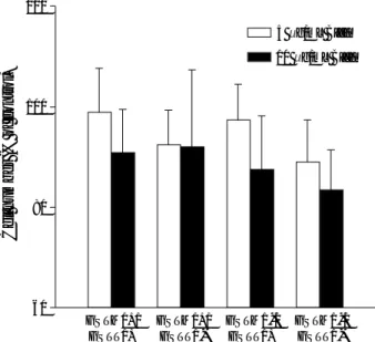
When the combinations of polymorphisms of GSTM1 and GSTT1 were analyzed, the highest induction of DPC coeffi-cient was observed with the GSTM1-null/GSTT1-null geno-type at 10 g/mL B[a]P (Fig. 4). At a lower B[a]P concen-tration of 5 g/mL, the DPC coefficient was highest with the GSTM1-null/GSTT1-positive combination. However, no significant difference (p>0.05) was observed among the geno-typic combinations at either B[a]P concentration.
GSTM1 genotype Positive (+) 14 95.86±8.30 91.59±12.04 <0.05 Null (-) 16 93.36±8.58 85.64±9.22 <0.01 p for interaction� > 0.05 GSTT1 genotype Positive (+) 15 98.28±7.65 89.27±9.46 <0.01 Null (-) 15 90.78±7.58 87.56±12.39 <0.01 p for interaction� > 0.05 All donors 30 94.53±8.40 88.41±10.86 <0.01 No. of subjects Genotype
Table 1. Inhibition of cell proliferation with respect to the GSTM1 and the GSTT1 polymorphisms in cultured human lymphocytes
Cell number (% of control) 5 g/mL B[a]P 10 g/mL B[a]P p for trend*
*, Test for trend of cell proliferation at different concentrations of B[a]P using repeated measures ANOVA. �
, Test for interaction of trend of cell proliferation at different concentrations of B[a]P by genotype of GSTM1 or GSTT1 using repeated measures ANOVA.
Cell number (% of control)
120 100 80 60 GSTM1+/ GSTT1+ GSTM1+/ GSTT1-GSTM1-/ GSTT1+ GSTM1-/ GSTT1-5 g/mL B[a]P
Fig. 1. Inhibition of cell proliferation as induced by a 24 hr treat-ment with benzo[a]pyrene in the lymphocyte cultures of human donors with different GSTM1 and GSTT1 genotypes. Bars repre-sent mean and standard deviation.
10 g/mL B[a]P
DPC level (% of crosslinked DNA)
9 8 7 6 5 4 3 2 1 0 0 5 10 GSTM1-positive GSTM1-null B[a]P concentration ( g/mL)
Fig. 2. DNA-protein crosslinks formation with respect to the GSTM1 polymorphism in cultured human lymphocytes.
■ ■ ■ ◆ ◆ ◆ ◆ ■
DPC level (% of crosslinked DNA)
9 8 7 6 5 4 3 2 1 0 0 5 10 GSTT1-positive GSTT1-null B[a]P concentration ( g/mL)
Fig. 3. DNA-protein crosslinks formation with respect to the GSTT1 polymorphism in cultured human lymphocytes.
■ ■ ■ ◆ ◆ ◆ ◆ ■
DISCUSSION
Our results indicate that B[a]P, without external metabol-ic activation, induced a signifmetabol-icant dose-dependent increase in both cytotoxicity and DPC formation in cultured human lymphocytes (p<0.01). However, both GSTM1 and GSTT1 polymorphisms did not play significant roles in the modifi-cation of B[a]P-induced genetic damage. We could not observe any significant interactions of B[a]P concentrations and the GSTM1 or the GSTT1 for the effect of cytotoxicity and DPC formation.
Human lymphocytes used in this study were obtained from healthy, non-smoking hospital administrative workers who had not been exposed to PAH or other DNA damaging agents by their occupations. Therefore, the experiments were rela-tively well-controlled in terms of the subject environment for genotoxicity assay. In vitro lymphocyte culture has a recog-nized validity as an assay tool because the blood lymphocytes represent an integrated DNA adduct burden from exposure to genotoxins via diverse routes, such as ingestion, inhalation, and absorption (23). In addition, human lymphocytes are known to directly metabolize a variety of chemicals, which induce genotoxic responses (3).
Genetic polymorphisms in enzymes involved in carcinogen metabolism influence cancer susceptibility, and glutathione S-transferases are involved in the detoxification of metabolic electrophilic compounds by binding to them directly or cat-alyzing their conjugation (24). GSTM1 and GSTT1 polymor-phisms are associated with the metabolism of PAH, which is a recognized potential carcinogen (25-27). Deletion of the GSTM1 gene has also been shown to be associated with an
increase in the level of DNA-adduct (25) or sister chromatid exchange (26). It has also been associated with various can-cers, including lung cancer (27), malignant mesothelioma (28), and bladder cancer (29) in epidemiologic studies. Al-though GSTM1 is allegedly involved in the detoxification of a wide range of electrophilic compounds, such as active metabolites of PAH, our study did not show statistically sig-nificant modification of DPC formation after benzo[a]pyrene treatment by the GSTM1 polymorphism.
It is not known whether the GSTT1 deletion can modify the risks associated with exposure to toxic and carcinogenic chemicals. The GSTT1 deficiency has been shown to poten-tiate the induction of chromosomal damage, and the carriers of the null genotype may be at increased cancer risk (30). In contrast, it was also reported that the GSTT1 deficiency could be protective under some circumstances (31). Our study shows that there is no significant difference between the GSTT1 genotypes in terms of cytotoxicity and DPC formation. The GSTT1 polymorphism did not change the effect of benzo[a]-pyrene on the cytotoxicity and DPC formation, either. This negative result may be partly due to either the relatively minor role GSTT1 plays in the detoxification of B[a]P or to the low expression of GSTT1 in human lymphocytes (32).
The coordinated activities of GSTM1 and GSTT1 may further modulate the susceptibility to cancer. When both GSTM1 and GSTT1 genes were investigated for a possible association with lung cancer, Kelsey et al. found this gene interaction carried the risk for lung cancer (13). Our study also showed that the genotoxicity of B[a]P was the highest in the absence of both GSTM1 and GSTT1 genes. However, this association was not statistically significant and the small number of blood donors in our study limited the interpreta-tion of the interactive effects of the genetic polymorphisms.
In this study, we evaluated the genotoxicity by DPC forma-tion, since many established or suspected carcinogens, such as ionizing radiation, ultraviolet, and formaldehyde, and nickel and chromium compounds are known to induce covalent DPC (17-19, 33, 34). It is a significant DNA defect that impairs the gene expression and chromatin structure, and can also lead to a deletion of DNA sequences during DNA replication because of the high probability of resisting repair (35). In contrast to a previous result, which did not show the DPC formation after B[a]P treatment by alkaline elution anal-ysis (36), our results showed a significant DPC formation by B[a]P.
Our present study did not find the genetic modification effect of the GSTM1 and GSTT1 polymorphisms in the DPC formation. However, the DPC formation, which represents the DNA damage, was found to be increased in the case of the GSTM1-null and GSTT1-null genotypes at a high B[a]P concentration. This work suggests a possibility that DPC could be used as a biomarker of benzo[a]pyrene exposure, and in vitro lymphocyte culture assay could be a useful tool for evaluating genetic susceptibility to environmental cancers.
DPC coefficient 18 16 14 12 10 8 6 4 2 0 GSTM1+/ GSTT1+ GSTM1+/ GSTT1-GSTM1-/ GSTT1+ GSTM1-/ GSTT1-5 g/mL B[a]P
Fig. 4. DNA-protein crosslink formation induced by a 24 hr treat-ment with benzo[a]pyrene in lymphocyte cultures of human donors with different GSTM1 and GSTT1 genotypes. Bars represent mean and standard deviation.
REFERENCES
1. Grinberg-Funes RA, Singh VN, Perera FP, Bell DA, Young TL, Dicky C, Wang LW, Santella RM. Polycyclic aromatic hydrocar-bon-DNA adducts in smokers and their relationship to micronutri-ent levels and the glutathione-S-transferase M1 genotype. Carcino-genesis 1994; 15: 2449-54.
2. Ambrosone CB, Freudenheim JL, Graham S, Marshall JR, Vena JE, Brasure JR, Laughlin R, Nemoto T, Michalek AM, Harrington A, Ford TD, Shields PG. Cytochrome P4501A1 and glutathione S-trans-ferase (M1) genetic polymorphisms and postmenopausal breast can-cer risk. Cancan-cer Res 1995; 55: 3483-5.
3. Salama SA, Abdel-Rahman SZ, Sierra-Torres CH, Hamada FA, Au WW. Role of polymorphic GSTM1 and GSTT1 genotypes on NNK-induced genotoxicity. Pharmacogenetics 1999; 9: 735-43. 4. Nelson HH, Wiencke JK, Christani DC, Cheng TJ, Zuo ZF, Schwartz
BS, Lee BK, Spitz MR, Wang M, Xu X, Kelsey KT. Ethnic differ-ences in the prevalence of the homozygous deleted genotype of glu-tathione S-transferase theta. Carcinogenesis 1995; 16: 1243-5. 5. Nebert DW. Role of genetics and drug metabolism in human cancer
risk. Mutat Res 1991; 247: 267-81.
6. Wolf CR. Metabolic factors in cancer susceptibility. Cancer Surv 1990; 9: 437-74.
7. Ryberg D, Skaug V, Hewer A, Phillips DH, Harries LW, Wolf CR, Ogreid D, Ulvik A, Vu P, Haugen A. Genotypes of glutathione trans-ferase M1 and P1 and their significance for lung DNA adduct levels and cancer risk. Carcinogenesis 1997; 18: 1285-9.
8. Zhong S, Wyllie AH, Barnes D, Wolf CR, Spurr NK. Relationship between the GSTM1 genetic polymorphism and susceptibility to blad-der, breast and colon cancer. Carcinogenesis 1993; 14: 1821-4. 9. Hirvonen A, Husgafvel-Pursiainen K, Attila S, Vainio H. The GSTM1
null genotype as a potential risk modifier for squamous cell carci-noma of the lung. Carcinogenesis 1993; 14: 1479-81.
10. Chen H, Sandler DP, Taylor JA, Shore DL, Liu E, Bloomfield CD, Bell DA. Increased risk for myelodysplastic syndromes in individuals with glutathione transferase theta 1 (GSTT1) gene defect. Lancet 1996; 347: 295-7.
11. Pemble S, Schroeder KR, Spencer SR, Meyer DJ, Hallier E, Bolt HM, Ketterer B, Taylor JB. Human glutathione S-transferase theta (GSTT1): cDNA cloning and the characterization of genetic poly-morphism. Biochem J 1994;300: 271-6.
12. Caporaso N, Land MT, Vinesis P. Relevance of metabolic polymor-phisms to human carcinogenesis: evaluation of epidemiologic evi-dence. Pharmacogenetics 1991; 1: 4-19.
13. Kelsey KT, Spitz MR, Zuo ZF, Wiencke JK. Polymorphisms in the glutathione S-transferase class mu and theta genes interact and in
-crease susceptibility to lung cancer in minority population (Texas, United States). Cancer Causes Control 1997; 8: 554-9.
14. Abdel-Rahman SZ, Anwar WA, Abdel-Aal WE, Mostafa HM, Au WW. GSTM1 and GSTT1 genes are potential risk modifiers for bladder cancer. Cancer Detect Prev 1998; 22: 129-38.
15. Geneste O, Camus AM, Castegnaro M, Petruzzelli S, Macchiarini P, Angeletti CA, Giuntini C, Bartsch H. Comparison of pulmonary DNA adduct levels measured by 32P-postlabelling and aryl
hydro-carbon hydroxylase activity in lung parenchyma of smokers and ex-smokers. Carcinogenesis 1991; 12: 1301-5.
16. Voitkun V, Zhitkovich A. Analysis of DNA-protein crosslinking activi-ty of malondialdehyde in vitro. Mutat Res 1999; 424: 97-106. 17. Cress AE, Kurath KM, Stea B, Bowden GT. The crosslinking of nu
-clear protein to DNA using ionizing radiation. J Cancer Res Clin On
-col 1990; 116: 324-30.
18. Casanova M, Deyo DF, Heck HD. Covalent binding of inhaled formaldehyde to DNA in the nasal mucosa of Fisher 344 rats: anal-ysis of formaldehyde and DNA by high-performance liquid chroma
-tography and provisional pharmacokinetic interpretation. Fundam Appl Toxicol 1989; 12: 397-417.
19. Sugiyama M, Wang XW, Costa M. Comparison of DNA lesions and cytotoxicity induced by calcium chromate in human, mouse, and ham-ster cell lines. Cancer Res 1986; 46: 4547-51.
20. Norppa H. Cytogenetic markers of susceptibility: influence of poly-morphic carcinogen-metabolizing enzymes. Environ Health Perspect 1997; 105 (Suppl. 4): 829-35.
21. Zhitkovich A, Costa M. A simple, sensitive assay to detect DNA-pro-tein crosslinks in intact cells and in vivo. Carcinogenesis 1992; 13: 1458-89.
22. Hong YC, Ha EH, Park HS. Influence of genetic susceptibility on uri-nary excretion of 8-OH-dG of firefighters. Occup Environ Med 2000; 57: 370-5.
23. Nia AB, Maas LM, Brouwer EM, Kleinjans JC, Van Schooten FJ. Comparison between smoking-related DNA adduct analysis in induced sputum and peripheral blood lymphocytes. Carcinogenesis 2000; 21: 1335-40.
24. Mannervik B, Danielson UH. Glutathione transferases-structure and catalytic activity. CRC Crit Rev Biochem 1988; 23: 283-337. 25. Ketterer B, Harris JM, Talaska G, Meyer DJ, Pemble SE, Taylor JB,
Lang NP, Kadlubar FF. The human glutathione S-transferse super-gene family, its polymorphism, and its effects on susceptibility to lung cancer. Environ Health Perspect 1992; 98: 87-94.
26. van Poppel G, de Vogel N, van Balderen PJ, Kok FJ. Increased cyto-genic damage in smokers deficient in glutathione S-transferase isozyme . Carcinogenesis 1992; 13: 303-5.
27. McWilliams JE, Sanderson BJS, Harris EL, Richert-Boe KE, Hen-ner WD. Glutathione S-transferase M1 (GSTM1) deficiency and lung cancer risk. Cancer Epidemiol Biomarkers Prev 1995; 4: 589-94.
28. Hirvonen A, Pelin K, Tammilehto L, Kajalainen A, Mattson K, Lin-nainmaa K. Inherited GSTM1 and NAT2 defects as concurrent risk modifiers in asbestos-related human malignant mesothelioma. Can-cer Res 1995; 55: 2981-3.
29. Katoh T, Inatomi H, Nagaoka A, Sugita A. Cytochrome P4501A1 gene polymorphism an homozygous deletion of the glutathione S-transferase M1 gene in urothelial cancer patient. Carcinogenesis 1995; 16: 655-7.
30. Jahnke V, Matthias C, Fryer A, Strange R. Glutathione S-transferase and cytochrome P-450 polymorphism as risk factors for squamous cell carcinoma of the larynx. Am J Surg 1996; 172: 671-3. 31. Kim H, Kim WJ, Lee HL, Lee MS, Kim CH, Kim RS, Nam HM. A
N-acetyl-transferase 2 and glutathione S-N-acetyl-transferase mu and theta on the risk of bladder cancer. Korean J Prev Med 1998; 31: 275-84.
32. Sasiadek M, Hirvonen A, Noga L, Paprocka-Borowicz M, Norppa H. Glutathione S-transferase M1 genotype influences sister chromatid exchange induction but not adaptive response in human lymphocytes treated with 1,2-epoxy-3-butane. Mutat Res 1999; 439: 207-12. 33. Shaham J, Bomstein Y, Meltzer A, Kaufman Z, Palma E, Ribak J.
DNA-protein crosslinks, a biomarker of exposure to formaldehyde in vitro and in vivo studies. Carcinogenesis 1996; 17: 121-5.
34. Patiermo SR, Costa M. DNA-protein crosslinks induced by nickel compounds in intact cultured mammalian cells. Chemico-Biol Inter-act 1985; 55: 75-91.
35. Costa M, Zhitkovich A, Toniolo P. DNA-protein cross-links in weld
-ers: molecular implications. Cancer Res 1993; 53: 460-3. 36. Cosma GN, Jamasbi R, Marchok AC. Growth inhibition and DNA
damage induced by benzo[a]pyrene and formaldehyde in primary cultures of rat tracheal epithelial cells. Mutat Res 1988; 201: 161-8.
![Fig. 4. DNA-protein crosslink formation induced by a 24 hr treat- treat-ment with benzo[a]pyrene in lymphocyte cultures of human donors with different GSTM1 and GSTT1 genotypes](https://thumb-ap.123doks.com/thumbv2/123dokinfo/5078612.73797/4.892.87.429.136.440/protein-crosslink-formation-induced-lymphocyte-cultures-different-genotypes.webp)
