Korean J Gastroenterol Vol. 60 No. 2, 128-131 http://dx.doi.org/10.4166/kjg.2012.60.2.128
IMAGE OF THE MONTH
Korean J Gastroenterol, Vol. 60 No. 2, August 2012 www.kjg.or.kr
내시경 점막하 박리절제술로 제거한 용종성 낭종성 위염
박혜정, 조인래, 김연희
1,
이상길
연세대학교 의과대학 내과학교실, 소화기병연구소, 병리학교실1
Gastritis Cystica Polyposa Treated with Endoscopic Submucosal Dissection
Hye Jung Park, In Rae Cho, Yon Hee Kim1 and Sang Kil Lee
Department of Internal Medicine and Institute of Gastroenterology, Department of Pathophysiology1, Yonsei University College of Medicine,
Seoul, Korea
CC This is an open access article distributed under the terms of the Creative Commons Attribution Non-Commercial License (http://creativecommons.org/licenses/ by-nc/3.0) which permits unrestricted non-commercial use, distribution, and reproduction in any medium, provided the original work is properly cited.
교신저자: 이상길, 120-752, 서울시 서대문구 연세로 50, 연세대학교 의과대학 내과학교실
Correspondence to: Sang Kil Lee, Department of Internal Medicine, Yonsei University College of Medicine, 50 Yonsei-ro, Seodaemun-gu, Seoul 120-752, Korea. Tel: +82-2-2228-1996, Fax: +82-2-393-6884, E-mail: sklee@yuhs.ac
Financial support: None. Conflict of interest: None.
Fig. 1. Endoscopic findings of the lesion. (A) A 4 cm sized polypoid mass was noted in the greater curvature of the midbody. (B) Distict ulceration and hyperemia were noted on the surface of the lesion.
증례: 57세 남자 환자가 검사 5일 전부터 발생한 흑색변을
주소로 외부 병원 방문하여 받은 상부위장관 내시경검사에서,
궤양을 동반한 위의 종괴가 발견되어 본원으로 전원되었다.
환자는 과거력 및 가족력에서 특이 소견은 없었으며, 위절제
술을 포함한 수술력도 없었다. 내원 당시 혈압은 120/80
mmHg, 맥박은 분당 65회, 호흡수는 분당 18회, 체온 36.6
oC
이었으며, 신체 검진에서 흉부 및 복부의 이상소견은 없었다.
말초혈액검사에서 백혈구 4,140/mm
3, 혈색소 11 g/dL, 혈소
판 229,000/mm
3이었다. 혈청생화학검사에서 혈청 총단백 7
g/dL, 알부민 4.5 g/dL, AST/ALT 22/19 IU/L, 총빌루리빈
0.4 mg/dL, BUN 14.0 mg/dL, 크레아티닌 1.19 mg/dL로
이상 소견은 없었다.
외부 병원에서 시행한 상부위장관 내시경검사에서 위체부
대굽이에 40 mm 크기의 돌출형 종괴가 관찰되었는데, 용종
의 표면에 출혈의 흔적을 동반한 궤양이 동반되어 있었다
(Fig. 1). 본원에서 시행한 내시경초음파에서 위체부 대굽이에
점막하층과 고유근층 사이에서 기원하는 24×16 mm 크기의
낭성, 저음영의 균질한 등에코성을 보이는 병변이 관찰되었다
(Fig. 2). 복부 전산화단층촬영에서는 경계가 뚜렷하며 저음영
을 보이는 2.3 cm 크기의 병변이 관찰되었으며, 그 외 림프절
종대나 원격전이 소견은 보이지 않았다(Fig. 3). 상기 소견을
종합해 볼 때, 중복 낭종(duplication cyst), 또는 림프관종
Park HJ, et al. Gastritis Cystica Polyposa Treated with Endoscopic Submucosal Dissection
129
Vol. 60 No. 2, August 2012
Fig. 2. Endoscopic ultrasound find-ings. (A) Hypoechoic and polypoid lesion (2.4×1.6 cm) was origninated from the junction between submuco-sa and proper muscle. (B) The mass showed low and homogeneous inter-nal echogeneicity.
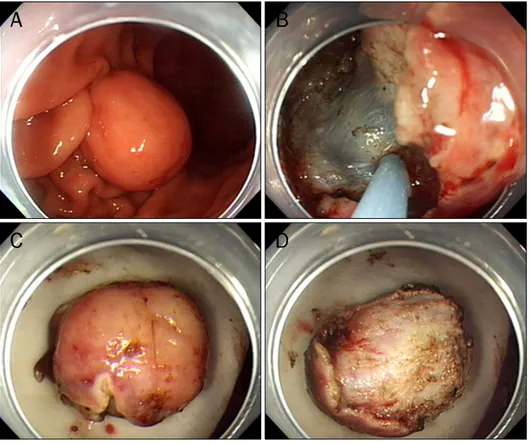
Fig. 4. Endoscopic resection (ESD) of the lesion. (A) 2.5 cm sized polypoid lesion, which was covered with normal gastric mucosa was seen at the greater curvature of body. (B) ESD was done by IT-knife. (C, D) Resected mass was well capsulated and easily detached from the base.
130
박혜정 등. 내시경 점막하 박리절제술로 제거한 용종성 낭종성 위염The Korean Journal of Gastroenterology
Fig. 5. Microscopic findings (H&E). (A, B) There was a dilated cyst lined by cell layer of the submucosa (×1.25). (C) A normal-appearing lamina propria surrounded the focal area of the cyst (×40). (D) The lining epithelium consisted of foveolar and mucin secreting cells (×100).
등의 양성 상피하 종양의 가능성이 높으나, 표면의 궤양과 출
혈을 동반한 경우로 조직 확진이 필요할 것으로 판단되었다.
시술 당시 외부 병원에서 3개월 전 시행한 상부위장관 내
시경검사에서 관찰되었던 용종 표면의 출혈의 흔적을 동반한
궤양은 관찰되지 않았고, 정상 위 점막으로 덮인 2.5 cm 크기
의 병변이 확인되었으며, 내시경 점막하 박리절제술을 통해
성공적으로 제거되었다(Fig. 4). 조직 검사 결과 점막하층까지
용종성 과증식 소견 관찰되어 용종성 낭종성 위염(gastritis
cystica polyposa)으로 진단되었다(Fig. 5). 이후 출혈이나 천
공 등의 합병증 없이 퇴원하였고, 추가적인 치료 없이 현재까
지 1년 이상 외래 추적 관찰 중이며, 재발이나 새로운 병변은
발견되지 않았다.
진단: 용종성 낭종성 위염
용종성 낭종성 위염은 위점막의 용종성 과증식이 특징인
드문 질환이다. 1972년 Littler와 Gleibermann
1에 의해 처음
기술된 이 질환은 흔히 위 절제술을 받은 환자에서 위장문합
부에서 발병되며, 드물게 수술병력이 없이 발병되기도 한
다.
1-5병리기전이 명확히 밝혀지지는 않았지만 만성 염증에
의해 발병하는 것으로 여겨지고 있는데, 수술 후 봉합사에 의
한 위 점막의 미란, 만성 허혈, 담즙 역류 및 만성 위축성 위염
등이 가능한 원인으로 보고되고 있다.
3-5반복적인 위 점막 손
상에 의한 미란과 재생성은 위선을 포함한 상피조직을 점막하
층까지 이주시켜 용종성 낭종성 위염을 일으킬 뿐만 아니라
상피이형성이나 위선암종과 같은 악성 병변으로의 변화도 일
으킬 수 있다.
4그러므로 용종성 낭종성 위염은 전암 병변으로
여겨져 대부분 절제술을 시행해왔다.
6-8용종성 낭종성 위염은 내시경에서 점막하 종양이나 단순
용종처럼 큰 규모의 위 주름의 모양을 하고 있으며,
6내시경초
음파에서 점막하층에 균일하고, 저에코의 다발성의 용종 소견
을 보인다.
6,7조직 검사에서는 점막하층까지 위선의 낭종 확
Park HJ, et al. Gastritis Cystica Polyposa Treated with Endoscopic Submucosal Dissection
131
Vol. 60 No. 2, August 2012
장 및 과립오목의 증식과 길이 확장의 소견을 보인다.
9이번 증례는 출혈의 흔적을 동반한 궤양이 있던 자리에 용
종성 낭종성 위염이 발생한 사례로서, 궤양이 생성되고 치유
되는 과정에서 위 점막의 만성 염증, 반복적인 미란, 상처 치
유 및 재생화를 거치며 용종성 낭종성 위염이 발생했을 것으
로 추측된다. 이는 그동안 드물게 보고되어 온 비수술적 환자
에서의 용종성 낭종성 위염이 위궤양의 생성과 치유 과정에서
도 발생될 수 있다는 가능성을 시사한다. 저자들이 아는 바로
는 국내에서 보고된 용종성 낭종성 위염의 사례 중 궤양을
동반했던 사례는 이번 증례가 처음이다. 용종성 낭종성 위염
은 전암 병변으로 여겨지기 때문에, 수술, 내시경 점막절제술,
내시경 점막하 박리절제술 등의 방법으로 치료되어 왔다.
6,8REFERENCES
1. Littler ER, Gleibermann E. Gastritis cystica polyposa. (Gastric mucosal prolapse at gastroenterostomy site, with cystic and in-filtrative epithelial hyperplasia). Cancer 1972;29:205-209. 2. Wu MT, Pan HB, Lai PH, Chang JM, Tsai SH, Wu CW. CT of gastritis
cystica polyposa. Abdom Imaging 1994;19:8-10.
3. Oberman HA, Lodmell JG, Sower ND. Diffuse heterotopic cystic malformation of the stomach. N Engl J Med 1963;269:909-911. 4. Iwanaga T, Koyama H, Takahashi Y, Taniguchi H, Wada A. Diffuse
submucosal cysts and carcinoma of the stomach. Cancer 1975; 36:606-614.
5. Yamagiwa H, Matsuzaki O, Ishihara A, Yoshimura H. Heterotopic gastric glands in the submucosa of the stomach. Acta Pathol Jpn 1979;29:347-350.
6. Park JS, Myung SJ, Jung HY, et al. Endoscopic treatment of gas-tritis cystica polyposa found in an unoperated stomach. Gast-rointest Endosc 2001;54:101-103.
7. Tuncer K, Alkanat M, Musoğlu A, Aydin A. Gastritis cystica poly-posa found in an unoperated stomach: an unusual case treated by endoscopic polypectomy. Endoscopy 2003;35:882. 8. Park CH, Park JM, Jung CK, et al. Early gastric cancer associated
with gastritis cystica polyposa in the unoperated stomach treat-ed by endoscopic submucosal dissection. Gastrointest Endosc 2009;69:e47-e50.
9. Okada M, Iizuka Y, Oh K, Murayama H, Maekawa T. Gastritis cyst-ica profunda presenting as giant gastric mucosal folds: the role of endoscopic ultrasonography and mucosectomy in the diag-nostic work-up. Gastrointest Endosc 1994;40:640-644.