Distal hyperintense vessels on FLAIR
An MRI marker for collateral circulation in acute stroke?
K.Y. Lee, MD, PhD L.L. Latour, PhD M. Luby, PhD A.W. Hsia, MD J.G. Merino, MD, MPhil S. Warach, MD, PhD ABSTRACT
Background:Hyperintense vessels (HV) on fluid-attenuated inversion recovery imaging are fre-quently observed in acute ischemic stroke patients. However, the exact mechanism and clinical implications of this sign have not yet been clearly defined. The features of HV and its relevance to other imaging factors are presented here.
Methods:Prominence and location of HV were documented in 52 consecutive patients with mid-dle cerebral artery (MCA) territory infarction, before treatment with IV recombinant tissue plas-minogen activator. Pretreatment ischemic lesion volume, perfusion lesion volume, and vessel occlusion were determined in addition to recanalization status and ischemic lesion volume on follow-up imaging. NIH Stroke Scale (NIHSS) was used as a measure of clinical severity.
Results:HV distal to arterial occlusion was observed in 73% of patients; more frequent in proxi-mal than distal MCA occlusion patients. Among the 38 patients with proxiproxi-mal MCA occlusion, initial perfusion lesion volume was comparable among patients with different grade distal HV. However, patients with more prominent distal HV had smaller initial, 24-hour, and subacute isch-emic lesion volumes and lower initial NIHSS scores.
Conclusions:The presence of distal hyperintense vessels before thrombolytic treatment is asso-ciated with large diffusion–perfusion mismatch and smaller subacute ischemic lesion volumes in patients with proximal middle cerebral artery occlusion.Neurology®2009;72:1134–1139
GLOSSARY
DWI⫽ diffusion-weighted imaging; FLAIR ⫽ fluid-attenuated inversion recovery; GRE ⫽ gradient recalled echo; HV ⫽ hyper-intense vessels; MCA⫽ middle cerebral artery; MRA ⫽ magnetic resonance angiography; MTT ⫽ mean transit time; NIHSS ⫽ NIH Stroke Scale; PWI⫽ perfusion-weighted imaging; rt-PA ⫽ recombinant tissue plasminogen activator; TE ⫽ echo time; TI⫽ inversion time; TIMI ⫽ thrombolysis in myocardial infarction; TR ⫽ repetition time.
Hyperintense vessels (HV) on fluid-attenuated inversion recovery (FLAIR) images can be
observed in the hemisphere affected by an arterial occlusion in some patients, but not all.
1-6Although HV is thought to be similar to the hyperdense artery sign on CT scan or the
suscep-tibility vessel sign on gradient recalled echo (GRE) MRI, it has been shown to be superior to
both for the detection of arterial occlusion.
4,7-9HV usually has longer course than the other two
signs and this suggests that different mechanisms other than intraluminal thrombus cause HV.
While the mechanism of HV remains to be established, stationary blood and slow antegrade or
retrograde collateral circulation have been suggested as possible explanations for HV.
1,5,10-12There are only a few reports suggesting that HV is related to poor prognosis, or has no
specific prognostic meaning other than the knowledge of an arterial occlusion.
4,13However, the
stroke populations in previous studies were heterogeneous; patients with proximal and distal as
well as anterior and posterior circulation occlusions were considered together, potentially
con-founding tests for significance. In order to verify the clinical implication of HV on FLAIR, it
Address correspondence andreprint requests to Dr. Steven Warach, Section on Stroke Diagnostics and Therapeutics, National Institute of Neurological Disorders and Stroke, 10 Center Drive, Room B1D733, Bethesda, MD 20892-1063
warachs@ninds.nih.gov
e-Pub ahead of print on February 11, 2009, at www.neurology.org.
From the Section on Stroke Diagnostics and Therapeutics (K.Y.L., L.L.L., M.L., J.G.M., S.W.), National Institute of Neurological Disorders and Stroke, National Institutes of Health, Bethesda, MD; the Washington Hospital Center Stroke Center (A.W.H.), Washington, DC; and the Department of Neurology (K.Y.L.), Yonsei University College of Medicine, Seoul, Korea.
should be compared between similar groups
of patients, such as those having similar sites
of arterial occlusion or similar perfusion
le-sion volumes.
We speculate that HV may be the result of
beneficial collateral arterial flow beyond the
site of arterial occlusion, and those patients
with HV will have a better prognosis and
smaller infarction size than those patients
without HV.
METHODSSubjects. For this analysis, we considered
con-secutive acute stroke patients enrolled in a natural history of stroke study approved by the Institutional Review Board at the National Institute of Neurological Disorders and Stroke and Suburban Hospital in Bethesda, MD, and at Washington Hos-pital Center in Washington, DC. We included patients who presented with acute middle cerebral artery (MCA) territory ischemic stroke within 3 hours after onset and were treated with IV recombinant tissue plasminogen activator (rt-PA). We ex-cluded patients with no prethrombolysis brain MRI, no follow-up MRI after treatment, or lacunar infarction. Neuro-logic deficit was evaluated with the NIH Stroke Scale (NIHSS) on admission and 5 days after admission. All patients, or their legally authorized representative, signed informed consent.
MRI protocol. Patients were imaged with either a 1.5 T (GE
Medical Systems, Milwaukee, WI) or 3.0 T (Philips Medical Systems, Best, the Netherlands) clinical MRI system using com-mercially available hardware and software. The baseline and follow-up imaging protocols were standardized and included co-localized diffusion-weighted imaging (DWI), T2-FLAIR, GRE, bolus tracking perfusion-weighted imaging (PWI), as well as in-tracranial and exin-tracranial contrast enhanced magnetic reso-nance angiography (MRA), and intracranial time-of-flight MRA. FLAIR images were acquired with commercially available 2D sequences, balanced (roughly) across field strength for con-spicuity of chronic ischemic parenchyma, with the following rel-evant parameters at 1.5 T: repetition time (TR)/echo time (TE)⫽ 9,000/145 msec, inversion time (TI)⫽ 2,200 msec, 20 contigu-ous slices with 0.9⫻ 0.9 ⫻ 7 mm resolution (zero filled) for a total acquisition of 2 minutes 25 seconds; at 3T: TR/TE⫽ 9,000/120 msec, TI⫽ 2,600 msec, 20 contiguous slices with 1⫻ 1 ⫻ 4 mm resolution (SENSE R ⫽ 1.75), for a total acqui-sition of 2 minutes 15 seconds.
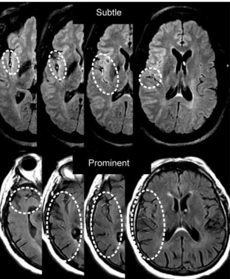
Imaging analysis. HV was defined as a linear or serpentine
appearing hyperintensity on FLAIR imaging corresponding to a typical arterial course. Presence of HV was categorized as proxi-mal or distal in relation to arterial branching of the MCA. HV was described as proximal when it was present to or within the Sylvian fissure, i.e., corresponding to the MCA M1 or M2 seg-ments, and it was graded as either absent or present. HV was described as distal when it was present distal to the Sylvian fis-sure, i.e., corresponding to MCA M3 or distal segments, and was graded as 1) absent, 2) subtle, or 3) prominent (figure 1). Subtle distal HV was defined as presence of HV less than 1/3 of the perfusion lesion and prominent distal HV was defined as pres-ence of HV more than 1/3 of the perfusion lesion. Two readers graded the FLAIR imaging independently with discordance set-tled by a separate consensus reading. Initial ischemic lesion vol-ume and perfusion lesion volvol-ume were measured on initial
pretreatment DWI and mean transit time (MTT) maps calcu-lated from PWI, 24-hour lesion volume measured on DWI, and subacute ischemic lesion volume measured on 3- to 7-day follow-up FLAIR imaging. All volumes were measured by a third experienced reader, blinded to the HV grading. Initial diffusion– perfusion mismatch was calculated by the equation [(initial per-fusion lesion volume on MTT⫺ initial ischemic lesion volume on DWI)/initial perfusion lesion volume on MTT]⫻ 100. Arte-rial occlusion site and recanalization status was read indepen-dently from the other readings. Arterial occlusion site was determined using MRA and PWI. We defined proximal MCA occlusion as M1 or M2 segment occlusion solely based on the MRA. M1 occlusion was defined as a main MCA trunk occlu-sion before the bifurcation and M2 occluocclu-sion was defined as a branch occlusion after the bifurcation. M3 distal occlusion was defined as distal branch occlusion without a perfusion lesion in the insular area on PWI. If there was no definite occlusion site visible on MRA but a perfusion lesion was visible on PWI, the patient was considered to have a M3 distal occlusion. Recanali-zation status was categorized by a modified grading system based on thrombolysis in myocardial infarction (TIMI) grade using both MRA and PWI.14TIMI 0 and 1 was regarded as poor
recanalization and TIMI 2 and 3 as successful recanalization.
Statistical analysis. Statistical analysis was performed with
SPSS 14.0 software for Windows. Mann–Whitney U test and Kruskal-Wallis H test was used to compare acute DWI ischemic lesion volume, acute perfusion lesion volume on MTT, diffu-sion–perfusion mismatch, 24-hour and subacute ischemic lesion volumes, and NIHSS score. Correlation between HV grade and recanalization status was analyzed by2test. Regression
anal-ysis was performed using 5-day NIHSS score as an outcome variable. Interobserver variability of HV grading was analyzed by k statistics. A two-tailed value of p⬍ 0.05 was considered significant.
RESULTSFrom 111 patients who were treated with IV rt-PA and consented to natural history study, 59 patients were excluded because of no pre rt-PA MRI (21), no follow-up MRI (10), unreadable FLAIR (5), and lacunar infarction or vertebrobasilar artery terri-tory infarction (23). Among the 52 patients who met the inclusion criteria, 62% of patients were women, mean age was 68.9 ⫾ 15.4 years, and the median initial NIHSS score was 8 (range 1–28). Twenty-two patients were determined to have MCA M1 occlu-sion (including 6 patients with an internal carotid artery T occlusion), 16 patients with MCA M2 oc-clusion, and 11 patients with MCA M3 distal occlu-sion. The remaining three patients did not have any MRA or PWI abnormalities.
All 52 patients had interpretable prethrombolysis FLAIR images; 19 of those patients were imaged at 3.0 T. Proximal HV was seen in 77% of patients (k⫽ 0.833). Prominent distal HV was seen in 46% of patients and subtle distal HV was seen in 27% (k⫽ 0.661) (table 1).
In the group of 38 patients having MCA M1 or M2 occlusion, ischemic lesion volume could be de-termined on initial DWI in all patients and on
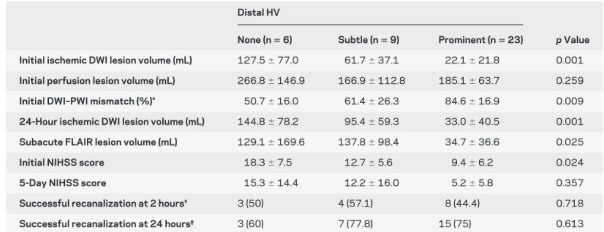
24-hour DWI in 36 patients; initial perfusion lesion volume on MTT was available in 32 and subacute lesion volume on FLAIR in 27. Patients with distal HV had smaller initial, 24-hour and subacute lesion volumes, and had larger diffusion–perfusion mis-match at baseline than did patients without distal HV. The initial perfusion lesion volume on MTT
was comparable among the three groups (figure 2, table 2). DWI-positive lesions were usually distrib-uted in the area without HV in patients with large diffusion–perfusion mismatch.
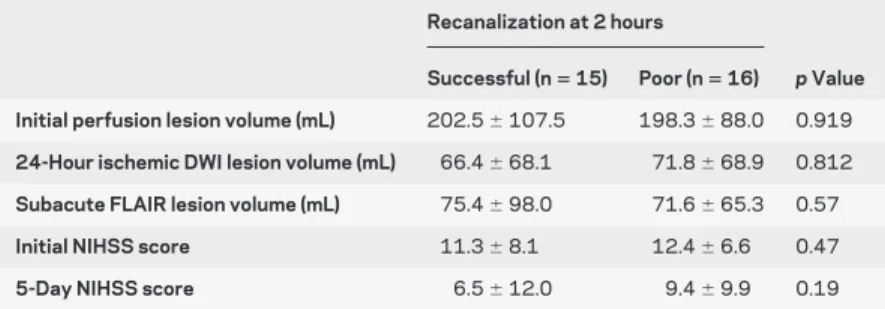
Successful recanalization was achieved in 48% (15 out of the 31 with available MRA) at 2 hours after thrombolysis and 74% (25 out of the 34 with available MRA) at 24 hours after thrombolysis in pa-tients with M1 or M2 occlusion. There was no dif-ference in recanalization rate among patients with different grade distal HV. Also, 24-hour and sub-acute infarction volume and 5-day NIHSS score did not show significant differences based on 2-hour re-canalization status (table 3). However, patients with prominent distal HV who recanalized at 2 hours had smaller subacute ischemic lesion volumes on FLAIR compared with patients who did not recanalize (21.3 vs 53.0 mL; p⫽ 0.108). NIHSS score was obtained in all patients with M1 or M2 occlusion at the time of initial imaging, but only in 29 of 38 patients at 5 days. Initial and 5-day NIHSS scores were lower in patients with high grade distal HV on initial imaging than in patients without, though only initial NIHSS score reached significance. Regression analysis using age, recanalization status, HV grade, and initial NIHSS score as independent variables demonstrated that only initial NIHSS score was a predictor of 5-day NIHSS score (B⫽ 0.666, p ⫽ 0.025).
DISCUSSION This study demonstrates that the presence of distal HV on initial FLAIR imaging is associated with smaller ischemic lesion volume on MRI and milder clinical severity as measured by the NIHSS. While all patients with proximal MCA oc-clusion had similar perfusion lesion volumes and some degree of diffusion–perfusion mismatch on ini-tial MRI, patients with prominent distal HV had smaller initial ischemic lesion volumes, larger diffu-sion–perfusion mismatch, relatively smaller lesion growth, and smaller subacute ischemic lesion vol-umes than did patients without it. The mechanism and causality cannot be definitively established in this study, but it is reasonable to speculate that prom-inent distal HV may be reflective of good collateral blood flow distal to the site of arterial occlusion and less ischemic injury to tissue supplied by occluded artery.
We categorized the HV in MCA occlusion as oc-curring proximally and distally. Contrary to proxi-mal HV, which frequently appears as a single, linear structure with limited curvature and with proximity to the M1–M2 segments of the MCA, distal HV appears as a varying number of serpentine vessel-like structures distal to the M2 segment and covers an area variable in size. Therefore, we implemented a
Figure 1 Example of distal hyperintense vessels (HV) grading
Both patients have middle cerebral artery M1 occlusion and demonstrate different levels of HV on fluid-attenuated inversion recovery imaging.
Table 1 Frequency of HV based on middle
cerebral artery occlusion site
M1 (nⴝ 22) M2(nⴝ 16) M3 distal or none (nⴝ 14) Proximal HV 22 (100) 13 (81) 5 (36) Distal HV Prominent 15 (68) 8 (50) 1 (7) Subtle 4 (18) 5 (31) 5 (36) None 3 (14) 3 (19) 8 (57) Values are n (%).
HV⫽ hyperintense vessels; M1 ⫽ middle cerebral artery M1 portion; M2⫽ middle cerebral artery M2 portion; M3 distal⫽ middle cerebral artery M3 and distal portion.
grading system for distal HV based on the size of the area involved. Distal HV is frequently observed at the MCA M3 branch level, originating from an
MCA M2 segment coursing anterior to posterior in the Sylvian fissure. The M3 branches in this area are still of relatively large vessel caliber and track
pre-Figure 2 Representative cases of different grade distal hyperintense vessels (HV)
(A) Initial diffusion- and perfusion-weighted MRI shows large left middle cerebral artery ischemic and perfusion lesions. No distal HV is seen on initial fluid-attenuated inversion recovery (FLAIR) imaging. Five-day follow-up FLAIR imaging shows a large cerebral infarction. (B) Initial diffusion- and perfusion-weighted MRI shows a small ischemic lesion with a large perfusion lesion. Prominent distal HV is noted on initial FLAIR imaging (arrows). Three-day follow-up FLAIR shows a small cerebral infarction. DWI⫽ diffusion-weighted imaging; PWI ⫽ perfusion-weighted imaging; MTT ⫽ mean transit time.
Table 2 Comparison of ischemic lesion volume, perfusion lesion volume, clinical outcome, and recanalization rate by distal hyperintense vessels (HV) grade in patients with middle cerebral artery M1 or M2 occlusion
Distal HV
None (nⴝ 6) Subtle (nⴝ 9) Prominent (nⴝ 23) p Value Initial ischemic DWI lesion volume (mL) 127.5⫾ 77.0 61.7⫾ 37.1 22.1⫾ 21.8 0.001
Initial perfusion lesion volume (mL) 266.8⫾ 146.9 166.9⫾ 112.8 185.1⫾ 63.7 0.259
Initial DWI–PWI mismatch (%)* 50.7⫾ 16.0 61.4⫾ 26.3 84.6⫾ 16.9 0.009
24-Hour ischemic DWI lesion volume (mL) 144.8⫾ 78.2 95.4⫾ 59.3 33.0⫾ 40.5 0.001
Subacute FLAIR lesion volume (mL) 129.1⫾ 169.6 137.8⫾ 98.4 34.7⫾ 36.6 0.025
Initial NIHSS score 18.3⫾ 7.5 12.7⫾ 5.6 9.4⫾ 6.2 0.024
5-Day NIHSS score 15.3⫾ 14.4 12.2⫾ 16.0 5.2⫾ 5.8 0.357
Successful recanalization at 2 hours† 3 (50) 4 (57.1) 8 (44.4) 0.718
Successful recanalization at 24 hours‡ 3 (60) 7 (77.8) 15 (75) 0.613
Data are mean⫾ SD or n (%).
*Calculated by [(initial perfusion lesion volume on MTT⫺ initial ischemic lesion volume on DWI)/initial perfusion lesion vol-ume on MTT] x 100.
†Recanalization rate was evaluated in 7 subtle and 18 prominent distal HV patients. ‡Recanalization rate was evaluated in 5 none and 20 prominent distal HV patients.
DWI⫽ diffusion-weighted imaging; PWI ⫽ perfusion-weighted imaging; FLAIR ⫽ fluid-attenuated inversion recovery; NIHSS⫽ NIH Stroke Scale.
dominantly in the plane of the axial FLAIR MRI slice, allowing for easy detection. More distal MCA branches are smaller and have an inferior–superior orientation that appear as bright dots in the trans-verse imaging and are therefore more difficult to detect.
Proximal HV, which is frequently observed prox-imal to or within the Sylvian fissure, may have differ-ent implications in comparison to distal HV. In contrast to distal HV, 92% of patients with proximal MCA occlusion had proximal HV, regardless of the initial ischemic lesion volume, lesion volume pro-gression, and clinical severity. Proximal HV was not a useful prognostic indicator in this study. Proximal HV may be used as a marker for arterial occlusion, presumably the result of the thrombus inside the ar-terial lumen.4,7 Distal HV is more likely related to
either slow, anterograde flow at the site of the occlu-sion or retrograde collateral flow from arteries unaf-fected by occlusion, both having a relative delay in transit time with the latter owing to a more circui-tous route of delivery. In patients with similar perfu-sion leperfu-sion volumes, prominent distal HV may provide a mechanism for discriminating tissue kept viable for extended periods by way of a well-developed collateral network from tissue rapidly evolving as the result of marginalized flow distal to the site of the occlusion.
We did not find any association between distal HV and successful recanalization at 2 and 24 hours after IV thrombolysis. A previous study using cerebral angiography suggested that both good collateral flow and complete recanalization were independently re-lated to good clinical outcome and small infarction volume.15However, we could not find a significant
difference in infarction volume and 5-day NIHSS score based on the 2-hour recanalization status. We speculated that the small sample size of our study and different outcome evaluation methods (discharge modified Rankin Scale vs 5-day NIHSS) explain
these apparently discrepant findings. Subgroup anal-ysis using the patient with prominent distal HV showed that successful recanalization at 2 hours showed trend of smaller subacute ischemic lesion vol-umes on FLAIR than patients with poor recanaliza-tion. This may suggest that successful recanalization, independent of collateral blood flow, remains an im-portant prognostic factor in rt-PA–treated patients.
Multivariate regression analysis demonstrated that only initial NIHSS score was a significant pre-dictor of 5-day NIHSS score. We speculated that the clinical significance of HV to predict good clinical outcome was already reflected to low initial NIHSS score as shown in table 2. Therefore, HV grading showed no significance in predicting 5-day NIHSS score.
Good collateral blood flow in acute ischemic stroke is known to influence prognosis and infarct volume.15-17The pial collaterals assessed by cerebral
angiography have been reported to have prognostic significance, including an association with smaller infarct volumes and good clinical outcomes in acute ischemic stroke.15Collateral flow can prolong tissue
viability and maximize the volume of salvageable tis-sue. Thus the information of collateral blood flow has potential clinical applications for making treat-ment decisions and predicting outcome after acute ischemic stroke. Cerebral angiography is the gold standard used to evaluate collateral blood flow, but has the limitation of invasiveness, relatively long ac-quisition and procedural times, and low accessibility for general use in acute ischemic stroke.18,19 Other
imaging modalities like MRI, CT, and transcranial Doppler can be used to evaluate collateral blood flow.20-22Based on our findings and reported cases,1,12
we suggest that distal HV in patients with MCA oc-clusion results from collateral blood flow originating in neighboring arterial territories, especially via pial collaterals.
A limitation of this study is the inability to estab-lish a direct association between distal HV on FLAIR imaging and an independent measure of collateral blood flow. We hope that direct comparison of HV on FLAIR and collateral blood flow on conventional angiography may help to define the mechanism of HV in future studies. In addition, we could not es-tablish the relationship between presence of HV and outcome, although there was a trend of lower 5-day NIHSS score in patients with prominent distal HV than without. A larger number of patients and an-other outcome measurement, such as 90-day modi-fied Rankin Scale score, are needed to prove the clinical significance of HV.
In our cohort, patients with prominent distal HV presented with small acute ischemic lesions and
sub-Table 3 Comparison of ischemic lesion volume, perfusion lesion volume, and
clinical outcome by recanalization status in patients with middle cerebral artery M1 or M2 occlusion
Recanalization at 2 hours
Successful (nⴝ 15) Poor (nⴝ 16) p Value Initial perfusion lesion volume (mL) 202.5⫾ 107.5 198.3⫾ 88.0 0.919
24-Hour ischemic DWI lesion volume (mL) 66.4⫾ 68.1 71.8⫾ 68.9 0.812
Subacute FLAIR lesion volume (mL) 75.4⫾ 98.0 71.6⫾ 65.3 0.57
Initial NIHSS score 11.3⫾ 8.1 12.4⫾ 6.6 0.47
5-Day NIHSS score 6.5⫾ 12.0 9.4⫾ 9.9 0.19
DWI⫽ diffusion-weighted imaging; FLAIR ⫽ fluid-attenuated inversion recovery; NIHSS ⫽ NIH Stroke Scale.
acute infarct volumes despite proximal MCA occlu-sion and large perfuocclu-sion leocclu-sions. If prominent distal HV on acute evaluation is predictive of prolonged tissue viability, it can be used to identify patients who will benefit from treatment beyond the current 3-hour thrombolysis time window. Further study is needed to confirm our findings and prove this hy-pothesis.
AUTHOR CONTRIBUTIONS
Statistical analysis was performed by K.Y. Lee, MD, PhD.
Received August 3, 2008. Accepted in final form December 29, 2008.
REFERENCES
1. Noguchi K, Ogawa T, Inugami A, et al. MRI of acute cerebral infarction: a comparison of FLAIR and T2-weighted fast spin-echo imaging. Neuroradiology 1997; 39:406–410.
2. Cosnard G, Duprez T, Grandin C, Smith AM, Munier T, Peeters A. Fast FLAIR sequence for detecting major vascu-lar abnormalities during the hyperacute phase of stroke: a comparison with MR angiography. Neuroradiology 1999; 41:342–346.
3. Makkat S, Vandevenne JE, Verswijvel G, et al. Signs of acute stroke seen on fluid-attenuated inversion recovery MR imaging. AJR Am J Roentgenol 2002;179:237–243. 4. Schellinger PD, Chalela JA, Kang DW, Latour LL,
Warach S. Diagnostic and prognostic value of early MR imaging vessel signs in hyperacute stroke patients imaged ⬍3 hours and treated with recombinant tissue plasmino-gen activator. AJNR Am J Neuroradiol 2005;26:618–624. 5. Kamran S, Bates V, Bakshi R, Wright P, Kinkel W, Miletich R. Significance of hyperintense vessels on FLAIR MRI in acute stroke. Neurology 2000;55:265–269.
6. Maeda M, Koshimoto Y, Uematsu H, et al. Time course of arterial hyperintensity with fast fluid-attenuated inversion-recovery imaging in acute and subacute middle cerebral arterial infarction. J Magn Reson Imaging 2001;13:987– 990.
7. Assouline E, Benziane K, Reizine D, et al. Intra-arterial thrombus visualized on T2* gradient echo imaging in acute ischemic stroke. Cerebrovasc Dis 2005;20:6–11. 8. Flacke S, Urbach H, Keller E, et al. Middle cerebral artery
(MCA) susceptibility sign at susceptibility-based perfusion MR imaging: clinical importance and comparison with hy-perdense MCA sign at CT. Radiology 2000;215:476–482. 9. Chalela JA, Haymore JB, Ezzeddine MA, Davis LA, Warach S. The hypointense MCA sign. Neurology 2002; 58:1470.
10. Noguchi K, Ogawa T, Inugami A, et al. Acute subarach-noid hemorrhage: MR imaging with fluid-attenuated in-version recovery pulse sequences. Radiology 1995;196: 773–777.
11. Toyoda K, Ida M, Fukuda K. Fluid-attenuated inversion re-covery intraarterial signal: an early sign of hyperacute cerebral ischemia. AJNR Am J Neuroradiol 2001;22:1021–1029. 12. Liebeskind DS. Location, location, location: angiography
discerns early MR imaging vessel signs due to proximal arterial occlusion and distal collateral flow. AJNR Am J Neuroradiol 2005;26:2432–2433.
13. Girot M, Gauvrit JY, Cordonnier C, et al. Prognostic value of hyperintense vessel signals on fluid-attenuated inversion recovery sequences in acute cerebral ischemia. Eur Neurol 2007;57:75–79.
14. The thrombolysis in myocardial infarction (TIMI) trial: phase I findings: TIMI study group. N Engl J Med 1985; 312:932–936.
15. Christoforidis GA, Mohammad Y, Kehagias D, Avutu B, Slivka AP. Angiographic assessment of pial collaterals as a prognostic indicator following intra-arterial thrombolysis for acute ischemic stroke. AJNR Am J Neuroradiol 2005; 26:1789–1797.
16. Bozzao L, Fantozzi LM, Bastianello S, Bozzao A, Fieschi C. Early collateral blood supply and late parenchymal brain damage in patients with middle cerebral artery occlu-sion. Stroke 1989;20:735–740.
17. Roberts HC, Dillon WP, Furlan AJ, et al. Computed to-mographic findings in patients undergoing intra-arterial thrombolysis for acute ischemic stroke due to middle cere-bral artery occlusion: results from the PROACT II trial. Stroke 2002;33:1557–1565.
18. Qureshi AI. New grading system for angiographic evalua-tion of arterial occlusions and recanalizaevalua-tion response to intra-arterial thrombolysis in acute ischemic stroke. Neu-rosurgery 2002;50:1405–1415.
19. Higashida RT, Furlan AJ, Roberts H, et al. Trial design and reporting standards for intra-arterial cerebral throm-bolysis for acute ischemic stroke. Stroke 2003;34:e109– 137.
20. Lee KH, Cho SJ, Byun HS, et al. Triphasic perfusion com-puted tomography in acute middle cerebral artery stroke: a correlation with angiographic findings. Arch Neurol 2000; 57:990–999.
21. Liebeskind DS. Collaterals in acute stroke: beyond the clot. Neuroimaging Clin N Am 2005;15:553–573. 22. Hendrikse J, Klijn CJ, van Huffelen AC, Kappelle LJ, van
der Grond J. Diagnosing cerebral collateral flow patterns: accuracy of non-invasive testing. Cerebrovasc Dis 2008; 25:430–437.