저작자표시-비영리-변경금지 2.0 대한민국 이용자는 아래의 조건을 따르는 경우에 한하여 자유롭게 l 이 저작물을 복제, 배포, 전송, 전시, 공연 및 방송할 수 있습니다. 다음과 같은 조건을 따라야 합니다: l 귀하는, 이 저작물의 재이용이나 배포의 경우, 이 저작물에 적용된 이용허락조건 을 명확하게 나타내어야 합니다. l 저작권자로부터 별도의 허가를 받으면 이러한 조건들은 적용되지 않습니다. 저작권법에 따른 이용자의 권리는 위의 내용에 의하여 영향을 받지 않습니다. 이것은 이용허락규약(Legal Code)을 이해하기 쉽게 요약한 것입니다. Disclaimer 저작자표시. 귀하는 원저작자를 표시하여야 합니다. 비영리. 귀하는 이 저작물을 영리 목적으로 이용할 수 없습니다. 변경금지. 귀하는 이 저작물을 개작, 변형 또는 가공할 수 없습니다.
A Doctoral Dissertation
Wound healing activities of TMF/Glycitin
and BMM, and their action mechanisms
Ga Young Seo
(Supervised by professor Moonjae Cho)
Department of Medicine
Graduate School
Jeju National University
Wound healing activities of TMF/Glycitin
and BMM, and their action mechanisms
Ga Young Seo
Department of Medicine
Graduate School
LIST OF ABBREVIATIONS
α-SMA Alpha-smooth muscle actin
BMM (1E,2E)-1,2-bis((6-bromo-2H-chromen-3-yl)methylene)hydrazine) Cyr61 Cysteine-rich angiogenic inducer 61
ECM Extracellular matrix
EMT Epithelial–mesenchymal transition FAK Focal adhesion kinase
FSP-1 Fibroblast specific protein 1
Glycitin 4'-hydroxy-6-methoxyisoflavone-7-D-glucoside
KRT Keratin
MMP Matrix metalloproteinase
NOX Nicotinamide adenine dinucleotide phosphate oxidase PDGF Platelet-derived growth factor
ROS Reactive oxygen species
TGF-β Transforming growth factor beta
TMF 4',6,7-trimethoxyisoflavone
CONTENTS
LIST OF ABBREVIATIONS ... 5 LIST OF FIGURES ... 7Overview...
9 PART I ... 10 1. ABSTRACT ... 11 2. INTRODUCTION... 123. MATERIALS AND METHODS ... 15
4. RESULTS ... 20
5. DISCUSSION ...41
PART II ... 46
1. ABSTRACT ... 47
2. INTRODUCTION... 48
3. MATERIALS AND METHODS ... 50
4. RESULTS ... 55 5. DISCUSSION ... 74 REFERENCES ... 77 CONCLUSION ... 77 ABSTRACT IN KOREAN ... 84 ACKNWOLEGEMENT ... 86
LIST OF FIGURES
Part I . TMF and glycitin act synergistically on keratinocytes and fibroblasts to promote wound healing and anti-scarring activity
Figure 1 : The chemical structure of TMF and Glycitin………..……….21
Figure 2 : Glycitin and TMF act synergistically to promote the proliferation and migration of HaCaT keratinocytes and human dermal fibroblasts in culture…...23
Figure 3 : A 1:1 mixture of glycitin and TMF increases invasive ability of dermal fibroblasts and HaCaT cells in co-culture conditions via induction of a secreted factor...………26
Figure 4 : Co-treatment with glycitin and TMF promotes differentiation and proliferation in co-culture condition via secretion of TGF-β...………..30
Figure 5 : A 1:1 mixture of glycitin and TMF accelerates wound closure and protects against scar formation in an in vivo excisional wound model...………33
Figure 6 : A 1:1 mixture of glycitin and TMF reduces scarring an in vivo burn wound model...……….…..37
Supplementary 1 : Result of TMF and glycitin treatment on excisional wound healing...……….…..39
Part II . A novel synthetic material, BMM, accelerates wound repair by stimulating re-epithelialization and fibroblast activation
Figure 1 : Scheme and chemical structure of BMM...………..56
Figure 2 : BMM increases basal layer human keratinocytes (HaCaTs) migration but not proliferation and has no effect on proliferation and migration of dermal fibroblasts (Fbs)...………..57
Figure 3 : BMM promotes HaCaT cell migration by inducing an EMT-like phenotype and FAK/Src pathway and stimulates differentiation...…...……….61
Figure 4 : HaCaTs stimulated by BMM differentiate into myofibroblasts via the Cyr61/NOX2,4 pathway...………...………65
Figure 5 : In co-culture, BMM further stimulates the migratory ability of HaCaT cells through the secretion of TGF-beta and Cyr61 from HaCaT and Fbs, respectively...………...………68
Figure 6 : BMM accelerates wound healing in an in vivo excisional wound model..………....……….71
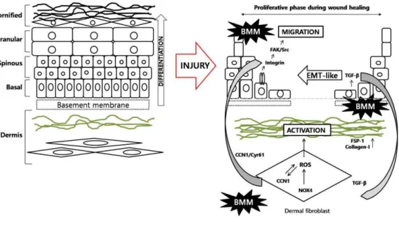
Figure 7 : Schematic representation of mechanisms of BMM in the wound healing processes...………73
Overview
Skin is the outer tissue and consists of epidermis and dermis. Epidermis has several stratified layers, such as cornified, granular, spinous, and basal layer, and functions to barrier against external environment. Dermis is the layer of skin tissue beneath the epidermis and composed of extracellular matrix, collagen fibrils, sweat glands, lymphatic vessels, sebaceous glands, and blood vessels.
Wound healing is the trauma repair process and complex biological process that has serial steps; Inflammation, Proliferation, Maturation. But there are several common impediments to wound repair like hypoxia, infection, and metabolic disorders.
In the past, wounds have been cured using topical medications that act on speeding repair, preventing from infection, removing debris, or reducing pain. Because of the complexity of the skin layer, it is believed that the raw materials of the extract form were mainly used for wound healing agent and more effective than single-substances, but most substance of the extract is not completely understood. As a typical example, Madecassol® is used of treatment for skin injury. It is a healing ointment made from an extract of the medicinal herb, Centella asiatica, which contains antibiotic and anti-inflammatory properties. It has been shown to promote collagen synthesis and regeneration and to prevent scar formation.
As the development of novel wound treatment using a natural substances has been reached the limit, a single ingredient in a new substance is expected to change the market for external application.
Through screening experiment using flavonoids or synthetic materials on skin cells, we discovered some effective compounds and studied their mechanisms to act for wound repair.
PART I
TMF and glycitin act synergistically on
keratinocytes and fibroblasts to promote wound
healing and anti-scarring activity
1. ABSTRACT
Keratinocyte-fibroblast interactions are critical for skin repair after injury. During the proliferative phase of wound healing, proliferation, migration and differentiation of these cells are the major mechanisms leading to tissue remodeling. We have previously reported that glycitin, a major soy isoflavone, stimulates dermal fibroblast proliferation; and the phytochemical, 4′,6,7-trimethoxyisoflavone (TMF), induces migration of HaCaT keratinocyte cells. We therefore investigated whether these compounds display synergistic effects on skin cells during wound healing in vitro and in vivo. Co-treatment with TMF and glycitin synergistically promotes the proliferation and migration of both keratinocytes and dermal fibroblasts, with a 1:1 ratio of these compounds showing the greatest efficacy in our co-culture system. This keratinocyte-fibroblast interaction occurred via the secretion of TGF-β, and the induction of differentiation and proliferation was confirmed in both indirect and direct co-culture assays. In an excisional and burn wound animal model, mice treated with a 1:1 ratio of TMF and glycitin showed faster wound closure, regeneration and scar reduction than even the positive control drug. These data indicate that two isoflavones, TMF and glycitin, act synergistically to promote wound healing and anti-scarring and could potentially be developed together as a bioactive therapeutic for wound treatment.
2. INTRODUCTION
Flavonoids are a class of compounds comprised of more than 4000 phenylbenzopyrones, which are widely produced in edible plants. These have been reported to have diverse pharmacological activities, including antioxidative, antinflammatory, and anticancer properties [1-3]. Among them, the soy isoflavones in particular, including genistein, daidzein, and glycitein, have been found to exhibit chemo-preventive, cardio-protective, and anti-osteoporosis effects [4-6].
In my previous studies, I found that the soy isoflavone, glycitin (4’-Hydroxy-6-methoxyisoflavone-7-D-glucoside), protects skin from photoaging by increasing expression of collagen I in UV-exposed human dermal fibroblasts [7]. This compound can also promote cell viability and migration via the transforming growth factor-beta(TGF-β)pathway[8]. Another compound, TMF (4’,6,7-trimethoxyisoflavone), which is a chemically transformed product of an amphiisoflavone isolated from the roots of the medicinal plant, Amphimas pterocarpoides [9], was found to enhance migration of HaCaT keratinocytes through the activation of NADPH oxidase 2 (NOX2) [10].
Cutaneous wound repair is an intricate process that includes three phases: inflammation, proliferation, and remodeling. During the proliferation phase, various types of cells migrate to the wound site [11]. Keratinocytes, the most prevalent cell type in the epidermis, and fibroblasts, the predominant cell type in dermis, play important roles in the process of skin repair after injury, and their interactions are critical for this process [12]. Keratinocytes secrete both platelet-derived growth factor (PDGF) and TGF-β, which
induces expression of alpha-SMA (alpha-smooth muscle actin) in myofibroblast cells [15]. During the final phase of wound healing, known as remodeling, processes such as re-epithelialization, angiogenesis, and fibrosis, are stopped [16], and collagen synthesisis increased; this is then deposited on the wound siteto form a scar [17]. Over time, collagens are rearranged to form a cross stripe pattern, which is a feature of normal skin, and the scar becomes fainter. Various compounds have been shown to promote this process; clodronate liposomes, for example, decrease scar formation by reducing expression of both collagen and TGF-β [18].
A number of studies have utilized several types of co-culture systems to study the interactions between epidermal and dermal cells during wound healing. Zhe Wang et al. reported that co-culture in a transwell system with human fetal epidermal keratinocytes could promote the proliferation and migration of human fetal and adult dermal fibroblasts [19]. In this model, both migration and proliferation were enhanced through the induction of cyclin B1, phospho-CDK1, phospho-AKT, C-X-C chemokine receptor 4 (CXCR4), and matrix metalloproteinases (MMPs). Pierre Shephard et al. further demonstrated this effect with keratinocytes and fibroblasts that were simultaneously incubated on cell culture dishes in direct contact with one another [15]. These, and numerous other recent studies, have highlighted the importance of synergistic interaction and communication between these cell types.
Here, I show that treatment with a combination of TMF and glycitin promotes wound repair processes, both in cell culture and in vivo, and this activity is most pronounced when the compounds are mixed in a 1:1 ratio. The effects of the combined glycitin-TMF treatment on keratinocytes and fibroblasts were confirmed in both an indirect and direct-contact culture system, and the combined synergistic activity was mediated, at least in part, by elevated levels of secreted TGF-β. To confirm these results in vivo, we tested the
burn wound models. my results suggest that co-treatment with TMF and glycitin accelerates skin regeneration as well as reduces scar formation after injury.
3. MATERIALS AND METHODS
Materials
4'-Hydroxy-6-methoxyisoflavone-7-D-glucoside (glycitin) and 4',6,7-trimethoxyisoflavone (TMF) were purchased from Indofine Chemical Co. (Hillsborough, NJ, USA). These were used without further purification, and purities were determined using high-performance liquid chromatography [10].The TMF and glycitin powder was dissolved in DMSO. For in vivo experiments, butylene glycol was used as a carrier to treat the wound site of mice.
Cell culture
Primary human dermal fibroblasts and the human keratinocyte cell line (HaCaT) were stabilized and cultured in Dulbecco's Modified Eagle Medium (DMEM, Gibco) that was supplemented with 10% fetal bovine serum (FBS, Omega) and 1% penicillin/streptomycin (PAA). Cells were incubated in a humidified atmosphere at 37°C in 5% CO2.
MTT (3-(4,5-dimethyltiazol-2yl)-2,5-diphenyltetrazolium bromide) assay
HaCaT keratinocyte cells were seeded in 96-well plates at a density of 2ⅹ103cell/well. After 24 h, cells were treated with one of the following :dimethyl sulfoxide (DMSO), glycitin:TMF=1:1 (10μM:10μM), glycitin:TMF=1:2 (6.7μM:13.3μM), glycitin:TMF=2:1 (13.3μM:6.7μM),or conditioned media from fibroblasts treated with the above conditions. MTT solution (Sigma) was then added to each well, and cells were incubated for an additional 4h. Subsequently, the medium was removed and replaced with 150 μL DMSO,
performed with primary human dermal fibroblast cells using the glycitin and TMF ratios specified above and conditioned media from HaCaT cells treated with same conditions.
Scratch wound healing assay
HaCaT keratinocyte cells were seeded in 48-well plates at a density of 3×10⁴ cell/well for 24 h. As cratch was made on the monolayer by drawing a sterile pipette tip across the well, and the culture medium was supplemented with DMSO, glycitin:TMF=1:1 (10 μM:10 μM), glycitin:TMF=1:2 (6.7 μM:13.3 μM), glycitin:TMF=2:1 (13.3 μM:6.7 μM),or conditioned media from fibroblasts treated with the same conditions. At time 0 and 24 h post-treatment, wound closure was captured at 40x magnification using an Olympus IX70 microscope equipped with a digital camera. Distance was measured using the Image J software, and the difference between the initial and final width of the scratch was calculated.
Invasion assay
For single culture assays, HaCaT cells or fibroblasts (7х104 cell/well) were seeded in the insert of a 12-well invasion assay kit (SPL), and the bottom portion was filled with media. For co-culture assays, one cell type was seeded in the insert, and the other type was seeded in the bottom, using equal numbers of each(e.g., HaCaT cells in the insert and fibroblasts in the bottom and vice versa). The membrane allows the exchange of media during incubation. After 24 h, serum free media was added to the insert to allow for chemotaxis, and cells in both the insert and bottom were treated with either DMSO control or G:T=1:1 for 48 h. Cells on the upper side of the insert were then removed using
Invasive ability was measured using the Image J program.
Reverse transcription polymerase chain reaction (RT-PCR)
TRIzol reagent (MRC) was used to isolate total cellular RNA according to the manufacturer’s instructions. The primer sequences were as follows: forward GTTCAAGCAGAGTAACACAGC-3’ and reverse
GTATTTCTGGTACAGCTCCACG-3’ for TGF-β; forward
5’-GAAGGTGAAGGTCGGAGTC-3’ and reverse 5’-GAAGATGGTGATGGGATTTC-3’ for GAPDH. The results were analyzed using Image J program.
In vivo excision wound model
Six-week old male ICR mice (n=6 for each group) were chosen for the experiment. All procedures were approved by the Animal Care and Use Committee Jeju National University (permission number 2015-0033). Prior to wounding, the fur was removed with an electronic hair clipper and removal cream. Dermal wounds were then placed on the middle of the back using a 5-mm punch instrument. Wounds of mice in the experimental group were treated daily with 200 μL of a 1:1 mixture of glycitin and TMF (200 μM:200 μM) dissolved with butylene glycol for 14 days. Madecassol® and fucidin® were utilized as positive controls and applied to wound sites at the same concentration and for the same duration of time.
The wounds were visualized every 2 days, and the rate of wound closure was calculated as relative percentage of the original wound area, using the image J program. At 14 days post-wound, skin tissue was isolated and fixed using a 4% formaldehyde solution. Paraffin-embedded tissues were cut into 3-mm sections and stained using hematoxylin-eosin and Masson’s trichrome.
In vivo burn wound model
Eight-week old female C57BL/6 mice (n=5 for each group) were chosen for this experiment. The fur was removed with an electronic hair clipper and removal cream, andburn wounds were placed on the rump using a red-hot iron nail (7mm in diameter). Burns in the experimental group were then treated with 200μL of TMF(200μM)or 200 μL of a 1:1 mixture of TMF and glycitin (200μM:200μM)daily for 8weeks; the control group was treated with the same amount of DMSO. Post-treatment wounded skin were collected from mice at each time points and fixed in 4% formaldehyde. Paraffin-embedded tissues were then cut into 3-mm sections, and all sections were stained using hematoxylin-eosin and Masson’s trichrome.
Western blot
HaCaT and fibroblast cells were seeded (1х105 each) on 100mm cell culture dishes and incubated for 24 h. These were treated with 10-20 µL of glycitin, 10-20 µL of TMF, or the following combinations of glycitin and TMF: glycitin:TMF=1:1 (10 μM:10 μM), glycitin:TMF=1:2 (6.7 μM:13.3 μM), and glycitin:TMF=2:1 (13.3 μM:6.7 μM) for 72 h. Total protein was extracted from treated cells using RIPA buffer, and protein concentration was determined using the BCA Protein Assay Kit (Thermo Scientific, Germany).To investigate the expression of secreted factors, conditioned media was harvested and concentrated using amicon centrifugal filter (Merck milipore), and total protein concentration was measured using the Bradford assay. Skin tissue isolated from mouse wound sites was lysed using T-PER Tissue Protein Extraction Reagent (Thermo, USA) and homogenized. Following centrifugation (14000rpm, 4℃, 20min), supernatants
contents were transferred to PVDF membranes, and these were blocked in 5% non-fat dry milk with Tris-buffered saline Tween 20 (TBST) buffer, followed by incubation with primary antibodies at 4℃ overnight. Cytokeratin 10 (sc-53252, 2nd Ab:rabbit),
Cytokeratin 14 (sc-17104, 2nd Ab:goat), and GAPDH (sc-25778, 2nd Ab:rabbit),antibodies were purchased from Santa Cruz Biotechnology (USA);S100A4 (ab27957, 2nd Ab:rabbit) antibody was purchased from ABcam (UK); TGF-β (#3711, 2nd Ab:rabbit) antibody was purchased from Cell Signaling Technology (USA); and β-actin (A5316, 2nd Ab:mouse) antibody was purchased from Sigma-Aldrich (USA).We used anti-mouse (K0211589, KOMABIOTECH), anti-rabbit (K021178, KOMABIOTECH), and anti-goat (AP1079P, Millipore). Proteins were detected using chemiluminescent reagent, ECL solution (W6002, Biosesang).
4. RESULTS
I previously reported that glycitin (4’-Hydroxy-6-methoxyisoflavone-7-D-glucose, Fig. 1A) induces the proliferation and migration of fibroblasts [7, 8],whereas TMF (4’,6,7-trimethoxyisoflavone, Fig. 1B) promotes HaCaT keratinocyte migration, but not proliferation [10]. In an excisional wound experiment, we found that TMF accelerates re-epithelialization, and appendages were formed along the epidermis at 15 days after the wounding event. By contrast, groups receiving other treatments had a thick epidermis and less differentiating appendages than those treated with TMF (Supplementary data 1). Because wound closure includes closure of the epidermis, known as re-epithelialization, as well as dermal closure, which requires both fibroblast cells and collagens, we hypothesized that a mixture of glycitin and TMF may enhance the wound healing process by synergistically stimulating cells in both the epidermis and dermis.
Figure1. The chemical structures of TMF and Glycitin. (A) TMF (4’,6,7-Trimethoxyisoflavone).
Treatment with a 1:1 ratio of glycitin and TMF most effectively stimulates proliferation, migration, and invasion ability in an indirect-co-culture system
To determine the effect of treatment with mixture of glycitin and TMF on keratinocytes and fibroblast cells, we mixed these compounds at various ratios: G:T=1:1, G:T=1:2, and G:T=2:1 (G: glycitin, T: TMF). I then measured the proliferation and migration of human dermal fibroblast cells that were either treated with the different combinations of the two compounds or incubated with conditioned media from HaCaT keratinocyte cells treated with the same stimuli. I observed increased proliferation of fibroblasts treated with G:T=1:1, 1:2, and 2:1 but did not see the same effect in cells treated with other combinations of glycitin and TMF or the HaCaT cell-conditioned media (Fig. 2A). Fibroblast migration was increased in all G:T treated groups, whereas treatment with conditioned media did not increase migration. I note that the migratory distance of fibroblasts treated with conditioned media actually decreased as compared to the untreated control group (Fig. 2B). This suggests that treatment with glycitin and TMF induces HaCaT cell to produce factors that inhibit fibroblast migration.
I next assessed the effect of glycitin and TMF treatment on HaCaT keratinocytes and observed both increased proliferation and migration distance in cells treated with G:T=1:1 (Fig. 2C and D). This synergistic effect was also present when HeCaT cells were incubated with conditioned media from fibroblast cells treated with G:T=1:1 (Fig. 2C and D).These data suggest that a 1:1 ratio of glycitin and TMF is most effective at inducing the proliferation and migration of both dermal fibroblasts and keratinocytes. Further, our results imply that these two types of skin cells can communicate with one another by secreting unidentified factors when stimulated with a mixture of glycitin and TMF.
Figure2. Glycitin and TMF act synergistically to promote the proliferation and migration of HaCaT keratinocytes and human dermal fibroblasts in culture.
(A) Proliferation of dermal fibroblasts treated with either glycitin and TMF, mixed in varying ratios,or conditioned media from HaCaT keratinocytes treated with same conditions for 24h, as measured by MTT assay.
(B) Scratch wound healing assay with dermal fibroblasts treated as described in (A)for24h. Migration distance was measured using the ImageJ program.
(C) Proliferation of HaCaT keratinocytes treated with either glycitin and TMF, mixed in varying ratios, or conditioned media from dermal fibroblasts treated with the same conditions for 24h, as measured by MTT assay.
(D) Scratch wound healing assay with HaCaT keratinocytes treated as described in (C)for24h. Migration distance was measured using the ImageJ program.
*, P<0.05 as compared to control, **, P<0.001 as compared to control, G:glycitin, T:TMF
A 1:1 mixture of glycitin and TMF promotes invasion by stimulating keratinocyte-fibroblast interaction
We next used transwell plates, which have 8.0µm pore polycarbonate membrane inserts and allow media to be shared simultaneously between different cell types, to assess the effect of glycitin and TMF on the 3D-invasion of fibroblasts and keratinocytes. In single culture conditions, HaCaT keratinocytes and fibroblasts were seeded alone and treated with DMSO control or glycitin:TMF=1:1 for 48 h(Fig. 3A and C, left diagram). For co-culture experiments, cells were incubated together, with one cell type in each half of the plate, and treated with the same stimuli (Fig. 3A and C, right diagram). In contrast to what was observed in our migration assays, we found that treatment of HaCaT and fibroblast cells individually with G:T=1:1 did not significantly affect the invasion (Fig. 3BandD, left). However, we did measure a significant increase in invasion for both the co-cultured fibroblast and keratinocyte cells stimulated with G:T=1:1, as compared to controls (Fig. 3B and D, right).
It should be noted that the co-culture system only allows the sharing of factors secreted through the media, whereas in tissues, fibroblasts and keratinocytes contact one another directly. We therefore cultured fibroblasts and HaCaT keratinocytes in the same insert plate to allow for direct contact, as well as media sharing (Fig. 3E). In this system, to distinguish these cell types, we developed a mCherry-labeled HaCaT cell line. Co-cultured cells were treated with the glycitin and TMF, and invasion was measured by fluorescence microscopy, for keratinocytes, and crystal violet staining, for total cells. We found that treatment with a mixture of glycitin and TMF enhanced the invasion of both cell types (Fig. 3F).Since the cell-to-cell contact did not show enhanced migration after the media change, we concluded that cell-to-cell contact may not be a main factor in the synergistic effect of TMF & glycitin on wound healing.
Figure3. A 1:1mixture of glycitin and TMF increases invasive ability of dermal fibroblasts and HaCaT cells in co-culture conditions via induction of a secreted factor.
(A, B) Transwell invasion assay with HaCaT keratinocytes treated with G:T=1:1 (10 μM:10 μM)for 48h;7х104 cells/well were seeded on the insert, and dermal fibroblasts were also seeded on bottom for co-culture. Invasive ability was measured as described in the Materials and Methods and is quantified in (B). (C, D) Transwell invasion assay with dermal fibroblasts treated with G:T=1:1 (10 μM:10 μM)for 48h;7х104 cells/well were seeded on the insert, and HaCaT keratinocytes were also seeded on bottom for co-culture. Invasive ability was measured as described in the Materials and Methods and is quantified in (D). (E) Diagram illustrating the direct co-culture system utilized for the invasion assays. Arrows and arrowheads indicate HaCaT keratinocytes and dermal fibroblasts, respectively.
(F) Invasion assay with HaCaT keratinocytes and dermal fibroblasts in direct contact, using HaCaT keratinocytes transfected with a vector expressing the mCherry fluorescent protein. Invasive ability was measured as described in the Materials and Methods.
*, P<0.05 as compared to control, **, P<0.001 as compared to control, G: glycitin, T:TMF
A combination of glycitin and TMF induces differentiation of keratinocytes and fibroblasts and increases secretion of TGF-β
Because the treatment with glycitin and TMF can synergistically promote migration and invasion of HaCaT cells in a co-culture system, we next tried to elucidate the mechanism underlying this process. After injury, in order to restore an epidermal barrier function, the epidermis begins to proliferate in the basal layer, followed by differentiation in the spinous and granular layers. Keratin 5 (KRT5)/Keratin 14 (KRT14)are often used as markers for proliferation in the basal layer, and Keratin 1 (KRT1)/Keratin 10 (KRT10)/involucrin are commonly utilized to distinguish differentiated suprabasal layers. Keratin expression induces a switch to re-epithelialization. We therefore performed direct contact co-culture assays with fibroblast and HeCaT cells treated with either glycitin, TMF, or a 1:1 mixture of the two compounds for 24hand measured expression of these marker proteins by western blot analysis (Fig. 4). We found that low-dose glycitin treatment could induce differentiation of keratinocytes, and in high doses, the expression of keratinocyte proliferation markers, KRT14, was increased (Fig.4A). Treatment with TMF alone mainly induced proliferation of keratinocytes, as evidenced by our previous research [10]. Interestingly, treatment of keratinocytes with a 1:1 mixture of glycitin and TMF treatment increased differentiation, while the proliferation was decreased, as compared to TMF treatment alone (Fig.4A).
In addition to the epidermis, differentiation in the dermis functions to contract and close the wound region, and fibroblasts in particular, which are the predominant cell type in the dermis, are activated to form myofibroblasts. Fibroblast-specific protein 1 (FSP1),a member of the S100 superfamily, is involved in inflammation, cell growth, cell
4A).
TGF-β plays important pleiotypic roles and enhances the migration and differentiation of keratinocytes and fibroblasts throughout the proliferation phase of wound healing. Using a scratch wound healing assay in HaCaT keratinocytes or fibroblasts, we determined that treatment with TGF- β (5 and 10 ng/ml) as well as treatment with TMF & glycitin increases the migratory ability of the cells (Supplementary data2). Therefore, we hypothesized that this protein might function as a secreted factor that mediates the interaction between keratinocytes and fibroblasts. To test this, we collected medium from co-cultured cells post-treatment and measured the expression of mature TGF-β1 protein using western blot analysis. TGF-β 1 was detected in all co-culture media and was significantly increased in supernatant from cells treated with G:T=1:1, but not from those stimulated with either G:T=1:2 or G:T=2:1 (Fig.4B).We also found that TGF-β 1 mRNA transcript levels were increased in both HaCaT keratinocytes and fibroblasts from co-culture assays (Fig. 4C). When tested during skin cell migration, no changes in the levels of TGF-β 2, 3, and KGF were observed (data not shown). The results suggest that keratinocytes and fibroblasts secrete TGF-β 1 as major mediators when stimulated by glycitin and TMF at a 1:1 ratio. To clarify whether TGF-β 1 affects keratinocytes and fibroblasts, we examined the expression of Smad2/3, which is downstream of the TGF- β pathway. The phosphorylation of Smad2/3 was significantly increased in co-cultured cells compared to single-cultured cells; it was activated more so by TMF&glycitin treatment, but not by Smad3 in fibroblasts (Fig. 4D).
From these experiments, we demonstrate that co-treatment with glycitin and TMF likely promotes the interaction between keratinocytes and fibroblasts via the secretion of TGF-β and promotes functions in both the epidermis and dermis related to wound repair.
Figure4. Co-treatment with glycitin and TMF promotes differentiation and proliferation in co-culture condition via secretion of TGF-β.
(A) HaCaT keratinocyte and dermal fibroblast cells (1х105 each) were seeded on 100mm cell culture dishes and incubated for 24 h. Protein lysates from cells treated with glycitin (G-10μM and G-20μM), TMF(T-10μM and T-20μM), or G:T=1:1 (10 μM:10 μM) were obtained using RIPA solution, and proteins were resolved by 10% SDS-PAGE. Western blot to measure the expression of differentiation and proliferation-related factors was performed, and in all cases, expression levels were normalized to GAPDH.
(B) Western blot assay to detect TFG-β expression in conditioned media from co-cultured cells treated with G:T=1:1(10 μM:10 μM), G:T=1:2(6.7 μM:13.3 μM), and G:T=2:1(13.3 μM:6.7 μM)for24h.TGF-β expression was measured by comparing with ponceuS.
(C) RT-PCR to measure TGF-β transcript levels in co-cultured cells from transwell assays; TGF-β expression levels were normalized to GAPDH.
(D) Western blot assay to detect phospho-Smad2 and phosphor-Smad3 in cell lysates from single-cultured and co-cultured cells after 24 h.
*, P<0.05 as compared to control, **, P<0.001 as compared to control, G: glycitin, T:TMF
Co-treatment with TMF and glycitin accelerates wound closure and collagen synthesis
To further verify that co-treatment with glycitin and TMF can promote wound healing, we assessed the effects of these compounds in an in vivo excision assay using ICR mice. We first made excisional wounds on their back using a 5mm diameter punch. Glycitin and TMF were mixed in a 1:1 ratio (200 µM each) in butylene glycol, and wound sites were treated with 200 µL of this mixture once a day, for 2 weeks. As a positive control, wounds were treated with Madecassol®, a healing ointment made from an extract of the medicinal herb, Centella asiatica, which contains antibiotic and anti-inflammatory properties. It has been shown to promote collagen synthesis and regeneration and to prevent scar formation. Fucidin®, an anti-infective ointment that contains the fusidic acid, was also utilized as a positive control. We observed that mice treated with G:T=1:1 showed faster wound closure, as compared with the Madecassol® and Fucidin® groups (Fig. 5A and B). These data are consistent with previous results, demonstrating that these compounds are most effective when used in a 1:1 ratio.
In addition, hematoxylin/eosin staining showed that combined treatment with glycitin and TMF effectively promoted the development of specialized appendages in the wound site, including hair follicles and sweat glands, which showed morphologies similar to those in normal skin (Fig. 5C). Skin damage in the combined treatment group was also ameliorated, as evidenced by small scar width (Fig. 5D)and relatively thin epidermis (Fig. 5E). Collagen deposition was observed through Masson’s trichrome staining (Fig. 5C and F), and Ki67-positive cells, which indicate proliferation, were present in the basal layer of the epidermis, as well as the adnexa (Data not shown), exclusively in glycitin and TMF
Figure5. A 1:1mixture of glycitin and TMF accelerates wound closure and protects against scar formation in an in vivo excisional wound model.
(A, B) Wound closure in our excisional wound model after2weeks. Photographs were taken every 2 days, and wounds were measured using the Image J program. ‘Control’ indicates the butylene glycol;*, P<0.05 as compared to Madecassol, #,
P<0.001 as compared to Fucidin.
(C) On the last day of treatment, skin tissues were isolated, fixed, and stained with hematoxylin/eosin and Masson’s Trichrome.
(D) Scar width,(E) epidermal thickness, and (F) collagen content were measured 14 days after wounding using the Image J program.
Co-treatment with TMF and glycitin promotes reorganization and inhibits wound fibrosis
I next assessed the effectiveness of glycitin/TMF treatment to prevent scarring using a murine burn wound model. Burns were made by searing the dorsal skin of C57BL/6 mice with a hot iron. These wounds were then treated using the same conditions as described for the excision wound model, except in this case, burns were treated for 8 weeks. As predicted, we observed that burns treated with the glycitin/TMF mixture showed faster healing than the other groups (Fig. 6A). After 8 weeks, we checked hair growth in the scar area, since this can indicate remodeling of skin, and found that the glycitin/TMF-treated group showed the most hair growth(Fig. 6B). We then removed the hair over the burn site and measured the size of the scars; we found that scars treated with glycitin/TMF were significantly reduced, as compared to those treated with the DMSO control (Fig. 6C and D). Specifically, whereas the scars from the positive controls and DMSO-treated groups showed thickened epidermis and excessive collagen deposition, both epidermal thickness and collagen deposition were reduced in the TMF and glycitin/TMF co-treated groups (Fig. 6E-H). The glycitin/TMF co-treated group, in particular, showed improved regeneration of appendages and a fully formed panniculus (Fig. 6E-F). Because glycitin has no effect on re-epithelialization but contributes to the synthesis of collagen, we excluded the group treated with glycitin only in the burn wound experiment.
Since TGF-β is highly expressed in fibroblasts derived from hypertrophic scars and in scar tissue [20], we collected scar tissues after 4 and 8 weeks of drug treatment and analyzed TGF-β expression in tissue using western blot. We found that the amount of TGF-β in glycitin/TMF-treated scars was significantly decreased, as compared to controls, with samples isolated at 4 (Fig. 6I) and 8 weeks (Fig. 6J) showing a similar trend. These
observations suggest that co-treatment with glycitin and TMF can promote wound healing and prevent scarring in an in vivo skin wound animal model.
Figure6.A 1:1mixture of glycitin and TMF reduces scarring anin vivo burn wound model.
(A) Burn wound closure after treatment for 10 days.
(B) Wound sites were treated every day for 8 weeks; hair was then removed, and the scars were photographed using a digital camera or (C) the skin testing machine.
(E-H) Skin tissues were isolated, fixed, and stained with both hematoxylin/eosin and Masson’s Trichrome. Epidermal thickness and collagen content were measured using the Image J program.
(I) Western blot analysis of skin tissue isolated from wound sites 4 weeks and (J) 8 weeks after wounding.
*, P<0.05 as compared to control, **, P<0.001 as compared to control, G: glycitin, T:TMF
Supplementary data 1. Result of TMF and glycitin treatment on excisional wound healing
Skin tissue was stained with Hematoxylin & Eosin and Masson’s trichrome at 15 days after the wounding event. An excisional wound was made on the backs of mice and 200 µM of Glycitin or 200 µM of TMF was applied to the wound every day.
Supplementary data 2. The effect of TGF-β and TMF &glycitin on skin cell migration
(A) Scratch wound healing assay with HaCaT keratinocytes and (B) fibroblasts treated with TGF-β (5 and 10 ng/ml) and TMF & Glycitin. Migration distance was
5. DISCUSSION
In this paper, I present evidence that treatment with glycitin and TMF can promote wound healing in vitro and in vivo, and a one-to-one ratio of these two compounds exerts synergistic effects on keratinocytes and fibroblasts that are mediated, at least in part, by secreted TGF-β.
Isoflavones are phytoestrogen compounds that are found in legumes, such as the soybean and kudzu vine [21]. In soybeans, these can be present in one of three aglycon forms, known as such as daizein, genistein, and glycitein, or in the corresponding glycoside forms, referred to as daidzine, genistin, and glycitin, respectively [22]. Whereas the glycosides are abundant in non-fermented soybean food, the aglycon forms are highly enriched in fermented food[23]. In any soy-based food, the specific isoflavone content is dependent on the type of soybean, the processing method, and the potential addition of other foods[24].Critically, isoflavones have demonstrated many health benefits and are thought to reduce the risk of hormone-related disease risk and protect against harmful elements such as UV radiation[25]. For example, genistein and daidzein can prevent osteoporosis by increasing bone mineral content and density and by stimulating the formation of bone tissue[26]. The antioxidant properties of isoflavones are also believed to protect against atherosclerosis, which is characterized by oxidation of the low density lipoprotein (LDL) component of cholesterol that blocks the blood flow[27]. Isoflavones can further activate estrogen receptors in the vagina, ovum, and lacteal gland by mimicking estrogens or functioning as anti-estrogens [28],and these have been shown to have an anti-oxidant ability, similar to vitamin E/C [29]. Among the isoflavones, genisteinin particular, can reduce cancer cell viability by inhibiting angiogenesis, which
promote fibroblast cell proliferation and migration but had no effect on keratinocytes. Conversely treatment with the amphiisoflavone-derivative, TMF, could induce the migration of keratinocytes, but not fibroblasts. Because the proliferation and migration of both keratinocytes and dermal fibroblasts are critical for effective skin wound repair, we tested the effect of a combined glycitin-TMF treatment on these cell types in a co-culture system and in two mouse models for skin repair.
Keratinocytes and dermal fibroblasts interact closely with one another, and they require cell-cell interactions in order to produce a cellular environment conductive to wound repair [31]. Critically, it has been shown that co-culturing keratinocytes in the presence of fibroblasts has profound effects on epithelial reorganization and differentiation. It is differ from what is observed when keratinocytes are cultured either alone or with few fibroblast cells [32].This epidermal reorganization appears to be mediated by a diffusible factor, as well as specific cellular matrix interactions that occur via the integrin pathway [33]. As the major cell-type that is located adjacent to epidermis, fibroblasts synthesize ECM components and exhibit the capability to secret factors to modulate the proliferation, migration, and differentiation of other cells, especially keratinocytes [34].
Here, we found that when keratinocytes and fibroblasts are cultured such that they do not contact one another but share medium, the invasive ability of fibroblasts remarkably increased by ten times, whereas migration was unaffected (Fig. 2, 4). Notably, under these same conditions, the proliferation, migration, and invasion of keratinocytes were all elevated, as compared to controls (Fig. 3, 4). Myofibroblasts are the activated form of dermal fibroblasts and are characterized by mesenchymal features. These cells
secretion [37]. Stromal myofibroblasts also form during cancer development and can drive invasive cancer growth [38]. In Figure 5A, we demonstrate that treatment with a mixture containing a 1:1 ratio of glycitin and TMF can induce expression of FSP1 in co-cultured cells, suggesting that this treatment can promote the activation and differentiation of fibroblasts.
The epidermal barrier protects against toxins, pathogens, and moisture loss [39]. During epidermis development, basal keratinocytes either differentiate, or migrate, to become suprabasal keratinocytes (spinous layer, granular layer), or divide disproportionally to produce proliferative cells, which remain in the basal layer[40]. The NF-κB, Notch, C/EBP, and MAPK signaling pathways are known to be involved in epidermal differentiation and barrier restoration [39]. Interestingly, we show that treatment with a 1:1 mixture of glycitin and TMF can promote keratinocyte differentiation, as evidenced by elevated KRT10 protein expression, and keratinocyte proliferation is correspondingly attenuated in response to this (Fig. 5). These data suggest that glycitin and TMF can induce keratinocyte differentiation, and modulate the proliferation and migration of both cell types.
Wound healing is a complex process that is aimed at restoring tissue integrity [41]. Several cell and tissue types, including keratinocytes, fibroblasts, blood vessels, macrophages, and neutrophils are involved in this process and communicate with one another via circulating the secreted factors[16]. Sander W.Spiekstra investigated the factors secreted from epidermal keratinocytes and dermal fibroblasts, using full-skin, as well as epidermal and dermal substitutes. Whereas the full-skin substitute produced numerous inflammatory and angiogenic factors, such interleukin (IL)-6, chemokine (C-X-C motif) ligand 8 ((C-X-CX(C-X-CL8 or IL-8), and (C-X-C(C-X-C chemokine ligands ((C-X-C(C-X-CLs), the epidermis and dermis produced these proteins but in lower amounts. The epidermal substitute, in
synergistically to mediate wound healing with factors secreted from the dermal substitute, such as TGF-β1, fibroblast growth factor (FGF)-2, and keratinocyte growth factor (KGF) [42].
The prevention of scarring after wound healing requires a lengthy period of medical intervention to reduce disfigurement, discomfort, and disability [43]. Hypertrophic scars are fibrotic lesions containing myofibroblasts that express α-SMA, collagen, and other ECM components [44], and these commonly form after injuries, such as burns, delayed epithelialization, or secondary damage [45]. The cytokine TGF-β, in particular, is responsible for fibrotic scarring [20]. During TGF-β pathway-mediated hypertrophic scar formation, there is an up-regulation of SMAD-2 and SMAD-3, as well as increased expression of TGF-β1, TβRI, and TβRII,which lead to positive feedback loop [46]. As a result, connective tissue growth factor (CTGF), a known downstream mediator of TGF-β, induces proliferation of myofibroblasts and expression of collagen types I and III[47]. Sisco et al. reported that inhibition of CTGF activity limited hypertrophic scarring via the suppression of myofibroblasts, collagen, and tissue inhibitor of metalloproteinase (TIMP)-1 expression [48]. The repression of apoptosis, mechanobiology, angiogenesis, and inflammatory responses can also contribute to a reduction in scarring. For example, increased levels of Bcl-2 and tissue transglutaminase, lead to decreased Fas, and p53 alteration inhibits apoptosis in hypertrophic scar-derived myofibroblasts. Further, mechanical tension can upregulate expression of genes related to matrix remodeling but not apoptosis genes, and when the angiopoietin1/angiopoietin2 ratio is decreased, microvessel density is increased in hypertrophic scars. In hypertrophic-derived fibroblasts, expression of IL-6, IL-8, TLR-4, and MCP-1 was increased, as well
acts on re-epithelialization and scarring. For example, increased expression of TGF-β induces re-epithelialization and contraction, whereas reduced expression of TGF-β contributes to reduced scarring. Collagen deposition is controlled by skin cells stimulated by TGF-β. During re-epithelialization, the dermis needs matrix proteins such as collagen to restore structure and replace granulation tissue, but during the maturation process, collagen deposition results in scar tissue formation. Thus, the balance of collagen regulated by TGF-β is important during the wound healing process. The ‘good’ wound healing can indicate faster wound closure and less scarring. We found that co-treatment with glycitin and TMF promotes TGF-β expression, reorganization, and fibroblast differentiation in normal wound model, whereas it decreases TGF-β and scar size in the impaired burn wound model.
In this study, we show that glycitin and TMF acts ynergistically to promote epidermal regeneration and dermal activation. Specifically, treatment with these compounds influences the interaction between keratinocytes and fibroblasts to promote wound repair
in vitro and in vivo, and TGF-β functions as a ‘critical mediator’ for this response.
Furthermore, long-term treatment helps to decrease scar size after burn injury by degrading matrix components, such as collagen, in a mouse burn wound model. In conclusion, our data suggest that glycitin and TMF act together on keratinocytes and fibroblasts to induce wound healing and scar reduction via the release of TGF-β. It is also strongly possible that these compounds may hold therapeutic potential for the development of improved treatments for cutaneous injury.
PART II
A novel synthetic material, BMM, accelerates
wound repair by stimulating re-epithelialization
1. ABSTRACT
Cutaneous wound repair is an intricate process whereby the skin reprograms itself after injury. In the mid-phase of wound repair, proliferation, migration, and differentiation of cells are the major mechanisms to lead remodeling. We investigated the effect of BMM, a novel synthetic material, on the migration and viability of keratinocytes or fibroblasts using the in vitro scratch wound healing, ECIS, invasion, and MTT assays. Cell migration-related factors were analyzed using western blot, and we found that treatment with BMM stimulated the EMT pathway and FAK/Src signaling. Differentiation of HaCaT and fibroblast cells was also stimulated by BMM and specifically, NOX2/4 contributed to the activation of fibroblasts for wound healing. Furthermore, BMM treated HaCaT and fibroblast-co-cultured cells increased migration and differentiation. TGF-β and Cyr61 were also secreted to a greater extent than that in single cultured cells. In vivo experiments showed that treatment with BMM promotes wound closure by promoting re-epithelialization. In this study, we demonstrate that a novel synthetic material, BMM, is capable of promoting wound healing via the stimulation of re-epithelialization in the epidermis and the activation of fibroblasts in the dermis, in particular, via the acceleration of the interaction between epidermis and dermis.
Key Word : Cyr61, Fibroplasia, NADPH oxidase, Re-epithelialization, TGF-beta, Wound healing
2. INTRODUCTION
Wound healing is a complex biological process of restoring damaged skin after injury and is composed of three major phases: inflammation, proliferation, and maturation [1], which are mediated by various cells derived from the epidermis and dermis [2].
In the proliferation phase, basal layer human keratinocytes (HaCaT) move toward the wound site from the edge [3]. HaCaT keratinocytes continue to divide and proliferate to cover the wound area and once complete, the cells cease division and begin to differentiate upward, forming spinous, granular, and cornified layers [4]. This process is known as ‘re-epithelialization’ and is crucial for rapid wound closure. In addition to differentiation, migration of HaCaT cells also contributes to wound closure through basement layer formation.
Furthermore, dermal fibroblasts (Fbs) crowd around wound sites after injury and form granulation tissue. Myofibroblasts are an activated form of Fbs and are elevated during wound healing [5]. These cells are able to contract by combining smooth muscle actin (SMA) and myosin, and this contraction contributes to wound repair. Extracellular matrix (ECM) components, such as collagens and gelatin, are synthesized from myofibroblasts. Initially, the ECM is composed of collagen-III, which is a weaker form of collagen that is later replaced by the longer formed-collagen-I as evidenced in scarring [6]. In the late phase of wound repair, the myofibroblasts are lost via the apoptosis pathway and expressed collagen-I is degraded by matrix metalloproteinases (MMPs) to balance the composition of the ECM [7], which is known as the cause of several fibrotic diseases
derived signaling [9]. NADPH oxidase is the one of ROS generating enzymes and has six types of subunits.
Several studies have reported a relationship between the epithelial–mesenchymal transition (EMT) and embryogenesis, wound healing, or metastasis. Epithelial cell adhesion molecules (ECAM) function to interlink epithelial cells and usually maintain a fixed form. However, when ECAM synthesis was blocked by Twist, Slug, or Snail transcription factors, cells lost their adhesive ability and transformed into spindle and motile mesenchymal cells types. These mechanisms are fundamentally relevant to the acquisition of cells’ migratory ability and are also related to wound healing [10].
Focal adhesion kinase (FAK) and C-Src form a dual kinase complex and phosphorylate down-stream factors such as paxillin of p130Cas. Integrin-regulated activation of FAK and Src functions to increase cell migration, cycle, and survival in normal cells or leads to tumor growth and metastasis in tumor cells [11].
In this study, we show that BMM, a novel synthetic material, has wound repair effects both in vitro and in vivo. BMM caused HaCaT keratinocytes to have an EMT-like phenotype and migratory effects via the FAK/Src pathway. Dermal fibroblasts were also activated by BMM through the generation of NOX2/4-ROS, and specific cytokines TGF-β or Cyr61 were related to these mechanisms. Specifically, we used a co-culture system that was recently developed in our previous studies to investigate whether BMM stimulates interaction between keratinocytes and dermal fibroblasts [12]. Our results suggest that BMM accelerates cutaneous wound repair by stimulating re-epithelialization and fibroblast activation.
3. MATERIALS AND METHODS
Generation and verification of BMM
Compound I (4-methoxybenzohydrazide) was purchased from Sigma-Aldrich and used without purification. Compound II was prepared according to previously described methods [13]. Compound I (166 mg, 1 mmol) and a catalytic amount of concentrated hydrochloric acid were added to a solution of compound II (237 mg, 1 mmol) in ethanol. The reaction mixture was refluxed at 90°C for 2 h. After completion of reaction, the reaction mixture was cooled to room temperature to allow precipitation. The solid was filtered and washed with cold ethanol to yield a pure compound III (yield: 50%, m.p: 238–240°C). The synthesis procedure for compound III, 1E,2E-1,2-bis((6-bromo-2H-chromen-3-yl)methylene)hydrazine, is shown in Fig. 1A and its structure and numbering is shown in Fig. 1B. The experimental methods for NMR spectroscopy and mass spectrometry to identify the compound followed a previously reported method [14]. Spectroscopic data are summarized as follows: 1H NMR (500 MHz, pyridine-d5) δ ppm: 8.34 (s, 2H, H-3a), 7.37 (dd, 2H, H-7, J = 2.4, 8.4 Hz), 7.35 (d, 2H, H-5, J = 2.4 Hz), 6.94 (s, 2H, H-4), 6.85 (d, 2H, H-8, J = 8.4), 5.31 (s, 4H, H-2); 13C NMR (125 MHz, pyridine-d5) δ ppm: 160.3 (C-3a), 155.1 (C-9), 133.8 (C-7), 131.6 (C-4), 130.9 (C-5), 130.6 (C-3), 124.8 (C-6), 118.3 (C-8), 113.8 (C-10), 65.0 (C-2); HRMS (m/z): calculated for (M+H)+: 472.9500.
BMM ([1E,2E-1,2-bis(6-bromo-2H-chromen-3-yl)methylene]hydrazine) was dissolved in dimethyl sulfoxide (DMSO) and stored at−20°C (Stock = 20 μM).
5% CO2.
MTT assay
Cells (2 × 10⁴ cell/ml) were seeded in 96-well plates. The following day, cells were treated with DMSO (control) and BMM. After incubation for 24 or 48 h with HaCaT or Fbs, 10 μl of MTT solution (10 mg/ml) was added to each well, and cells were incubated for an additional 4 h at 37 °C. The medium was removed and replaced with 150 μl DMSO, and the plate was incubated for 30 min at room temperature with shaking. Absorbance was measured at 570 nm using a spectrophotometer.
ECIS proliferation assay
ECIS is an experimental machine to analyze the proliferation or migration of cells in real time and show resistance values as a graph. The ECIS cell culture plate consists of 6 wells coated with metal on the bottom. As cells grow, resistance detected by metal is increased and is converted to impedance through a self-program. Two hundred microliters of electrode-stabilizing solution, composed of 10mM L-cysteine, were added to each well and incubated at room temperature. After 10 min, the wells were washed using medium and the cells were seeded at a density of 2 × 10⁴ cell/ml. The following day, 0–20 μl of BMM was added to the wells and the cells were incubated for 48 h. Using the ECIS program, we transferred the Excel data sheet and calculated normalized impedance.
Cell counting
The cell counting method used was one of the cell proliferation assays. A 6-well plate was used for the cell counting assay (2 wells per each time point) and 5× 10³ cells were seeded in each well. Cells were treated with 0–20 μl of BMM and incubated at 37 °C.
Collected cells were suspended and counted using a hemocytometer. Every two days, medium containing the diluted sample was carefully changed.
Scratch wound healing assay
Cells were seeded in 48-well plates at a density of 1×10⁵ cell/well for 24 h. A scratch was made on the cell monolayer by drawing a pipette tip across the well. The culture medium was supplemented with DMSO or BMM dose-dependently. At time 0 and 24 h post-treatment, wound closure was captured using an Olympus IX70 microscope equipped with a digital camera at 40× magnification. The distance migrated was measured using the Image J software, and calculated by comparing between the initial and final width of the scratch.
Matrigel invasion assay
We performed two types of Matrigel invasion assays: 1) single-culture or 2) co-culture. For single culture assays, 7 × 10⁴ cell/well of HaCaT or Fbs were seeded in the insert of a 12-well invasion assay kit (SPL), and the bottom chamber was filled with media. On the other hand, in case of the co-culture assays, HaCaT cells were seeded in the insert, and the Fbs were seeded into the bottom, using equal numbers of each. The membrane allows the exchange of media during incubation. After 24 h, media containing DMSO or BMM was added to the insert and bottom for 24 h. Cells on the upper side of the insert were removed using a cotton swab, and cells on the lower side of the insert were fixed with 4% formaldehyde. After washing with PBS, invaded cells were stained with a 1% crystal violet solution and photographs were acquired at 4× magnification using an Olympus
HaCaT, Fbs, or co-cultured cells were seeded at a density of 1 × 10⁵ each on 100mm cell culture dishes and incubated for 24 h. These were treated with DMSO or BMM in a time or dose dependent manner. Total protein was extracted from treated cells using RIPA buffer, and the protein concentration was determined using the BCA Protein Assay Kit. To investigate the expression of secreted factors, conditioned medium was harvested and concentrated using an Amicon centrifugal filter (Merck Millipore), and total protein concentration was measured using the Bradford assay. Equal amounts of protein (~30 μg/lane) were analyzed from each sample by resolving with 10% sodium dodecyl sulfate-polyacrylamide gel electrophoresis (SDS-PAGE). Protein loaded gels were transferred to NC membranes, and these were blocked in 5% skim milk with Tris-buffered saline Tween 20 (TBST) buffer, followed by incubation with primary antibodies at 4°C overnight. Cytokeratin 10 53252), Cytokeratin 14 17104), MMP-7 8832), Cyr61 271217), Collagen-I 25974),gp91-phox 130543), FAK 1688), p-FAK (sc-11765), C-Src (sc-130124),and GAPDH (sc-25778) antibodies were purchased from Santa Cruz Biotechnology (USA);S100A4 (ab27957) antibody was purchased from ABcam (UK); TGF-β (#3711), Slug (#9585), E-cadherin (#3195), Snail (#3879), p-Src (#2101), and MMP-2 (#4022) antibodies were purchased from Cell Signaling Technology (USA).NOX4 (NB110-58849) and MMP-1 (#444209) antibodies were purchased from Novus Biologicals and Calbiochem, respectively. We used HRP-conjugated anti-mouse (K0211589, KOMABIOTECH), anti-rabbit (K021178, KOMABIOTECH), and anti-goat (AP1079P, Millipore) as secondary antibodies. Proteins were detected using chemiluminescent reagent, ECL solution (W6002, Biosesang).
In vivo experiments
University (permission number 2015-0033). First, the fur was removed with an electronic hair clipper and removal cream. Then, wounds were made on the middle of the back using a 6-mm biopsy punch. Wounds of mice in the experimental group were treated daily with 200 μl of BMM (200 μM) dissolved in butylene glycol for 2 weeks. Madecassol® was used as a positive control and applied to wound sites in the same manner as BMM. Images were captured every 2 days to evaluate wound closure and the rate was calculated as a relative percentage of the original wound area, using the ImageJ program. At 7, 9, and 14 days post-wound, skin tissue was isolated and fixed using a 4% formaldehyde solution. Paraffin-embedded tissues were cut into 3-mm sections and stained using Hematoxylin-Eosin.
4. RESULTS
BMM increases migration of HaCaT cells but has no effect on Fbs
BMM is a DK223 derivate and an abbreviation of 1E,2E-1,2-bis(6-bromo-2H-chromen-3-yl)methylene]hydrazine (Fig.1). In a previous study, Manh Tin Ho et al. demonstrated that novel compound DK223 has wound healing effects via ROS production in HaCaTs and TGF-beta induction in Fbs [15]. We synthesized several derivatives of DK223, compared the wound healing activities, and selected BMM which is easier to produce and has a low cost unit price.
To investigate the effect of BMM on HaCaTs or Fbs, we examined migratory and proliferative effects using scratch wound healing and proliferation-related assays, respectively. Migration of HaCaTs and proliferation of Fbs are key biological processes during various steps of wound healing, including re-epithelialization and matrix composition. The effects of BMM on the proliferation of HaCaT and Fb cells, which were analyzed by MTT (Fig. 2A and D), ECIS (Fig. 2 B and E), and cell counting (Fig. 2 C and F) assays, did not show any significant dose-dependent change, and accordingly, it seems that BMM has no toxicity on HaCaTs and Fbs.
However, migration, which is a measurement of distance of mobile cells, and invasion, which is a measurement for penetrating the ability by forming lamellipodia, of HaCaT cells treated with BMM were maximally increased after treatment with 20 μM BMM (Fig. 2G and I) and thus we chose this as the optimum concentration for use in subsequent experiments. The effect of BMM on Fbs seemed to be lacking (Fig. 2H and I). These results indicate that BMM affects the migration of HaCaTs but not Fbs.
Figure1. Scheme and chemical structure of BMM (A) Scheme of BMM synthesis.
(1E,2E)-1,2-bis((6-bromo-2H-Figure2. BMM increases basal layer human keratinocytes (HaCaTs) migration but not proliferation and has no effect on proliferation and migration of dermal fibroblasts (Fbs)
(A) Viability of HaCaTs and (D) Fbs. Cells (2 × 10⁴ cell/ml) were seeded and treated with BMM (0–20 μM). After incubation for 24 and 48 h, viability of cells was measured using the MTT solution (5 mg/ml). N.S, not significant.
(B) Detection of real time proliferation of HaCaTs and (E) Fbs treated with BMM. Cells (2 × 10⁴ cell/ml) were seeded into 8 well plate pre-coated with 10 mM L-cysteine (Electrode-stabilizing solution). Cells were treated with 5–20 μM of BMM and measurement was started at 48 h by using the ECIS machine. Resistance was calculated with a ratio compared to 0 h. N.S, not significant.
(C) Cell counting assay of HaCaT and (F) Fbs treated with BMM (0–20 μM).Cells were seeded (5 ×10³) of on 6-well plates and the following day, cells were treated with various concentrations of BMM. After 2, 4, and 6 days, cells were collected using 1 × Trypsin and counted using a hemocytometer after centrifugation. N.S, not significant.
(G) Scratch wound healing assay with HaCaT and (H) Fbs treated as described in (A) 48 h post-treatment. Migration distance was measured using the ImageJ program.
(I) Transwell invasion assay with HaCaTs and Fbs treated with 20 μM BMM for 48 h; Invasive ability was measured as described in the Materials and Methods and is quantified under the graph.
HaCaTs have an EMT-like characteristic and promote MMP secretion by BMM treatment
Cell adherent molecules (CAM), such as E-cadherin, function to form tight junctions between cells and Snail, Twist, or Slug, which are transcription factors, blocks the synthesis of these molecules. As a result of changes of these molecules, the epithelial cells are transformed into a mesenchymal type and gain migratory ability; this is known as the EMT. Thus, we tried to analyze whether the EMT related to HaCaT migration is affected by BMM. When cells were treated with BMM in a dose-dependent manner, E-cadherin expression at 20 μM was down-regulated by 80% compared to that of the control, and levels of both Snail and Slug were significantly increased, especially at 20 μM (Fig. 3A). To confirm the mechanism of EMT action by BMM, we treated HaCaT cells time-dependently. Smad2/3 is known to be phosphorylated as a result of TGF-β stimulation through a canonical pathway and ERK phosphorylation is involved in the TGF-β-stimulated non-canonical pathway. Phosphorylation of Smad2/3 and ERK peaked until 1 and 6 h after treatment and then decreased (Fig. 3B). Down-regulated E-cadherin latterly might be affected by upstream stimulation, in both the canonical and non-canonical pathways.
Along with the EMT process, MMPs also contribute to cell migration [16]. It is known that MMPs degrade ECM components such as collagen and gelatin. The expression of MMP-2 and MMP-7, but not MMP-1 were dependently increased (Fig. 3C) and the detection of their secretion in conditioned medium was also doubly increased (Fig. 3D). These results indicate that BMM promotes HaCaT migration through EMT stimulation and MMP secretion.
BMM induces HaCaT migration through FAK/Src activation and promotes cell differentiation
It has been reported that there are various signaling pathways related to cell migration, including that of the FAK/Src pathway. FAK stimulated by several stimuli forms a complex with Src and activates the down-stream pathway by phosphorylation, for the regulation of cellular functions, especially migration and invasion [17]. As shown in Figure3E, phosphorylation of FAK peaked at 6h after BMM treatment and then decreased time-dependently. Phospho-Src was also increased and sustained until 24 h after treatment. These results showed that BMM promotes the migratory effect of HaCaTs through the FAK/Src pathway.
Next, we analyzed whether BMM affects the proliferation or differentiation of HaCaT cells. Keratin 10 is specifically expressed in the epidermal layer except the stratum basal and can be used as a marker of differentiated epidermal cells [18]. In Figure3F, BMM promotes differentiation of HaCaTs and surprisingly, keratin 14 expression, which is a marker of proliferation in the basal layer, was conflictingly decreased. Because a fully proliferated basal layer begins to differentiate for stratification upwards. From these experiments, we demonstrated that BMM induces differentiation of HaCaTs by ending basal layer proliferation and initiating stratification.