저작자표시-비영리-변경금지 2.0 대한민국 이용자는 아래의 조건을 따르는 경우에 한하여 자유롭게 l 이 저작물을 복제, 배포, 전송, 전시, 공연 및 방송할 수 있습니다. 다음과 같은 조건을 따라야 합니다: l 귀하는, 이 저작물의 재이용이나 배포의 경우, 이 저작물에 적용된 이용허락조건 을 명확하게 나타내어야 합니다. l 저작권자로부터 별도의 허가를 받으면 이러한 조건들은 적용되지 않습니다. 저작권법에 따른 이용자의 권리는 위의 내용에 의하여 영향을 받지 않습니다. 이것은 이용허락규약(Legal Code)을 이해하기 쉽게 요약한 것입니다. Disclaimer 저작자표시. 귀하는 원저작자를 표시하여야 합니다. 비영리. 귀하는 이 저작물을 영리 목적으로 이용할 수 없습니다. 변경금지. 귀하는 이 저작물을 개작, 변형 또는 가공할 수 없습니다.
A thesis
For the Degree of Master of Science in Medicine
Photo-protective effect of baicalein
against ultraviolet B-induced
oxidative stress
Min Chang Oh
Department of Medicine
Graduate School
Jeju National University
A thesis
For the Degree of Master of Science in Medicine
Photo-protective effect of baicalein
against ultraviolet B-induced
oxidative stress
Min Chang Oh
Department of Medicine
Graduate School
Jeju National University
자외선 B로 유도된
산화적 스트레스에 대한
baicalein의 광 보호효과
지도교수 현 진 원
오 민 창
이 논문을 석사학위 논문으로 제출함
2016년 8월
오민창의 의학 석사학위 논문을 인준함
심사위원장
위 원
위 원
제주대학교 대학원
2016년 8월
Photo-protective effect of baicalein against
ultraviolet B-induced oxidative stress
Min Chang Oh
(Supervised by Professor Jin-Won Hyun)
A thesis submitted in partial fulfillment of the requirement for the dgree
of Master of Sceince in Medicine
2016. 08.
This thesis has been examined and approved.
Department of Medicine
GRADUATE SCHOOL
JEJU NATIONAL UNIVERSITY
I
ABSTRACT
Baicalein (5,6,7-trihydroxy-2-phenyl-chromen-4-one) is a flavone, a type of flavonoid,
originally isolated from the roots of Scutellaria baicalensis. This study evaluated the
protective effects of baicalein against oxidative stress-mediated apoptosis induced by
ultraviolet B (UVB) radiation in a human keratinocyte cell line (HaCaT). Baicalein absorbed
light within the wavelength range of UVB. In addition, baicalein decreased the level of
intracellular reactive oxygen species (ROS) in response to UVB radiation. Baicalein
protected cells against UVB radiation-induced DNA breaks, 8-isoprostane generation and
protein modification in HaCaT cells. Furthermore, baicalein suppressed the apoptotic cell
death by UVB radiation. These findings suggest that baicalein protected HaCaT cells against
UVB-induced cell damage and apoptosis by absorbing UVB radiation and scavenging ROS.
II
CONTENTS
ABSTRACT……….…... I CONTENTS………... II LIST OF FIGURES……….…. IV 1. Introduction………... 12. Materials and Methods………... 3
2-1. Reagents
2-2. Cell culture and UVB radiation
2-3. Ultraviolet/visible absorption analysis
2-4. Cell viability
2-5. DPPH radical scavenging activity
2-6. Detection of hydroxyl radical
2-7. Detection of superoxide anion
2-8. Measurement of intracellular ROS
III 2-10. Lipid peroxidation assay
2-11. Protein carbonyl formation
2-12. Nuclear staining with Hoechst 33342
2-13. Statistical analysis
3. Results……….. 8
3-1. UVB absorption by baicalein 3-2. Baicalein reduces ROS generation 3-3. Baicalein protects DNA, lipids, and proteins against UVB-induced oxidative damage 3-4. Baicalein suppresses apoptosis induced by UVB radiation 4. Discussion……….. 21
5. Reference………... 23
6. Abstract in Korean………... 27
IV
LIST OF FIGURES
Figuer 1. Effect of baicalein on UVB absorption. ……….….. 8
Figure 2. Baicalein reduces ROS generation. ……… 10
Figure 3. Baicalein protects cells against UVB-induced DNA damage, lipid peroxidation,
and protein carbonylation. ………...…. 15
1
All the main contents and experimental data of the thesis have been published in the journal as “DOI: 10.4062/biomolther.2016.022, Biomolecules & therapeutics, June 2016”, entitled “Baicalein Protects Human Skin Cells against Ultraviolet B-Induced Oxidative
Stress”.
1. Introduction
Reactive oxygen species (ROS) are naturally produced in the body as a result of
environmental or exposure normal metabolism. At high concentrations, ROS may induce
oxidative stress to DNA, lipids, and proteins. Oxidation of these cellular substrates can cause
degenerative diseases (Lee et al., 2014; Dhumrongvaraporn and Chanvorachote, 2013).
Ultraviolet B (UVB) radiation can generate ROS including singlet oxygen, superoxide anion,
and hydroxyl radical. These ROS can damage and oxidize cellular lipids, proteins, and DNA,
leading to changes and photo-aging in skin (Palmer et al., 2010; Sklar et al., 2013). UVB
radiation also have deleterious effects on the skin, including solar erythema, inflammation,
premature aging, and carcinogenesis (Sime and Reeve, 2004; Narayanan et al., 2010; Lee
and Park, 2014).
Recently, natural compounds have attracted attention as antioxidant because many
synthetic compounds have toxic side effects; they show antioxidant effects via free radical
2
2015). Baicalein (5,6,7-trihydroxyflavone), a flavone compound, is originally isolated from the roots of Scutellaria baicalensis and the aglycone of baicalin. It is also reported in
Oroxylum indicum, also known as Indian trumpet flower. This flavonoid inhibits certain
types of lipoxygenases and acts as an anti-inflammatory agent (Deschamps et al., 2006;
Hsieh et al., 2007). It also has anti-proliferative effects on endothelin-1-induced proliferation
of pulmonary artery smooth muscle cells via inhibition of transient receptor potential
channel 1 expression (Lin et al., 2011). Our recent work showed that baicalein ameliorates
mitochondrial oxidative stress via induction of manganese superoxide dismutase (Lee et al.,
2011). Baicalein also protects cellular components against oxidative damage by scavenging
ROS, inhibiting apoptosis and attenuates oxidative damage-induced expression of matrix
metalloproteinase-1 (MMP-1) by regulating the mitogen-activated protein kinase pathway in
HaCaT cells (Kim et al., 2012A). In addition, baicalein reduces oxidative damage-induced
DNA damage by upregulating the DNA repair system (Kim et al., 2012B). However,
UVB-induced oxidative damage has not been researched. Therefore, this study investigated HaCaT
3
2. Materials and Methods
2-1. Reagents
Baicalein, 2',7'-dichlorodihydrofluorescein diacetate (DCF-DA), Hoechst 33342, N-acetyl
cysteine (NAC), 1,1-diphenyl-2-picrylhydrazyl (DPPH), 5,5-dimethyl-1-pyrroline-N-oxide
(DMPO) and [3-(4,5-dimethylthiazol-2-yl)-2,5-diphenyltetrazolium] bromide (MTT) were
purchased from Sigma-Aldrich Co. (St. Louis, MO, USA). Diphenyl-1-pyrenylphosphine
(DPPP) was obtained from Molecular Probes (Eugene, OR, USA). All other reagents and chemicals were of analytical grade.
2-2. Cell culture and UVB radiation
HaCaT cells were supplied by the Amore Pacific Company (Yongin, Republic of Korea)
and maintained in an incubator at 37°C in a humidified atmosphere containing 5% CO2 plus
95% air. The cells were incubated in RPMI 1640 medium including 10% fetal bovine serum
heated for 30 min at 56°C, penicillin (100 units/ml) and streptomycin (100 μg/ml). A
CL-1000M UV Crosslinker (UVP, Upland, CA) was used as the UVB source, which delivers a
UVB energy spectrum of 280–320 nm, and UVB dose is 30 mJ/cm².
2-3. Ultraviolet/visible absorption analysis
To observe the UVB absorption spectra of baicalein, which was diluted in DMSO (1:500),
the compound was scanned by UV at 220–520 nm using Biochrom Libra S22
ultraviolet/visible spectrophotometer (Biochrom Ltd., Cambridge, UK).
4
Cells were seeded in a 24well-plate at a density of 5 × 10⁴ cells/well. Sixteen hours after
seeding, cells were treated with baicalein at a concentration of 5, 10, 20, 30 μM or treating
the cells with 1 mM NAC, 20 μM baicalein for 16 h, was followed 1 h later by 30 mJ/cm2
of
UVB. After incubating for 24 h at 37°C, 10 μl of MTT solution (2 mg/ml) was added to each
well to yield a total volume of 500 μl and incubating cells for 4 h. Next, the plate was
centrifuged at 1500 rpm for 5 min and the supernatants were removed. The formazan crystals
were dissolved by 500 μl dimethyl sulfoxide (DMSO), and the absorbance at 540 nm was
measured by using aspectrophotometer.
2-5. DPPH radical scavenging activity
Baicalein (10, 20, 30 μM) and 1mM NAC was added to a solution of 0.1 mM DPPH in
methanol. The resulting reaction mixture was shaken vigorously. 3 h later, the amount of
residual, unreacted DPPH was detected at 520 nm by a scanning multi-well
spectrophotometer.
2-6. Detection of hydroxyl radical
Hydroxyl radical generated by the Fenton reaction (FeSO4+H2O2) was reacted with
DMPO. The resultant DMPO/∙OH adducts was detected using an ESR spectrometer (Li et al.,
2004). The ESR spectrum was measured 2.5 min after a phosphate buffer solution (pH 7.4)
was reacted with 20 μl each of 10 mM H2O2, 0.3 M DMPO and 20 μM baicalein. The ESR
spectrometer parameters were set as follows: power=1.00 mW; central magnetic field=336.8
mT; modulation width=0.2 mT; sweep width=10 mT; frequency=9.4380 GHz; sweep
time=0.5 min; amplitude=600; gain=200, time constant=0.03 sec; temperature=25oC.
5
Superoxide anion generated by the xanthine/xanthine oxidase system was reacted with DMPO, and the resultant DMPO/∙OOH adducts was detected using an ESR spectrometer.
ESR signaling was measured 2.5 min after 20 μl of xanthine oxidase (0.25 U/ml) was mixed with 20 μl each of 10 mM xanthine, 3 M DMPO and 20 μM baicalein. The ESR
spectrometer parameters were set as follows: power=1.00 mW; central magnetic field=336.8
mT; modulation width=0.2 mT; sweep width=10 mT; frequency=9.4380 GHz; sweep
time=0.5 min; amplitude=600; gain=500, time constant=0.03 sec; temperature=25oC.
2-8. Measurement of intracellular ROS
The DCF-DA method was determined to measure the levels of intracellular ROS
(Rosenkranz et al., 1992). Cells were seeded in a 96-well plate at 1 × 10⁴ cells per well and
at 16 h after plating, cells were treated with baicalein at the concentration of 5, 10, 20 and 30
μM/ml for 1 h. 1 mM H2O2 or UVB radiation (30 mJ/cm²) was then treated to the medium,
and cells were incubated for an additional 24 h at 37°C. After the treatment of 25 μM
DCF-DA for 10 min, the fluorescence of DCF-DCF-DA was detected using a Perkin-Elmer LS-5B
spectrofluorometer. The scavenging effect of ROS generation (in percent) was calculated as
[(fluorescence value of H2O2 or UVB-treated cells alone) − (fluorescence value of H2O2 or
UVB-treated cells with baicalein treatment) / (fluorescence value of H2O2 or UVB-treated
cells alone)] × 100. For imaging analysis of the generation of intracellular ROS, cells were
seeded on a four-well chamber slide at a density of 2 × 10⁵ cells/ml. At 16 h after plating,
cells were treated with baicalein at the concentration of 20 μM for 1 h. UVB (30 mJ/cm²)
was exposed to the plate and cells were incubated for an additional 24 h at 37°C. And then, 100 μM of DCF-DA was treated to each well, and the cells were incubated at 37°C. After 30
min, cells were washed by PBS, the stained cells were mounted on the chamber slide with
6
Laser Scanning Microscope 5 PASCAL software (Carl Zeiss).
2-9. Single-cell gel electrophoresis (Comet assay)
The degree of oxidative DNA damage was used in a Comet assay (Rajagopalan et al.,
2003). The cell mixture was mixed with 75 μl of 0.5% low-melting agarose (LMA) at 39°C
and the mixture was spread on a fully frosted microscopic slide pre-coated with 200 μl of 1%
normal melting agarose (NMA). After the agarose was solidified, the slide was covered with another 75 μl of 0.5% LMA and then immersed in a lysis solution (10% DMSO, 10 mM Tris,
2.5 M NaCl, 1% Trion X-100, 100 mM Na-EDTA and pH 10) for 1 h at 4°C. The slides
were subsequently placed in a gel electrophoresis apparatus containing 10 mM Na-EDTA
(pH 13) and 300 mM NaOH for 40 min to allow for the expression of alkali-labile damage
and DNA unwinding. An electrical field was then applied (300 mA, 25 V) for 20 min at 4°C
to draw the negatively charged DNA towards the anode. The slides were washed three times
for 5 min at 4°C in a neutralizing buffer (0.4 M Tris, pH 7.5), stained with 75 μl of propidium iodide (20 μg/ml) and observed under a fluorescence microscope and an image
analyzer (Kinetic Imaging, Komet 5.5, UK). The percentage of the total fluorescence in the
comet tails and the tail lengths of 50 cells per slide were recorded.
2-10. Lipid peroxidation assay
Lipid peroxidation was assayed by colorimetric detection of 8-isoprostane, a stable
end-product of lipid peroxidation, in the conditioned medium of HaCaT cells (Beauchamp et al.,
2002). A commercial enzyme immune assay (Cayman Chemical, Ann Arbor, MI, USA) was used according to the manufacturer’s instructions to detect 8-isoprostane levels. In addition,
lipid peroxidation was also estimated by use of diphenyl-1-pyrenylphosphine (DPPP), a
7
UVB (30 mJ/cm²) was exposed. 5 h later, 5 mM DPPP was added and incubated for 30 min in the dark. The DPPP fluorescence images were measured by a Zeiss Axiovert 200 inverted
microscope at an excitation wavelength of 351 nm and an emission wavelength of 380 nm.
2-11. Protein carbonyl formation
Cells were treated with 20 μM baicalein for 1 h, followed by the radiation of UVB (30
mJ/cm²) and further incubation for 36 h. The amount of protein carbonyl formation was
analyzed by using an Oxiselect™ protein carbonyl enzyme-linked immunosorbent assay (ELISA) kit (Cell Biolabs, San Diego, CA, USA) according to the manufacturer’s
instructions.
2-12. Nuclear staining with Hoechst 33342
Cells were treated with baicalein at a concentration of 20 μM or 1 mM NAC and exposed
to UVB radiation (30 mJ/cm²) into plate 1 h later. After 24 h incubation, the DNA specific fluorescent dye Hoechst 33342 (1.5 μl of a 10 mg/ml stock) was treated to each well and the
cells were incubated. After 10 min, the stained cells were observed under a fluorescence
microscope equipped with a Cool SNAP-Pro color digital camera. The degree of nuclear
condensation was measured and the apoptotic cells were quantified. The apoptotic index was
calculated as follows: (number of apoptotic cells in baicalein-treated group/total number of
cells in baicalein-treated group)/(number of apoptotic cells in control group/total number of
cells in control group).
2-13. Statistical analysis
All measurements were performed in triplicate and all values are expressed as the mean ± standard error. The results were subjected to an analysis of variance (ANOVA) using
8
Tukey’s test to analyze differences between means. In each case, a p value < 0.05 was
9
3. Results
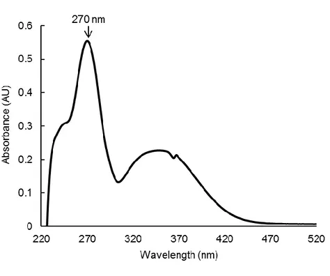
3-1. UVB absorption by baicalein
Absorption of UVB by baicalein was determined using an ultraviolet (UV)/visible
spectrophotometer. Baicalein showed peak absorption at 270 nm, which is close to the range of UVB (280–320 nm) (Fig. 1). Therefore, light absorption by baicalein might be closely
associated with its cytoprotective effect against UVB radiation.
10
the spectral range from 220 to 520 nm. The peak absorption is at 270 nm.
3-2. Baicalein reduces ROS generation
The MTT assay revealed that baicalein itself did not have cytotoxic effects on HaCaT cells
at any concentrations used up to 30 μM (Fig. 2A). Baicalein scavenged the DPPH radical in
a concentration-dependent manner; 1% of radicals were scavenged at 10 μM, 3% at 20 μM,
and 11% at 30 μM. By comparison, the well-known ROS scavenger N-acetyl cysteine (NAC)
scavenged 91% of radicals at a concentration of 1 mM (Fig. 2B). Moreover, we used
electron spin resonance spectrometry to measure the ability of baicalein to scavenge
hydroxyl radical and superoxide anion. In the FeSO4 + H2O2 system (Fe
2+
+ H2O2 → Fe
3+ +
∙OH + OH
-), DMPO/∙OH adducts yielded signals of 3,802 in the absence of baicalein and
1,376 in the presence of 20 μM baicalein (Fig. 2C). Similarly, in the xanthine/xanthine
oxidase system, DMPO/∙OOH yielded signals of 3,671 in the absence of baicalein and 2,045
in the presence of 20 μM baicalein (Fig. 2D), indicating that baicalein can scavenge
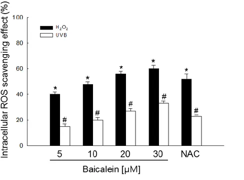
superoxide anion. Next, the intracellular ROS-scavenging activity of baicalein after H2O2
treatment or UVB radiation was determined using the DCF-DA assay (Fig. 2E).
Fluorescence spectrometry data revealed that the intracellular ROS-scavenging activity of
baicalein in H2O2-treated cells was 40% at 5 μM, 48% at 10 μM, 56% at 20 μM, and 60% at
30 μM, compared with 52% for 1 mM NAC, and in UVB-exposed cells was 12% at 5 μM,
14% at 10 μM, 19% at 20 μM, and 21% at 30 μM, compared with 18% for 1 mM NAC (Fig.
2E). Furthermore, confocal microscopy revealed that 20 μM baicalein ameliorated the
increase in intensity (green color) caused by UVB radiation (Fig. 2F), indicating that baicalein treatment reduces ROS generation and that this compound possesses
ROS-scavenging properties. Based on the results of these experiments, we chose to use the optimal dose of 20 μM baicalein for subsequent investigations.
13
Figure 2. Baicalein reduces ROS generation. (A) HaCaT cell viability was investigated using the MTT assay to determine the cytotoxic effects of baicalein. (B) The
radical-scavenging effects of baicalein were investigated using the DPPH assay. (C) The ability to
scavenge hydroxyl radical at 20 μM baicalein was estimated using the Fenton reaction
14
was evaluated using the xanthine/xanthine oxidase system. (E) Cells were treated with 5, 10, 20, or 30 μM baicalein or 1 mM NAC. One hour later, cells were irradiated with UVB or
treated with 1 mM H2O2. After 30 min, cells were stained with DCF-DA and intracellular
ROS were measured using a spectrophotometer. *,#Significantly different from control cells
of H2O2 or UVB radiation, respectively (p < 0.05). (F) Representative confocal images
illustrate that UVB radiation increased the fluorescence intensity of DCF (produced by ROS)
compared with the control, and baicalein treatment of UVB-exposed cells reduced the
15
3-3. Baicalein protects DNA, lipids, and proteins against UVB-induced oxidative damage
We next investigated whether baicalein can inhibit damage to macromolecules in
UVB-exposed cells. First, we monitored UVB-induced DNA damage using the comet assay. The
length of comet tails in microscopy images and the percentage of cellular fluorescence in
tails are shown in Figure 3A. After treatment of cells with UVB radiation, the comet tail
length was distinctly elongated, as well as the ratio of damaged DNA outside of nuclei.
Treatment of UVB-exposed cells with baicalein clearly reduces the percentage of damaged
DNA in comet tails from 52% to 30%. Second, we examined the level of 8-isoprostane, a
hallmark of lipid peroxidation, which is released by cells into the culture medium upon
oxidative damage. Cells exposed to UVB secreted a higher level of 8-isoprostane than
untreated cells; however, pretreatment of UVB-exposed cells with baicalein significantly
reduced the 8-isoprostane level (Fig. 3B). In addition, lipid peroxidation was investigated by
examining the fluorescent product DPPP oxide produced from DPPP. The intensity of DPPP
oxide was higher in UVB-exposed cells than in control cells (blue color). Pretreatment of
UVB-exposed cells with baicalein reduced the fluorescence intensity (Fig. 3C). Finally, we
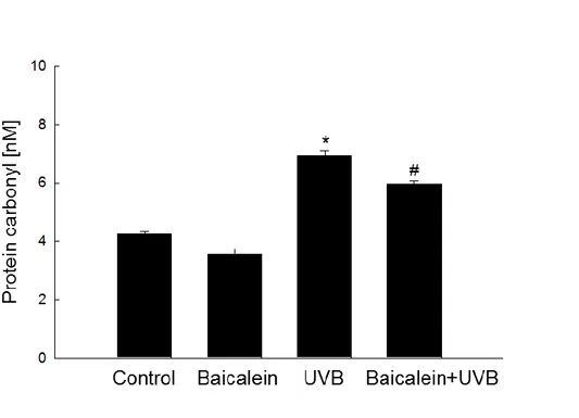
measured protein carbonylation, which is a reaction that occurs during the process of protein
oxidation to form carbonyl groups (Pirinccioglu et al., 2010). UVB irradiation obviously
increased the level of carbonyl moieties, whereas pretreatment of UVB-exposed cells with
baicalein notably suppressed the formation of protein carbonyls (Fig. 3D). Taken together,
these results indicate that baicalein effectively protects DNA, lipids, and proteins against oxidative damage induced by UVB radiation.
18
Figure 3. Baicalein protects cells against UVB-induced DNA damage, lipid peroxidation, and protein carbonylation. (A) The comet assay was performed to assess DNA damage.
Representative images and percentage of cellular fluorescence within comet tails are shown.
*Significantly different from control cells (p < 0.05) and #significantly different from cells
only exposed to UVB radiation (p < 0.05). (B) Cells were treated with 20 μM baicalein.
After 1 h, cells were exposed to UVB radiation. After incubation for a further 24 h, lipid
peroxidation was determined using an 8-isoprostane enzyme immunoassay kit.
*Significantly different from control cells (p < 0.05) and #significantly different from cells
only exposed to UVB radiation (p < 0.05). (C) Lipid peroxidation was detected using a
confocal microscope after DPPP staining. (D) Protein oxidation was assayed by measuring
the amount of carbonyl formation. *Significantly different from control cells (p < 0.05) and #
19
3-4. Baicalein suppresses apoptosis induced by UVB radiation
To elucidate the cytoprotective effect of baicalein against UVB-induced apoptosis, we examined the viability of HaCaT cells exposed to UVB, pretreated or not with 20 μM
baicalein. The viability of UVB-exposed cells was reduced to 75% relative to control cells;
however, pretreatment with baicalein increased viability to 85% compared to 95% of NAC
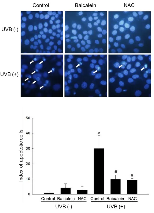
(Fig. 4A). Nuclei were stained with Hoechst 33342, and then cells were assessed by
microscopy. Normal nuclei were observed in control and baicalein-treated cells, whereas
significant nuclear condensation was observed in UVB-exposed cells (apoptotic index = 30).
However, when these cells were pretreated with baicalein or NAC, nuclear fragmentation
was reduced (baicalein, apoptotic index = 9.8; NAC, apoptotic index = 9.2) (Fig. 4B). These
20
Figure 4. Baicalein protects cells against apoptosis induced by UVB. (A) Cells were treated with 20 μM baicalein and exposed to UVB radiation 1 h later. After 24 h incubation, cell
21
viability was determined by the MTT assay and is expressed as a percentage of the control. 1 mM NAC was used as a positive control. *Significantly different from control cells (p < 0.05)
and #significantly different from cells only exposed to UVB radiation (p < 0.05). (B) Cells
were stained with Hoechst 33342 and detected by fluorescence microscopy. Apoptotic
bodies were quantitated. 1 mM NAC was used as a positive control. *Significantly different
from control cells (p < 0.05) and #significantly different from cells only exposed to UVB
22
4. Disccusion
In the present study, we focused on the protective effects of baicalein against UVB
radiation-induced oxidative stress. Our results indicated that baicalein absorbed UV photons,
that was mostly caused by due to its polyphenolic constituents. Spectral data of natural
phenols show a typical UV absorbance characteristic of benzene aromaticity at 270 nm. Baicalein is also natural phenol; therefore, its UV light absorbance (peak 270 nm) may be via
benzene aromaticity (Jean-Denis et al., 2006).
UVB radiation induces ROS generation such as singlet oxygen, superoxide anion,
hydroxyl radical and peroxy radical. In the present study, baicalein scavenged ROS such as
superoxide anion and hydroxyl radical in cell free system, and intracellular ROS induced by
treatment with H2O2 or UVB radiation. The distinct pathways by which flavonoid molecules
(ArOH) transfer their charge provide several mechanisms of their antioxidant action. The
representative one leads to the direct O-H bond breaking and proceeds by rapid donation of
the proton and electron to a radical form (ArOH + HO∙ → ArO∙ + HOH), while the second
one assumes indirect H atom abstraction (ArOH + HO∙ → ArOH+ + OH∙ → ArO∙ + HOH)
(Jasmina et al., 2011). Baicalein has O-H bonds, and it can scavenge radical by O-H bonds.
The UVB-induced ROS induce chemical modifications in DNA by the formation of
purine/pyrimidine dimers and strand breaks leading to mutagenesis and loss of normal
cellular metabolic functions. And they can damage lipids, producing lipid peroxides that are
converted to products such as 8-isoprostane, malondialdehyde and conjugated dienes
(Petrova et al., 2011; Schneider et al., 2006). Furthermore, UVB-induced oxidative stress
damages proteins by site-specific modifications of amino acids, aggregation of cross-linked
23
and oxidation of specific amino acids (Rajesh and Datta, 2015). Therefore, UVB-induced oxidative stress triggers cell damage. However, baicalein protects cellular components such
as DNA, lipids, and proteins against UVB-induced oxidative damage. The oxidative stress
by UVB radiation mediates apoptosis via the release of mitochondrial cytochrome c into the
cytosol and activation of apoptosis-related caspases (Pallela et al., 2010). However, baicalein
protects cells against UVB-induced apoptotic cell death.
In conclusion, baicalein protects against UVB-induced oxidative stress via scavenging
ROS, inhibiting apoptosis, and absorbing UV photons. In a future study, we will elucidate
24
5. References
Beauchamp, M. C., Letendre, E. and Renier, G. (2002) Macrophage lipoprotein lipase
expression is increased in patients with heterozygous familial hypercholesterolemia. J.
Lipid Res. 43, 215–222.
Deschamps, J. D., Kenyon, V. A. and Holman, T. R. (2006) Baicalein is a potent in vitro
inhibitor against both reticulocyte 15-human and platelet 12-human lipoxygenases.
Bioorg. Med. Chem. 14, 4295–4301.
Dhumrongvaraporn, A. and Chanvorachote, P. (2013) Kinetics of ultraviolet B
irradiation-mediated reactive oxygen species generation in human keratinocytes. J. Cosmet. Sci. 64,
207-217.
Hsieh, C. J., Hall, K., Ha, T., Li, C., Krishnaswamy, G. and Chi, D. S. (2007) Baicalein
inhibits IL-1β- and TNF-α-induced inflammatory cytokine production from human mast
cells via regulation of the NF-κB pathway. Clin. Mol. Allergy 5, 5.
Jasmina, M. D. M., Zoran, S. M., Tanja, P. B., Vesna, M. P. and Milka, B. J. (2011) Iron
complexes of dietary flavonoids: Combined spectroscopic and mechanistic study of
their free radical scavenging activity. Food Chem. 129, 1567-1577.
Jean-Denis, J. B., Pezet, R. and Tabacchi, R. (2006) Rapid analysis of stilbenes and
derivatives from downy mildew-infected grapevine leaves by liquid
chromatography-atmospheric pressure photoionisation mass spectrometry. J. Chromatogr. A 1112,
263-268.
25
expression of matrix metalloproteinase-1 by regulating the ERK/JNK/AP-1 pathway in human keratinocytes. Biomol. Ther. 20, 57–61.
Kim, K. C., Lee, I. K., Kang, K. A., Kim, H. S., Kang, S. S. and Hyun, J. W. (2012B)
Baicalein (5,6,7-trihydroxyflavone) reduces oxidative stress-induced DNA damage by
upregulating the DNA repair system. Cell Biol. Toxicol. 28, 421–433.
Lee, I. K., Kang, K. A., Zhang, R., Kim, B. J., Kang, S. S. and Hyun, J. W. (2011)
Mitochondria protection of baicalein against oxidative damage via induction of
manganese superoxide dismutase. Environ. Toxicol. Pharmacol. 31, 233–241.
Lee, K.O., Kim, S. N. and Kim, Y. C. (2014) Anti-wrinkle effects of water extracts of teas in
hairless mouse. Toxicol. Res. 30, 283–289.
Lee, S. J. and Park, J. W. (2014) Enhancement of UVB radiation-mediated apoptosis by
knockdown of cytosolic NADP+-dependent isocitrate dehydrogenase in HaCaT cells.
BMB. Rep. 47, 209-214.
Lin, Y. L., Lin, R. J., Shen, K. P., Dai, Z. K., Chen, I. J., Wu, J. R. and Wu, B. N. (2011)
Baicalein, isolated from Scutellaria baicalensis, protects against endothelin-1-induced
pulmonary artery smooth muscle cell proliferation via inhibition of TRPC1 channel
expression. J. Ethnopharmacol. 18, 373-381.
Li, L., Abe, Y., Kanagawa, K., Usui, N., Imai, K., Mashino, T., Mochizuki, M. and Miyata,
N. (2004) Distinguishing the 5,5-dimethyl-1-pyrroline N-oxide (DMPO)-OH radical
quenching effect from the hydroxyl radical scavenging effect in the ESR spin-trapping
method. Anal. Chim. Acta. 512, 121-124.
26
Int. J. Dermatol. 49, 978-986.
Okimoto, Y., Watanabe, A., Niki, E., Yamashita, T. and Noguchi, N. (2000) A novel
fluorescent probe diphenyl-1-pyrenylphosphine to follow lipid peroxidation in cell
membranes. FEBS. Lett. 474, 137–140.
Pallela, R., Na-Young, Y. and Kim, S. K. (2010) Anti-photoaging and photoprotective
compounds derived from marine organisms. Mar. Drugs 8, 1189-1202.
Palmer, D. M. and Kitchin, J. S. (2010) Oxidative damage, skin aging, antioxidants and a
novel antioxidant rating system. J. Drugs Dermatol. 9, 11-15.
Petrova, A., Davids, L. M., Rautenbach, F. and Marnewick, J. L. (2011) Photoprotection by
honeybush extracts, hesperidin and mangiferin against UVB-induced skin damage in
SKH-1 mice. J. Photochem. Photobiol. B. 103, 126-139.
Pirinccioglu, A. G., Gökalp, D., Pirinccioglu, M., Kizil, G. and Kizil, M. (2010)
Malondialdehyde (MDA) and protein carbonyl (PCO) levels as biomarkers of oxidative
stress in subjects with familial hypercholesterolemia. Clin. Biochem. 43, 1220–1224.
Rajagopalan, R., Ranjan, S. and Nair, C. K. (2003) Effect of vinblastine sulfate on
gamma-radiation-induced DNA single-strand breaks in murine tissues. Mutat. Res. 536, 15–25.
Rajesh, P. R. and Datta, M. (2015) UV-Induced Oxidative Stress in Cyanobacteria: How life
is able to survive? Biochem. Anal. Biochem. 4, 1000173.
Rosenkranz, A. R., Schmaldienst, S., Stuhlmeier, K. M., Chen, W., Knapp, W. and Zlabinger,
G. J. (1992) A microplate assay for the detection of oxidative products using
27
Schneider, L. A., Bloch, W., Kopp, K., Hainzl, A., Rettberg, P., Wlaschek, M., Horneck, G. and Scharffetter-Kochanek, K. (2006) 8-Isoprostane is a dose-related biomarker for
photo-oxidative ultraviolet (UV) B damage in vivo: a pilot study with personal UV
dosimetry. Br. J. Dermatol. 154, 1147-1154.
Sen, S., Chakraborty, R., Sridhar, C., Reddy, Y. S. R. and De, B. (2010) Free radicals,
antioxidants, diseases and phytomedicines: current status and future prospect. Int. J.
Pharm. Sci. Rev. Res. 3, 91-100.
Sime, S. and Reeve, V. E. (2004) Protection from inflammation, immunosuppression and
carcinogenesis induced by UV radiation in mice by topical Pycnogenol® . Photochem.
Photobiol. 79, 193-198.
Sklar, L. R., Almutawa, F., Lim, H. W. and Hamzavi, I. (2013) Effects of ultraviolet
radiation, visible light, and infrared radiation on erythema and pigmentation: a review.
Photochem. Photobiol. Sci. 12, 54-64.
Vera Saltos, M. B., Naranjo Puente, B. F., Milella, L., De Tommasi, N., Dal Piaz, F. and
Braca, A. (2015) Antioxidant and free radical scavenging activity of phenolics from
28
6. Abstract in Korean
Baicalein (5,6,7-trihydroxy-2-phenyl-chromen-4-one) 은 하나의 플라본이자 플라보노이드의 한 타입으로, 황금 (Scutellaria baicalensis) 의 뿌리로부터 추출되었다. 이 연구는 인간 각질세포 (HaCaT) 에서 자외선 B 의 조사에 의해 유도된 산화적 스트레스 매개 세포사멸에 대한 baicalein 의 보호효과를 평가하였다. Baicalein 은 자외선 B 의 파장 범위 내의 빛을 흡수했다. 또한, baicalein 은 자외선 B 의 조사에 반응하여 세포 내 활성산소종 (ROS) 의 수준을감소시켰다. Baicalein 은 HaCaT 세포에서 자외선 B 의 조사에 유도된 DNA 손상,
8-isoprostane 발생과 단백질 변형에 대하여 세포를 보호했다. 게다가, baicalein 은
자외선 B 조사에 의한 세포사멸을 감쇠시켰다. 이러한 연구결과는 baicalein 이
자외선 B 로 유도된 세포손상과 세포사멸을 자외선 B 조사를 흡수하고 ROS 를
29
7. Acknowledgement
짧고도 긴 2 년이라는 시간. 화장품 연구원이라는 꿈을 품고 좀 더 전문적인 공부를 하기 위해서 생명대학교에서 의학전문대학원으로 건너와 아무것도 몰랐던 저를 처음부터 따뜻하게 대해주시고 환영해주신 현진원 교수님! 언제나 해박한 지식으로 저에게 지식을 전수해주시고 격려를 아끼지 않으셨습니다. 덕분에 다른 석사연구생보다 더 많은 공부를 하고 갑니다. 정말 감사합니다. 그리고 학사 시절 항상 반겨주시고 4 학년 생활을 함께했던, 석사 공부를 응원해주셨던 김동순 교수님, 언제나 저에게 웃어주시고 제 진로를 지도해주셨던 현해남 교수님, 지금은 독일에 계신 항상 열정적이신 전용철 교수님 모두 감사합니다. 또한, 바쁘신 와중에도 불구하고 저의 학위 논문 심사에 참여해주시고 아낌없는 조언 해주신 고영상 교수님과 유은숙 교수님께도 감사 드립니다. 꼼꼼하고 날카로운 질문으로 정말 많은 도움이 되었던 강희경 교수님, 조문제 교수님께도 감사의 말씀을 드립니다. 저의 통계학 지식을 쌓게 해 주신 배종면 교수님, 김수영 교수님 정말 감사합니다. 또 저의 영어 공부에 큰 힘이 되어주시고 정말 많은 조언을 해주신 양진혁 선생님! 감사 드립니다. 그리고 우리 생화학실 최고로 존경하는 박미경 박사님! 언제나 가장 앞에서 저희를 위해 앞장서시고, 지도해주셔서 정말 감사합니다. 지금은 미국에 계신 저에게 대부분의 실험을 가르쳐주신 김기천 선배님 감사 드립니다. 또 힘들 때면 옆에서 힘이 되어주고 섬세하게 실험을 가르쳐줬던지원이도 고맙다. 먼저 졸업한 Han Xia, Susara, 계속 연락하면서 지내고 싶고
one of my friend, Madushan! I hope to go to Sri Lanka. See you soon! 항상
30 후배 유진, 이제 시작될 석사과정 힘들겠지만 멋지게 졸업하고 내 뒤를 잇길 기대하마. 그 외에도 도와주신 분들이 많이 남았지만 한 분 한 분 감사의 말씀을 전하지 못하여 죄송합니다. 감사의 말씀을 전하지 못한 분들께도 진심으로 감사하다는 말씀 드리고 싶습니다. 계속해서 성장하는 오민창 되겠습니다. 감사합니다!