https://doi.org/10.20307/nps.2021.27.1.49
49
Phenolic Constituents from Balanophora laxiflora with their Anti-inflammatory
and Cytotoxic Effects
Nguyen Thi Hong Anh1, Nguyen Thuy Duong2, Pham Duc Vinh2, and Do Thi Ha1,*
1National Institute of Medical Materials, Hanoi, 111000, Vietnam
2Department of Pharmacology, Hanoi University of Pharmacy, 13 Le Thanh Tong, Hanoi, 11021, Vietnam
Abstract Balanophora laxiflora Hemsl. (Balanophoraceae) is a traditional medicinal plant with a diverse array of biological activities. In our exploration of new bioactive constituents from B. laxiflora, we isolated five compounds, including a new lignan, balanophorone (5), and four known phenolic compounds (1–4). The chemical structures of these compounds were determined by extensive spectroscopic analyses, including 1D and 2D NMR, HR-ESI-MS, and CD. In addition, we evaluated the effects of each of the isolates (1–5) on the messenger RNA expression levels of tumor necrosis factor (TNF)-α and cyclooxygenase (COX)-2 in lipopolysaccharide (LPS)-stimulated RAW 264.7 macrophages and cytotoxicity against MCF-7 and MDA-MB-231 breast cancer cells. Compound 2 showed significant inhibition of LPS-induced COX-2 and TNF-α expression in RAW 264.7 macrophages, while compound 4 showed moderate cytotoxicity against MCF-7 and MDA-MB-231 breast cancer cells, with IC50 values of 18.3 and 30.7 μM, respectively. No significant effects on
the viability of normal mammary epithelial cells were observed.
Keywords Balanophora laxiflora, Balanophorone, Anti-inflammation, COX-2, TNF-α, Cytotoxicity
Introduction
Balanophora laxiflora Hemsl. belongs to the Balano-phoraceae family and grows in tropical and subtropical regions across Vietnam, China, Thailand, and Laos. In Vietnam, B. laxiflora is a dioeciously parasitic plant, found primarily in the forests of the Ninh Binh, Kontum, Lao Cai, and Yen Bai provinces.1 In Vietnamese folk medicine, this plant is used for the treatment of limb aches and colic, and as a tonic for elderly and postnatal women.2 Chemical investigations of B. laxiflora have identified a diverse variety of compounds, including phenylpropanoids, tannins, lignans, and triterpenoids.3 Earlier studies demonstrated that the crude extract and compounds isolated from B. laxiflora exhibit DPPH radical-scavenging activity,4 hypouricemic effect,5 cytoto-xicity against certain cancer cell lines,6 and anti-inflammatory actions.7 Continuing the prior research on the chemical composition of the ethyl acetate of B. laxiflora, 8 this paper reports our recent chemical investigation on the ethyl acetate extract of B. laxiflora,
collected in Sapa - Lao Cai province together with the anti-inflammatory and cytotoxicity activities to elucidate biochemical and bioactive significance as well as extend the use of B. laxiflora.
Experimental
Plant materials The plant samples were collected in Sapa, Lao Cai, Vietnam in November 2015, and botanically identified as Balanophora laxiflora Hemsl. by Dr. Tran The Bach - Institute of Ecology and Biological Resource. A voucher specimen (BSp2) was deposited at the National Institute of Medicinal Materials.
General experiment procedures Optical rotations were measured on a JASCO V-550 UV/Vis spectrometer (JASCO International Co. Ltd., Tokyo, Japan). Circular dichroism (CD) measurements were carried out on a Chirascan spectrometer (UK). The NMR [1H (500 MHz), 13C (125 MHz)] experiments were performed on a Bruker Advance 500 spectrometer (Bruker, Billerica, MA, USA). The chemical shift was reported in ppm downfield from MS, with J in Hz. Mass spectra were obtained with an AGILENT 1200 series LC-MSD Ion rap (Agilent Tech-nologies, Palo Alto, CA, USA). Analytical thin-layer chromatography (TLC) was performed on Kieselgel 60
*Author for correspondence
Do Thi Ha, Department of Analytical Chemistry and Standardiza-tion, National Institute of Medicinal Materials.
mm layer thickness) and RP-18 F254 TLC plate (Merck, Darmstadt, Germany). UV spots were visualized using ultraviolet irradiation (at 254–366 nm) and by spraying with 10% H2SO4 in 96% ethanol, followed by heating with a heat gun. Column chromatography (CC) was performed on silica gel 70–230 and 230–400 mesh (Merck, Darmstadt, Germany).
Extraction and isolation Air-dried and pulverized samples of B. laxiflora (4.0 kg) were extracted by refluxing in an aqueous solution of 80% ethanol for 3 hours. The extraction process was repeated three times. The extracts were then combined and evaporated at 50 ºC under low pressure, yielding 650.12 g of residue. The total residue was suspended in water and successively extracted with n-hexane and EtOAc. The solvents were then evaporated in vacuo to obtain the corresponding n-hexane (BLH, 60.2 g), EtOAc (BLEA, 124.1 g), and water layer (BLW, 180.7 g) fractions. The BLEA fraction (124.0 g) was separated on a silica gel column and eluted with a gradient solvent system of n-hexane/EtOAc (100/1–1/10, v/v) to afford 15 fractions (E1–E15). Fraction E5 (590 mg) was further separated on an RP-C18 column eluted with methanol/water (2/3, v/v) to give four fractions (E5.1–E5.4). Fraction E5.1 (112 mg) was then separated by column chromatography (CC) using an n-hexane/ EtOAc (1/1, v/v) eluent to give compounds 1 (11.3 mg), 2 (4.8 mg), 3 (1.4 mg), and 4 (2.8 mg). Fraction E10 (1.2 g) was separated by CC and eluted with n-hexane/EtOAc (1/ 1, v/v) to yield compound 5 (6.0 mg).
p-Hydroxybenzaldehyde (1): White amorphous powder; ESI-MS: m/z 123.05 [M+H]+ (C 7H6O2, M = 122); 1 H-NMR (500 MHz, CD3OD): δH 9.78 (1H, s, H-7), 7.79 (2H, d, J = 8.5 Hz, H-2, H-6), 6.94 (2H, d, J = 8.5 Hz, H-3, H-5); 13C-NMR (125 MHz, CD 3OD): δC 130.3 (C-1), 133.4 (C-2, C-6), 116.9 (C-3, C-5), 165.2 (C-4), 192.9 (C-7). Piceol (2): White amorphous powder; ESI-MS: m/z 137.10 [M+H]+ (C 8H8O2, M = 136); 1H-NMR (500 MHz, CD3OD): δH 7.90 (2H, d, J = 9.0 Hz, H-2, H-6), 6.86 (2H, d, J = 9.0 Hz, H-3, H-5), 2.54 (3H, s, H-8); 13C-NMR (125 MHz, CD3OD): δC 130.2 (C-1), 132.1 (C-2, C-6), 116.2 (C-3, C-5), 163.9 (C-4), 199.5 (C-7), 26.3 (C-8).
Trans-p-coumaryl aldehyde (3): White amorphous powder; ESI-MS: m/z 149.15 [M+H]+ (C 9H8O2, M = 148); 1H-NMR (500 MHz, CD 3OD): δH 7.57 (2H, d, J = 8.5 Hz, H-2, H-6), 6.87 (2H, d, J = 8.5 Hz, H-3, H-5), 7.61 (1H, d, J = 16.0 Hz, H-7), 6.64 (1H, d, J = 8.0, 16.0 Hz, H-8), 9.59 (1H, d, J = 8.0 Hz, H-9); 13C-NMR (125 MHz, CD 3OD): δC 127.1 (C-1), 132.0 (C-2, C-6), 117.1 (C-3, C-5), 162.3 (C-4), 156.0 (C-7), 126.5 (C-8), 196.2 (C-9). MS: m/z 149.15 [M+H]+ (C 9H8O2, M = 148); 1H-NMR (500 MHz, CD3OD): δH 7.28 (1H, d, J = 2.0 Hz, H-2), 6.88 (1H, d, J = 8.0 Hz, H-5), 7.19 (1H, dd, J = 2.0, 8.0 Hz, H-6), 7.61 (1H, d, J = 15.5, H-7), 6.68 (1H, dd, J = 8.0, 15.5, H-8), 9.59 (1H, d, J = 8.0, H-9), 3.93 (3H, s, 3-OCH3); 13C-NMR (125 MHz, CD3OD): δC 126.6 (C-1), 109.5 (C-2), 149.0 (C-3), 147.0 (C-4), 115,0 (C-5), 124.0 (C-6), 153.2 (C-7), 126.3 (C-8), 193.7 (C-9), 56.0 (3-OCH3). Balanophorone (5): Yellow amorphous powder; HR-ESI-MS (molecular ion peak): m/z 415.1395 [M-H] (Calcd for C22H23O8, 415.1393); 1H-NMR (500 MHz, CD3OD): δH 7.62 (1H, dd, J = 2.0, 8.0 Hz, H-6), 7.60 (1H, d, J = 2.0 Hz, H-2), 7.08 (1H, d, J = 2.0 Hz, H-2'), 6.92 (1H, d, J = 8.0 Hz, H-5), 6.87 (1H, dd, J = 2.0, 8.5 Hz, H-6'), 6.80 (1H. d. J = 8.5 Hz, H-5'), 4.61 (1H, d, J = 9.0 Hz, H-7'), 4.28 (1H, m, H-9), 4.22 (1H, t, J = 8.5 Hz, H-8), 4.19 (1H, d, J = 6.5 Hz, H-9'), 4.11 (1H, dd, J = 5.5, 8.5 Hz, H-9), 3.94 (3H, s, 3'-OCH3), 3.91 (3H, s, 3-OCH3), 3.00 (1H, m, H-8'), 1.82 (3H, s, COOCH3); 13C-NMR (125 MHz, CD 3OD): δC 130.0 1), 112.4 (C-2), 149.4 (C-3), 153.8 (C-4), 1116.0 (C-5), 125.1 (C-6), 199.6 (C-7), 51.1 (C-8), 72.0 (C-9), 56.4 (3-OCH3), 132.8 1'), 111.5 2'), 149.2 3'), 147.8 4'), 116.0 (C-5'), 121.1 (C-6'), 86.0 (C-7'), 51.2 (C-8'), 64.7 (C-9'), 56.5 (3'-OCH3), 20.5 (COOCH3), 172.5 (COOCH3).
Cell culture The RAW 264.7 macrophage, MCF-7, and MDA-MB-231, and MCF-10A cell lines were purchased from the American Type Culture Collection (ATCC, Rockville, MD, USA). RAW 264.7 macrophages, MDA-MB-231, and MCF-7 cells were routinely cultured in Dulbecco's Modified Eagle Medium (DMEM) supple-mented with 10% (v/v) fetal bovine serum (FBS) and 1% (v/v) penicillin-streptomycin, while MCF-10A cells were maintained in Mammary Epithelial Basal Medium (MEBM) containing 100 ng/ml cholera toxin.9 Cells were kept in a 37 ºC incubator with a humidified atmosphere of 5% CO2.
Cell viability assay Cells were seeded in 96-well plates at a density of 2×10⁴ cells/well. After overnight incubation, the cells were treated with increasing concentrations of compounds 1-5 for 72 h. Finally, the cells were incubated with 20 µL of 3-(4,5-dimethylthiazol- 2-yl)-5-(3-carboxymethoxyphenyl)-2-(4-sulfophenyl)-2H-tetrazolium (MTS) (Promega Corporation) for 2 h at 37oC. The number of viable cells was examined by measuring the absorbance of the resultant formazan dye at 490 nm using a microplate reader (Spark™ 10M multimode microplate reader, Tecan, Mannedorf, Switzerland).
RNA isolation, Reverse Transcription (RT), and Quantitative PCR (qPCR) RAW 264.7 macrophages were seeded in 35-mm dishes at a density of 1×10⁶ cells/ dish. After overnight incubation, cells were treated with 10 µM of compounds 1-5 for 18 h, followed by stimulation with lipopolysaccharide (LPS) (100 ng/ml) for a further 6 h. The RT-qPCR assay was performed as previously described.10 Briefly, total RNA was isolated using Qiagen lysis reagent (Qiagen, MD, USA) according to the manufacturer’s instructions. Complementary DNA (cDNA) was then synthesized from one microgram of total RNA using the Go Script reverse transcription system (Promega Corporation, Madison, WI, USA). Quantitative real-time PCR amplification was performed with a Roche LightCycler 2.0 (Mannheim, Germany) using the absolute qPCR SYBR green capillary mix AB gene system (Thermo Scientific, UK). The primer sequences of target genes used in this study as follows: TNFα: Forward 5′-GGTGCCTATGTCTCAGCCTCTT-3′, Reverse 5′-GCCA TAGAACTGATGAGAGGGAG-3′; COX-2: Forward 5′-CCACTTCAAGGGAGTCTGGA-3′, Reverse 5′-AGTC ATCTGCTACGGGAGGA-3′; GAPDH: Forward 5′-CAT CACTGCCACCCAGAAGACTG-3′, Reverse 5′-ATGCC AGTGAGCTTCCCGTTCAG-3′.
Statistical analysis Data were presented as mean ± standard of error (SEM) from three independent experi-ments. All statistical analyses were carried out using GraphPad Prism 5.01 software. P values were determined based on the one-way analysis of variance (ANOVA), followed by Tukey’s post hoc multiple comparison tests. P values of lower than 0.05 were considered being statistically significant. The half-maximal inhibitory con-centration (IC50) was estimated by nonlinear regression with a sigmoidal dose-response model.
Results and Discussion
The ethanol extract of B. laxiflora was suspended in water and successively divided into n-hexane, EtOAc, and water-soluble fractions. A new compound (5), and four
known compounds (1–4) were isolated via separation on silica gel and/or C18 reversed-phase silica gel columns, or by thin layer chromatography (TLC) of the EtOAc fraction of B. laxiflora. The chemical structures of the known compounds were identified using spectroscopic analyses (1H- and 13C- NMR and MS) and by comparing these data with previously reported data from p-hydroxybenzaldehyde (1),11 piceol (2),12 trans-p-coumaryl aldehyde (3),13 and ferulic aldehyde (4).14
Compound 5 was isolated as a yellow, amorphous powder. The molecular formula of 5 was C22H24O8, as determined by the molecular ion in its HR-ESI-MS spectrum at m/z 415.1395 [M-H], indicating 12 degrees of unsaturation (calcd. for C22H23O8, 415.1393). The 1 H-NMR spectrum of 5 contained signals corresponding to six protons involved in two ABX spin-spin interactions, i.e., at δH 7.62 (1H, dd, J = 2.0, 8.0 Hz), 7.60 (1H, d, J = 2.0 Hz), and 6.92 (1H, d, J = 8.0 Hz) and δH 7.08 (1H, d, J = 2.0 Hz), 6.80 (1H, d, J = 8.5 Hz), and 6.87 (1H, dd, J = 2.0, 8.5 Hz). Signals corresponding to two methoxy groups at δH 3.91 (3H, s) and 3.94 (3H, s), and a methyl group at δH 1.82 (3H, s), were also observed. The 13C NMR spectrum of 5 contained 22 carbon signals that were classified using HSQC spectra as 8 non-protonated carbons, 9 methines, 2 methylenes, and 3 methyl carbon atoms. Two deshielded carbon signals at δC 199.6 and 172.5 were assigned to one ketone and one carbonyl group, respectively. Two carbon signals at δC 56.5 and 56.4 were assigned to two methoxy groups, and a signal at δC 20.5 was assigned to a methyl group. The COSY
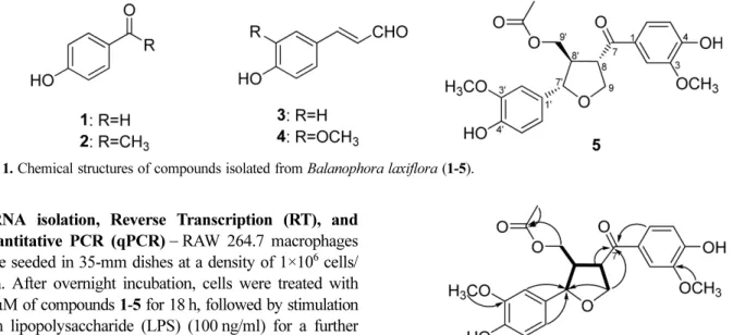
Fig. 1. Chemical structures of compounds isolated from Balanophora laxiflora (1-5).
Fig. 2. 1H-1H COSY (─) and key HMBC (→) correlations of
Fig. 3. The effects of compounds 1–5 on the mRNA expression levels of TNF-α and COX-2 in RAW 264.7 macrophages. RAW 264.7 macrophages were treated with compounds 1–5 (10 µM) for 18 h, followed by stimulation with 100 ng/ml of LPS for 6 h. The mRNA levels of (A) TNF-α and (B) COX-2 were determined by RT-qPCR, as described in the Methods section. * denotes P < 0.05 compared to cells treated with LPS alone.
Fig. 4. The effects of compounds 1–5 on the viability of (A) MCF-7 and (B) MDA-MB-231 breast cancer cells are shown with increasing concentrations of compounds 1–5 (10–150 µM) for 72 h. Cell viability was examined by MTS assay, as described in the Methods section. IC50 values were calculated by nonlinear regression with a sigmoidal dose-response model, and are presented in the lower panel of each
figure. (C) MCF-10A mammary epithelial cells were treated with compounds 1–5 (100 µM) for 72 h. The cell survival rate was determined by MTS assay.
spectrum showed H interactions between 7' and H-8', H-8' and H-9', H-8' and H-8, and H-8 and H-9, corresponding to a furan ring. HMBC correlations between H-9 (δH 4.28, 4.11), and C-7' (δC 86.0) indicated the presence of an ether bridge between C-9 and C-7'. HMBC interactions between the methoxy proton at δH 3.91 to C-3 (δC 149.4) and δH 3.94 to C-3' (δC 149.2) indicated two methoxy groups linked at C-3 and C-3'.
The location of the acetyl group at C-9' was determined based on the HMBC correlations between H-9' (δH 4.19) and the carboxyl carbon atom of the acetoxy group (δC 172.5). The HMBC correlations between H-8 (δH 4.22)/ H-9 (δH 4.11, 4.28)/H-6 (δH 7.62) and C-7 (δC 199.6) indicated a ketone group at C-7.
Relative configuration of 5 was examined by NOESY analysis. NOESY interactions between H-7' (δH 4.61) and H-8 (δH 4.22) indicated H-7' and H-8 adopt a cis confi-guration. Comparisons with reference data 15 the chemical shift of H-7' at 4.61 suggested that H-7' and H-8' were trans-oriented, and this was supported by a NOESY correlation between H-2' and H-8'.
The absolute configurations of 5 at chiral centers 7', C-8', and C-8 were confirmed by circular dichroism (CD) measurements, as described in a previous report.16 The CD spectrum of 5 observed Cotton effects (Δε: -0.41 (304 nm)) similar to those of wikstrone ([θ]: -1.9 (320 nm) for 7'S,8'R,8S configurations),16 indicating 7'S,8'R,8S-con-figurations. Thus, compound 5 was identified as a new furan lignan and named (7'S,8'R,8S)-9'-acetyl-3,3′-dimethoxy-4,4′-dihydroxy-7',9-epoxylignan-7′-one (Balanonophorone).
Compounds (1–5) were evaluated for anti-inflammatory activity via inhibition of TNFα and COX-2 mRNA expression in LPS-stimulated RAW 264.7 cells. Com-pounds 1 and 3–5 were weak inhibitors at 10 µM (Fig. 3). In contrast, compound 2 drastically decreased the mRNA expression levels of TNF-α and COX-2 in LPS-stimulated RAW 264.7 cells. The cytotoxic effects of the isolated compounds (1–5) were also evaluated using MTS assays. Notably, cytotoxicity against MCF-10A mammary epithelial cells was not observed with any of the isolated
com-pounds (Fig. 4C). However, compound 4 exhibited significant cytotoxicity against MCF7 and MDA-MB-231 breast cancer cells (IC50 of 18.30 and 32.97 µM, res-pectively), as shown in Figs. 4A–B. This result shows (at least in the part) compound 2 and compound 4 may contribute to the anti-inflammatory and cytotoxic effect of B. laxiflora, and would suggest to further investigate their anti-inflammatory and cytotoxic effect.
References
(1) Nguyen, M. K. List of medicinal plants in Vietnam; Science and Technics Publishing House: Vietnam, 2016, p 628.
(2) Do, T. L. Vietnamese medicinal plants and herbs; Medical publisher: Vietnam, 2014, p 114.
(3) Wang, X.; Liu, Z.; Qiao, W.; Cheng, R.; Liu, B.; She, G. Chem. Cent. J. 2012, 6, 79.
(4) Ho, S. T.; Tung, Y. T.; Cheng, K. C.; Wu, J. H. Food Chem. 2010, 122, 584-588.
(5) Ho, S. T.; Tung, Y. T.; Huang, C. C.; Kuo, C. L.; Lin, C. C.; Yang, S. C.; Wu, J. H. Evid. Based Complement. Alternat. Med. 2012, 2012, 1-7.
(6) Quang, D. N.; So, T. C.; Thanh, N. T. P.; Hoa, L. T. P.; Dien, P. H.; Luong, T. M.; Tung, N. Q.; Long, L. D.; Dai, T. D.; Tien, N. Q. Nat. Prod. Res. 2018, 32, 767-772.
(7) Chiou, W. F.; Shen, C. C.; Lin, L. C. J. Food Drug Anal. 2011, 19, 502-508.
(8) Bui, H. T.; Nguyen, T. H. A.; Nguyen, T. C.; Pham, H. Y.; Nguyen, X. N.; Tran, H. Q.; Do, T. H.; Dan, T. T. H.; Nguyen, T. H.; Chau, V. M.; Phan, V. K. Nat. Prod. Commun. 2019, 14, 1-6.
(9) Mosmann, T. J. Immunol. Methods. 1983, 65, 55-63.
(10) Pham, D. V.; Raut, P. K.; Pandit, M.; Chang, J. H.; Katila, N.; Choi, D. Y.; Jeong, J. H.; Park, P. H. Cancers 2020, 12, 613.
(11) Jang, D. S.; Han, A. R.; Park, G.; Jh on, G. J.; Seo, E. K. Arch. Pharm. Res. 2004, 27, 386-389.
(12) Masi, M.; Mubaiwa, B.; Cimmino, A.; van Otterlo, W. A. L.; Green, I. R.; Evidente, A. S. Afr. J. Bot. 2017, 114, 37-39.
(13) Stange, R. R.; Sims, J. J.; Midland, S. L.; McDonald, R. E. Phytochemistry 1999, 52, 41-43.
(14) Tran, D. D.; Nguyen, Q. T.; Nguyen, N. T.; Nguyen, Q. A.; Truong, T. T. N.; Trinh, T. T.; Nguyen, T. T.; Dang, N. Q. Vietnam J. Chemistry 2017, 55, 48-51.
(15) Jung, K. Y.; Kim, D. S.; Oh, S. R.; Park, S. H.; Lee, I. S.; Lee, J. J.; Shin, D. H.; Lee, H. K. J. Nat. Prod. 1998, 61, 808-811.
(16) Liao, S. G.; Wu, Y.; Yue, J. M. Helv. Chim. Acta. 2006, 89, 73-80.
Received January 21, 2021 Revised February 24, 2021 Accepted March 1, 2021