Original Article
Association of pancreatic adenocarcinoma up-regulated
factor expression in ovarian mucinous adenocarcinoma
with poor prognosis
Sang Kyum Kim1, Si Young Song2, Sunghoon Kim3, Nam Hoon Cho1, Ga Won Yim3, Sang Wun Kim3, Young Tae Kim3, Eun Ji Nam3
1Department of Pathology, Yonsei University College of Medicine, Seoul, South Korea; 2Division of
Gastroenterol-ogy, Department of Internal Medicine, Yonsei University College of Medicine, Seoul, South Korea; 3Department of
Obstetrics and Gynecology, Yonsei University College of Medicine, Seoul, South Korea
Received June 2, 2014; Accepted July 16, 2014; Epub July 15, 2014; Published August 1, 2014
Abstract: Pancreatic adenocarcinoma up-regulated factor (PAUF) expression is elevated in both ovarian tumors and pancreatic adenocarcinoma. However, PAUF expression in ovarian tumors according to histologic subtype and grade has not been investigated. In this study, we examined various clinicopathologic features of 24 patients with mucinous cystadenoma (MCA), 36 with mucinous borderline tumors (MBTs), and 46 with mucinous adenocarcino-mas (MACs) according to PAUF expression status assessed using immunohistochemistry. We found that MACs more frequently stained positive for PAUF than did MCAs and MBTs (P < 0.0001). Although there was no significant differ-ences with respect to other clinicopathologic characteristics of MACs according to PAUF expression status, patients with PAUF-weakly positive and PAUF-strongly positive MACs tended to have a shorter overall survival (OS) than those with PAUF-negative MAC, determined using a Kaplan–Meier analysis (P = 0.1885). After adjusting for various clini-copathologic parameters, PAUF positivity of MACs was a significant predictive factor for disease-free survival (DFS) (negative vs. weakly positive: P = 0.045, hazard ratio [HR] = 57.406, 95% confidence interval [CI]: 1.090-3022.596; and negative vs. strongly positive: P = 0.034, HR = 97.890, 95% CI: 1.412-6785.925). In conclusion, PAUF was more frequently expressed in MAC than in its benign and borderline counterparts, and was associated with a poor OS and DFS in MAC patients. Therefore, we suggest that PAUF may be a practical biomarker for histopathological categoriza-tion and a prognostic marker for patients with an ovarian mucinous tumor.
Keywords: Mucinous tumor, ovary, PAUF, prognostic factor
Introduction
Ovarian mucinous neoplasm is a common ovar-ian epithelial tumor. It is categorized as benign mucinous cystadenoma (MCA), mucinous bor-derline tumor (MBT) of intermediate grade, and malignant mucinous adenocarcinoma (MAC) based on its biological behavior [1, 2]. Of these, MCA is the most common ovarian mucinous neoplasm, accounting for 14% of all ovarian tumors and approximately 80% of all primary ovarian mucinous tumors [3]. MBTs, which are associated with an excellent prognosis and have a survival rate of > 90%, are the most common type of borderline ovarian tumor in Asia and the second most common in North America and Europe [4-8]. In contrast, ovarian
MAC accounts for 3-4% of all primary ovarian carcinomas and is known to have an aggressive behavior, often recurring early and showing resistance to chemotherapy and radiotherapy [9, 10]. It is therefore important to distinguish between these tumor subtypes; however, there are no suitable biomarkers to that can help to differentiate between intermediate and malig-nant mucinous tumors.
Pancreatic adenocarcinoma up-regulated fac-tor (PAUF) is a novel, tumor-specific protein [11]. It is known to play an important role in cancer progression and metastasis in pancreatic duc-tal adenocarcinoma [11, 12]. In addition, Kim et
al. demonstrated that PAUF was also highly
Intestinal - 30 (83.33) Endocervical - 6 (16.67) -Invasion pattern (%) Expansile - - 37 (80.43) Infiltrative - - 9 (19.57) PAUF expression (%) < 0.0001 Negative 16 (66.67) 27 (75.00) 10 (21.74) Weakly positive 7 (29.17) 5 (13.89) 19 (41.30) Strongly positive 1 (4.17) 4 (11.11) 17 (36.96) SD, standard deviation.
Northern blot analysis [11]. However, the expression profiles of PAUF in ovarian neo-plasms according to histologic subtype or malignant potential are not known. In this study, we evaluated PAUF expression status accord-ing to the histologic grade of ovarian mucinous neoplasms and investigated its potential as a prognostic biomarker.
Materials and methods
Case selection and tumor samples
The study cohort consisted of 24 patients with MCA, 36 patients with MBT, and 46 patients with MAC who had undergone surgery and were diagnosed at Yonsei University Medical Center between 2001 and 2012. This retrospective study was approved by the institutional review board of Yonsei University Medical Center (IRB no. 4-2014-0034). The following clinical param-eters were recorded: age at diagnosis, tumor stage, follow-up duration, and survival. We grouped cases of mucinous neoplasms using the International Federation of Gynecology and Obstetrics (FIGO) staging criteria at diagnosis. These groups were a localized stage including FIGO stages IA and IB, a regional stage includ-ing FIGO stages IC and II, and a distant stage including FIGO stages III and IV.
Tissue samples were fixed in 10% buffered
for-sues stained with hematoxylin and eosin (H&E) were reviewed by two obstetrics and gynecolo-gy pathologists (SK Kim and NH Cho). A repre-sentative area was selected on an H&E-stained slide and the corresponding area was marked on the formalin-fixed paraffin-embedded (FFPE) tissue block to make a tissue microarray (TMA), with 5 mm tissue cores. The pathologic param-eters included the histologic subtype of MBT, invasion patterns of MAC, PAUF expression, estrogen receptor (ER) expression, and Ki-67 labeling index (LI).
Immunohistochemistry
We used an anti-recombinant human PAUF (antirhPAUF) polyclonal antibody (pAb) which was kindly provided by Dr. Sun A Kim [11]. Immunohistochemistry was performed using 5-μm-thick sections cut from a TMA block using a microtome. These were transferred onto adhesive slides and dried at 70°C for 30 min. Immunohistochemistry with anti-rhPAUF pAb (diluted 1:200), ER antibody (Thermo Scientific, diluted 1:100), and Ki-67 (Abcam, diluted 1:1000) was performed using a Dako Envision Kit following the manufacturers’ instructions.
Interpretation of immunohistochemical stain-ing
We divided ovarian mucinous tumors into three groups based on the immunostaining intensity
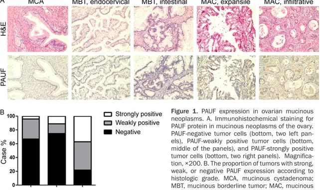
Figure 1. PAUF expression in ovarian mucinous neoplasms. A. Immunohistochemical staining for PAUF protein in mucinous neoplasms of the ovary. PAUF-negative tumor cells (bottom, two left pan-els), PAUF-weakly positive tumor cells (bottom, middle of the panels), and PAUF-strongly positive tumor cells (bottom, two right panels). Magnifica-tion, ×200. B. The proportion of tumors with strong, weak, or negative PAUF expression according to histologic grade. MCA, mucinous cystadenoma; MBT, mucinous borderline tumor; MAC, mucinous adenocarcinoma.
of PAUF: 0 (negative), 1 (weakly positive) and 2 (strongly positive). The proportion of PAUF-stained cells was scored as 0 (negative), 1 (< 10%) and 2 (> 10%). The final score for each tumor was calculated by multiplying the immu-nostaining intensity by the proportion of stained cells, and was then categorized as follows: 0-1, negative; 2, weakly positive; and 4, strongly positive.
A cut-off value of > 1% of nuclei that were strongly stained was used to define ER expres-sion [13]. To establish the cutoff Ki-67 LI, we analyzed the Ki-67 LI of ovarian mucinous tumors using receiver operator characteristic (ROC) curves according to PAUF positivity and determined that 1.167% (sensitivity: 70.4%, 1 - specificity: 84.8%) was the optimal cutoff value.
Statistics
Statistical analyses were performed using GraphPad Prism 5 software, version 5.01 (GraphPad Software, Inc., La Jolla, CA, USA) and SPSS for Windows, version 12.0 (SPSS Inc., Chicago, IL, USA). For the analysis of age at diagnosis, a significant difference between means was determined by analysis of varianc-es (ANOVA). Follow-up durations of patients
with MBT and MAC were compared using a
t-test. FIGO stage, histologic subtypes, invasion
patterns, and the expressions of PAUF, ER, and Ki-67 LI were compared using the chi-square and Fisher’s exact test. Kaplan-Meier survival curves and log-rank statistics were employed to evaluate overall survival (OS) and disease-free survival (DFS). Univariate and multivariate regression analyses were performed using the Cox proportional hazards model. All reported
P-values are two-sided, and P-values <0.05
were considered statistically significant. Results
Clinicopathologic features of ovarian mucinous neoplasms
Ovarian mucinous neoplasms are categorized as benign, intermediate, or malignant based on their pathologic features, and are referred to as MCA, MBT, and MAC, respectively [2]. We selected 24 cases of MCA, 36 cases of MBT, and 46 cases of MAC and compared the clinico-pathologic features of these ovarian mucinous neoplasms (Table 1).
There was no significant difference in the medi-an age at diagnosis of patients with MCA (49.25 ± 3.632 years), MBT (44.36 ± 2.502 years),
Histologic subtype 0.1466 Intestinal 24 4 2 Endocervical 3 1 2 ER status 0.1466 Negative 24 4 2 Positive 3 1 2 Ki-67 LI *0.0863 < 1.167% 4 1 3 ≥ 1.167% 23 4 1
SD, standard deviation. *We calculated P-value using Fisher’s exact test after grouping PAUF weakly positive and strongly posi-tive MBTs together as PAUF-posiposi-tive MBTs and compared them with PAUF-negaposi-tive MBTs, because the chi-square calculation is not valid when all expected values are < 1.
Table 3. Clinicopathologic features of 46 MACs according to PAUF expression status
PAUF expression
P-value
Negative Weakly positive Strongly positive
Number of cases 10 19 17
Age at diagnosis (years, mean ± SD) 40.40 ± 3.933 39.58 ± 2.838 49.06 ± 3.957 0.1095 Follow-up (months, mean ± SD) 69.10 ± 12.96 53.74 ± 8.538 37.53 ± 5.829 0.0696 Overall survival (%) 10/10 (100.000) 14/19 (73.684) 13/17 (76.471) 0.1885 Disease-free survival (%) 9/10 (87.500) 13/19 (60.014) 12/17 (63.995) 0.3732 Stage 0.2205 Localized 3 10 9 Regional 3 4 7 Distant 4 5 1 Invasion pattern (%) 0.5464 Expansile 8 14 15 Infiltrative 2 5 2 SD, standard deviation. and MAC (43.26 ± 2.122, P = 0.2951). We grouped cases of MBTs and MACs based on FIGO stage and categorized them as being localized, regional, or distant. Most MBT cases were at a localized stage, whereas most MCA cases were at a regional or distant stage (P < 0.0001).
For a more detailed analysis, we subdivided MBTs into two groups according to the histo-logic features of their epithelial component:
intestinal type or endocervical type [2]. According to the most recent World Health Organization classification of female reproduc-tive organs, endocervical-type MBTs are con-sidered a subset of seromucinous tumors, referred to as seromucinous borderline tumors [14]. However, to date, seromucinous border-line tumors have been reported as endocervi-cal-type MBTs and compared with other intesti-nal-type MBTs. Therefore, we included endocervical-type MBTs (seromucinous
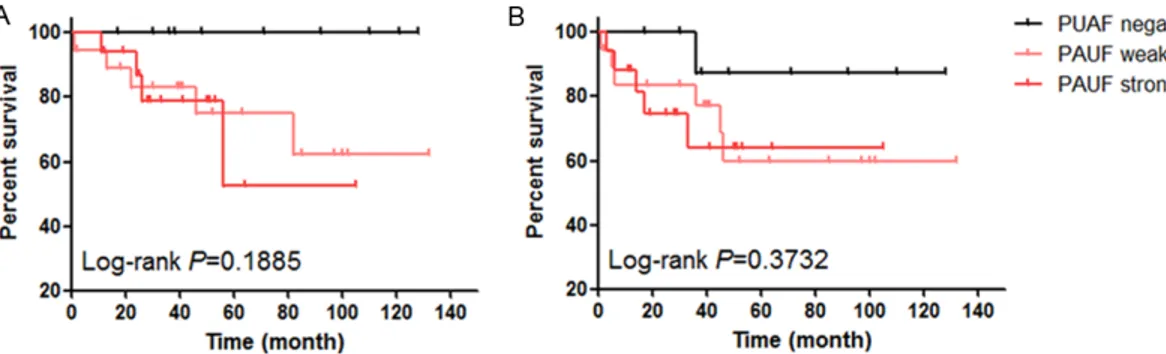
border-Figure 2. Survival analysis of patients with ovarian mucinous adenocarcinoma according to PAUF expression. A. Overall survival of MAC patients according to PAUF expression status. B. Disease-free survival of MAC patients ac-cording to PAUF expression status.
line tumors) in this study to clarify the clinico-pathological features of intestinal-type MBTs. Thirty of the 36 MBTs (83.33%) had an epithe-lial component resembling intestinal epitheli-um, and the other six cases (16.67%) exhibited mucinous epithelial cells resembling endocervi-cal epithelium. This finding is similar to those of previous reports, in which the intestinal type accounted for 85-90% of MBTs and the endo-cervical type for 10-15% [7, 15-17].
MACs were subdivided into expansile and infil-trative types according to their invasion pat-terns, rather than a grading system, because it is well established that the current grading sys-tem for mucinous carcinoma can predict nei-ther tumor behavior nor treatment response [15, 16, 18]. However, infiltrative stromal inva-sion has proved to be more biologically aggres-sive than expansile invasion [2]. We found infil-trative invasion growth in nine out of 46 MACs (19.57%) and an expansile invasion pattern in 37 out of 46 MACs (80.43%).
PAUF expression status according to subtypes of ovarian mucinous tumors
We performed immunohistochemical staining with antirhPAUF pAb on FFPE tissue of ovarian mucinous neoplasms, and interpreted PAUF reactivity as negative, weakly positive, or strongly positive as described above (Figure 1A). Interestingly, MACs most frequently dem-onstrated weak PAUF positivity (19 of 46 cases, 41.30%) and strong PAUF positivity (17 of 46 cases, 36.96%), in contrast to MCAs (weakly positive: 29.17%, strongly positive: 4.17%) and MBTs (weakly positive: 13.89%, strongly posi-tive: 8.33%; Table 1 and Figure 1B, P < 0.0001).
Thus, strongly positive PAUF staining was more common in ovarian mucinous tumors with more aggressive behavior.
Clincopathologic features of MBTs according to PAUF expression status
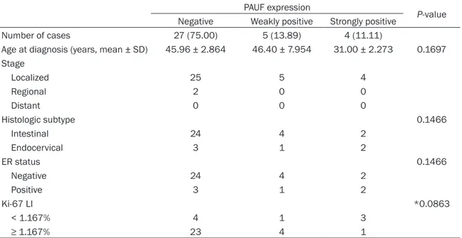
We next compared the clinicopathologic fea-tures of MBTs according to their PAUF expres-sion status (Table 2). Although only four of 36 MBTs were strongly positive for PAUF, these cases tended to occur 10 years earlier (31.00 ± 2.273 years) than their PAUF negative (45.96 ± 2.864 years) or weakly positive PAUF (46.40 ± 7.954 years) equivalents, although these differ-ences were not statistically different (P = 0.1697). Differences in tumor stage according to PAUF expression status could not be statisti-cally analyzed because most MBTs were at a localized stage, except for two cases at a regional stage.
Chi-square calculations are usually only valid when all expected values are > 1, and this con-dition was not met when we assessed the pathologic features of MBTs according to PAUF expression status. Therefore, we combined MBTs that were either weakly or strongly posi-tive for PAUF as a single PAUF-posiposi-tive group (n = 9) and compared their pathologic parameters with those of the PAUF-negative group (n = 27) using Fisher’s exact test. This revealed no sta-tistically significant differences with respect to histologic subtype or ER status. However, all of the endocervical MBTs were ER positive, as they are known [19]. There were also no signifi-cant differences between PAUF-positive and negative MBTs with respect to the Ki-67 LI, a marker of proliferative activity.
PAUF 0.960 0.425
Negative vs. weakly positive 0.948 0.230 3.655 0.440-30.369 Negative vs. strongly positive 0.947 0.197 4.134 0.480-35.618
Table 5. Multivariate analysis for overall survival and disease-free survival
Parameter Overall survival Disease-free survival
P-value Hazard ratio 95% CI P-value Hazard ratio 95% CI Age 0.042 1.116 1.004-1.240 0.044 1.083 1.002-1.170 Stage 0.024 0.080 Localized vs. regional 0.378 0.313 0.024-4.131 0.882 1.159 0.164-8.184 Localized vs. distant 0.021 23.365 1.605-340.079 0.037 9.941 1.142-86.496 Invasion Expansile vs. infiltrative 0.640 0.626 0.088-4.450 0.823 1.218 0.216-6.884 PAUF 0.809 0.099
Negative vs. weakly positive 0.941 0.045 57.406 1.090-3022.596 Negative vs. strongly positive 0.937 0.034 97.890 1.412-6785.925
MAC PAUF expression is associated with shorter OS and DFS
As described above, MACs more frequently stain positive for PAUF than other ovarian muci-nous tumors. We therefore compared the clini-copathologic features of MACs according to PAUF expression (Table 3). There was no signifi-cant difference in age at diagnosis, tumor stage, and invasion pattern in MACs with respect to PAUF expression.
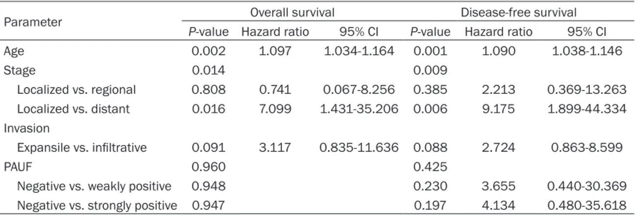
When we compared the OS and DFS of MAC patients according to PAUF expression status using a Kaplan-Meier analysis, patients with MACs staining weakly or strongly for PAUF tend-ed to have a shorter OS than those negative for PAUF (Figure 2, P = 0.1885). We conducted a statistical analysis to identify predictive factors for survival of ovarian MAC patients using a Cox regression assay (Table 4). In univariate analy-sis, age at diagnosis (P = 0.002, hazard ratio [HR] = 1.097, 95% confidence interval [CI]: 1.034-1.164), tumor stage (localized vs.
dis-tant, P = 0.016, HR = 7.099, 95% CI: 1.431-35.206), and invasion pattern (expansile vs. infiltrative, P = 0.091, HR = 3.117, 95% CI: 0.835-11.636) affected the OS of MAC patients. Age at diagnosis (P = 0.001, HR = 1.090, 95% CI: 1.038-1.146), tumor stage (localized vs. distant, P = 0.006, HR = 9.175, 95% CI: 1.899-44.334), and invasion pattern (expansile vs. infiltrative, P = 0.088, HR = 2.724, 95% CI: 0.863-8.599) also affected the DFS of MAC patients. There was a tendency towards a shorter DFS in patients with MACs that stained strongly for PAUF than that in those with PAUF-negative MACs (P = 0.197, HR = 4.134, 95% CI: 0.480-35.618). When we adjusted various parameters by multivariate analysis, older age at diagnosis and a distant tumor stage were associated with a shorter OS and DFS amongst MAC patients (Table 5). In particular, PAUF expression in MACs was an independent predictive factor for DFS (negative vs. weakly positive PAUF staining, P = 0.045, HR = 57.406, 95% CI: 1.090-3022.596 and negative vs. strongly positive PAUF staining, P =
0.034, HR = 97.890, 95% CI: 1.412-6785.95), although there was no statistical difference in DFS according to PAUF expression status using Kaplan-Meier survival analysis.
Discussion
PAUF, a novel tumor-specific protein, is known to be associated with cancer progression and metastasis in pancreatic ductal adenocarcino-ma and ovarian tumors [11, 12]. MBTs are the most common type of borderline ovarian tumor and generally have an excellent prognosis. In contrast, MACs, although far less common, show an aggressive behavior [4-10]. We there-fore hypothesized that PAUF expression status might be different between MBTs and MACs. As expected, we found that MACs were more frequently positive for PAUF expression than other ovarian mucinous tumors, with an increasing frequency of strong PAUF staining in MCA, MBT, and MAC tumor types. This sug-gests that PAUF could be a sensitive and spe-cific biomarker for distinguishing between inter-mediate and malignant mucinous tumors of the ovary.
We also failed to find any significant differences in PAUF expression status of MBTs with respect to a range of clinicopathological features, which might reflect the generally low PAUF expression in these tumors. However, for MACs, PAUF expression tended to be higher in patients with a shorter OS. When we adjusted for possible confounders using multivariate Cox regression analysis, weak or strong PAUF expression in MAC was associated with a shorter DFS. Our findings indicate that PAUF expression can help categorize ovarian mucinous tumors, and predicts shorter OS and DFS amongst MAC patients. Thus, PAUF could act as a prognostic and diagnostic biomarker for patients with an ovarian mucinous tumor.
Acknowledgements
This study was supported by grants from the National Research Foundation (NRF) of Korea Grant funded by the Korean Government (No. 2011-0010800 and 2014-002926; EJN) and the Basic Science Research Program through the NRF of Korea funded by the Mid-career Researcher Program by the MEST (No. 2012R1A2A4A01006435; CHO).
Disclosure of conflict of interest None.
Address correspondence to: Dr. Eun Ji Nam, Department of Obstetrics and Gynecology, Yonsei University College of Medicine, 50-1 Yonsei-ro, Seodaemun-gu, Seoul, South Korea (postal code: 120-752). Tel: +82-2-2228-2250; Fax: +82-2-313-8357; E-mail: NAHME6@yuhs.ac
References
[1] Zaloudek CF. Diagnostic Histopathology of Tu-mors. Philadelpiha, USA: Elsevier; 2007. [2] Lee KR, Tavassoli FA, Prat J, Dietel M, Gersell
DJ, Karseladze AI, Hauptmann S, Rtugers JKL, Russell P, Buckley CH, Pisani P, Schwartz PE, Goldgar DE, Silva EG, Caduff R and Kubik-Huch R. Tumours of the Breast and Female Genital Organs. Lyon, France: International Agency for Research on Cancer (IARC); 2003. [3] Hart WR. Mucinous tumors of the ovary: a
re-view. Int J Gynecol Pathol 2005; 24: 4-25. [4] Khunamornpong S, Settakorn J, Sukpan K,
Su-prasert P and Siriaunkgul S. Mucinous tumor of low malignant potential (“borderline” or “atypical proliferative” tumor) of the ovary: a study of 171 cases with the assessment of in-traepithelial carcinoma and microinvasion. Int J Gynecol Pathol 2011; 30: 218-230.
[5] Kaern J, Trope CG and Abeler VM. A retrospec-tive study of 370 borderline tumors of the ova-ry treated at the Norwegian Radium Hospital from 1970 to 1982. A review of clinicopatho-logic features and treatment modalities. Can-cer 1993; 71: 1810-1820.
[6] Russell P and Merkur H. Proliferating ovarian “epithelial” tumours: a clinico-pathological analysis of 144 cases. Aust N Z J Obstet Gyn-aecol 1979; 19: 45-51.
[7] Siriaunkgul S, Robbins KM, McGowan L and Silverberg SG. Ovarian mucinous tumors of low malignant potential: a clinicopathologic study of 54 tumors of intestinal and mullerian type. Int J Gynecol Pathol 1995; 14: 198-208. [8] Kehoe S and Powell J. Long-term follow-up of
women with borderline ovarian tumors. Int J Gynaecol Obstet 1996; 53: 139-143.
[9] Schiavone MB, Herzog TJ, Lewin SN, Deutsch I, Sun X, Burke WM and Wright JD. Natural his-tory and outcome of mucinous carcinoma of the ovary. Am J Obstet Gynecol 2011; 205: 480, e1-8.
[10] Zaino RJ, Brady MF, Lele SM, Michael H, Greer B and Bookman MA. Advanced stage muci-nous adenocarcinoma of the ovary is both rare and highly lethal: a Gynecologic Oncology Group study. Cancer 2011; 117: 554-562.
pression. Oncogene 2010; 29: 56-67.
[13] Hammond ME, Hayes DF, Dowsett M, Allred DC, Hagerty KL, Badve S, Fitzgibbons PL, Fran-cis G, Goldstein NS, Hayes M, Hicks DG, Lester S, Love R, Mangu PB, McShane L, Miller K, Os-borne CK, Paik S, Perlmutter J, Rhodes A, Sa-sano H, Schwartz JN, Sweep FC, Taube S, Tor-lakovic EE, Valenstein P, Viale G, Visscher D, Wheeler T, Williams RB, Wittliff JL and Wolff AC. American Society of Clinical Oncology/Col-lege Of American Pathologists guideline rec-ommendations for immunohistochemical test-ing of estrogen and progesterone receptors in breast cancer. J Clin Oncol 2010; 28: 2784-2795.
[14] Kobel M, Bell DA, Carcangiu ML, Oliva E, Prat J, Shihl M, Soslow R and Vang R. WHO Classifica-tion of Tumours of Female Reproductive Or-gans. Lyon, France: International Agency for Research on Cancer (IARC); 2014.
[17] Rutgers JL and Scully RE. Ovarian mullerian mucinous papillary cystadenomas of border-line malignancy. A clinicopathologic analysis. Cancer 1988; 61: 340-348.
[18] Hoerl HD and Hart WR. Primary ovarian muci-nous cystadenocarcinomas: a clinicopatholog-ic study of 49 cases with long-term follow-up. Am J Surg Pathol 1998; 22: 1449-1462. [19] Vang R, Gown AM, Barry TS, Wheeler DT and
Ronnett BM. Immunohistochemistry for estro-gen and progesterone receptors in the distinc-tion of primary and metastatic mucinous tu-mors in the ovary: an analysis of 124 cases. Mod Pathol 2006; 19: 97-105.