Theoretical Estimation of Cannulation Methods for Left
Ventricular Assist Device Support as a Bridge to Recovery
Left ventricular assist device (LVAD) support under cannulation connected from the left atrium to the aorta (LA-AA) is used as a bridge to recovery in heart failure patients because it is non-invasive to ventricular muscle. However, it has serious problems, such as valve stenosis and blood thrombosis due to the low ejection fraction of the ventricle. We theoretically estimated the effect of the in-series cannulation, connected from ascending aorta to descending aorta (AA-DA), on ventricular unloading as an alternative to the LA-AA method. We developed a theoretical model of a LVAD-implanted cardiovascular system that included coronary circulation. Using this model, we compared hemodynamic responses according to various cannulation methods such as LA-AA, AA-DA, and a cannulation connected from the left ventricle to ascending aorta (LV-AA), under continuous and pulsatile LVAD supports. The AA-DA method provided 14% and 18% less left ventricular peak pressure than the LA-AA method under continuous and pulsatile LVAD conditions, respectively. The LA-AA method demonstrated higher coronary flow than AA-DA method. Therefore, the LA-AA method is more advantageous in increasing ventricular unloading whereas the AA-DA method is a better choice to increase coronary perfusion. Key Words: Left Ventricular Assist Device (LVAD); Cannulation Methods; Bridge to Recovery; Ventricular Unloading
Ki Moo Lim1,*, Jeong Sang Lee2,*,
Jin-Ho Song1, Chan-Hyun Youn3,
Jae-Sung Choi2 and Eun Bo Shim1
1Department of Mechanical & Biomedical
Engineering, Kangwon National University, Chucheon;
2Department of Thoracic and Cardiovascular Surgery,
Seoul National University College of Medicine and SMG-SNU Boramae Hospital, Seoul; 3Department of
Information and Communications Engineering, Korea Advanced Institute of Science and Technology, Daejeon, Korea
*Ki Moo Lim and Jeong Sang Lee contributed equally to this work.
Received: 23 August 2011 Accepted: 17 October 2011 Address for Correspondence: Eun Bo Shim, PhD
Professor of Mechanical & Biomedical Engineering, Director of National Research Lab on Biosystems Engineering, Kangwon National University, Gangwondaehakgil 1, Chuncheon 200-701, Korea
Tel: +82.33-250-6318, Fax: +82.33-257-6595 E-mail: [email protected]
This work was supported by the NRL (National Research Lab) program of National Research Foundation of Korea (No. ROA-2008-000-20127-0).
http://dx.doi.org/10.3346/jkms.2011.26.12.1591 • J Korean Med Sci 2011; 26: 1591-1598
INTRODUCTION
Left ventricular assist devices (LVADs) have been widely used to help heart failure (HF) patients provide sufficient blood flow to peripheral organs, keeping patients alive as a bridge to trans-plantation. Additionally, LVADs unload failed ventricles and are used as a bridge to recovery.
Several studies have suggested how to increase the ventricu-lar unloading effect with regard to pumping types and cannula-tion locacannula-tions of the LVAD (1-4). Previously, we demonstrated that counter-pulsating LVAD is more beneficial in ventricular unloading and coronary perfusion, which are the main contrib-utors to cardiac recovery (5). Timms et al. (6) compared the ven-tricular unloading effect according to cannulation locations: can-nulation connected from the left ventricle to ascending aorta (LV-AA) and that from the left atrium to the ascending aorta (LA-AA), with continuous LVAD. They demonstrated experimental-ly that the LVAD support under LV-AA cannulation provided more ventricular unloading than that under LA-AA cannulation. They also showed that the LVAD support under LA-AA
cannu-lation developed an even higher afterload than that under the condition of HF without LVAD support (4), and suggested that the LV-AA cannulation method provided the best performance for unloading failed ventricles. However, when patients consid-er cardiac recovconsid-ery, it is bettconsid-er not to connect the LVAD directly to the diseased ventricle to keep the ventricular muscle intact un-til the LVAD is weaned out. Thus, any cannulation method con-necting the ventricle is unsuitable for LVAD therapy as a bridge to recovery. For this reason, the LA-AA cannulation method has been used alternatively in HF patients for whom cardiac recov-ery is expected with planned weaning off the LVAD in the future. However, as mentioned, LVAD support under the LA-AA method does not provide sufficient ventricular unloading, compared with that under LV-AA cannulation. Also, the ventricular ejection frac-tion in LVAD supported with LA AA cannulafrac-tion becomes very small or even zero. This may cause valve stenosis and blood thrombosis due to stagnated blood flow inside the ventricle (7). Shi et al. (8) suggested a new cannulation method that is con-nected in series to the failed heart and used a theoretical meth-od to show that LVAD support with the in-series cannulation
method provided good performance for pumping blood to pe-ripheral organs and ventricular unloading. However, they did not consider the effect on coronary perfusion during LVAD sup-port or provide a direct comparison between LA-AA and the in-series method in terms of ventricular unloading effect. Experi-mental methods to record ventricular pressure and volume and coronary perfusion are hampered by low spatiotemporal reso-lution. Computational modeling represents an alternative ap-proach to overcome this limitation. We have developed several computational models of cardiovascular system in order to ob-tain cardiovascular response according to various hemodynam-ic conditions (5, 9, 10).
In this study, the cardiovascular response during LVAD sup-port with the in-series cannulation was compared with that of LVAD support with the LA-AA cannulation method theoretical-ly. Here, the in-series cannulation represents a method in which LVAD is connected in-series with the ventricle from the ascend-ing aorta to the descendascend-ing aorta. We compared the ventricular unloading in terms of pressure and volume, ejection fraction, and coronary flow during LVAD support according to cannula-tion methods using a numerical model of a LVAD-implanted cardiovascular system. The model consisted of a time-varying elastance model of failed ventricle and atrium, a vascular sys-tem including coronary circulation, and LVAD functions. The effects of three different methods for cannulation were estimat-ed: LA-AA, AA-DA, and LV-AA; the last method was considered here as a reference.
MATERIALS AND METHODS
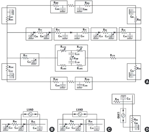
Lumped model of a LVAD-implanted cardiovascular system We modified Heldt’s lumped model of the cardiovascular sys-tem (11) and integrated it with left coronary circulation (12) and LVAD function. Right coronary circulation was neglected due to its low contribution to the total coronary flow compared with the left coronary circulation. The model was formulated with regard to an electric analog model, consisting of elements such as resistors, capacitors, and diodes. By mapping hemodynamic parameters into the electric elements, flow resistance to the elec-tric resistors, vessel compliance to the capacitors, and the func-tion of cardiac valves to the diodes, this model calculates the blood pressure and flow rate for each component of the body. Fig. 1 shows a schematic diagram of the cardiovascular system model (Fig. 1A) and three types of cannulation methods: LV-AA (Fig. 1B), LA-AA (Fig. 1C), and AA-DA (Fig. 1D).
The model includes the left atrium and ventricle, ascending aorta, brachiocephalic arteries, upper body arteries and veins, superior vena cava, right atrium and ventricle, pulmonary arter-ies and veins, descending aorta, lower body arterarter-ies and veins, inferior vena cava, left coronary arteries, left circumflex artery, left anterior descending artery, coronary veins, and LVAD com-ponent. The pumping action of four cardiac chambers, the two ventricles and atria, was implemented by time-varying elastance, based on data adapted from experimental results (11). Heart rate was applied as 70 beats per minute.
A
D C
B
Fig. 1. Schematic of the LVAD-implanted cardiovas-cular model. Cardiovascardiovas-cular model (A) and three types of cannulation methods: LV-AA (B), LA-AA (C), and AA-DA (D). R, resistance; C, compliance; LA, left atrium; MV, mitral valve; LV, left ventricle; AA, ascend-ing aorta; BA, brachiocephalic artery; UBA, upper body arteries; UBV, upper body veins; SVC, superior vena cava; RA, right atrium; TV, tricuspid valve; RV, right ventricle; PA, pulmonary arteries; PV, pulmonary veins; LCA, left coronary arteries; LCX, left circumflex; LAD, left anterior descending artery; CV, coronary veins; DA, descending aorta; LBA, lower body arter-ies; LBV, lower body veins; IVC, inferior vena cava, IMP, intra-myocardial pressure.
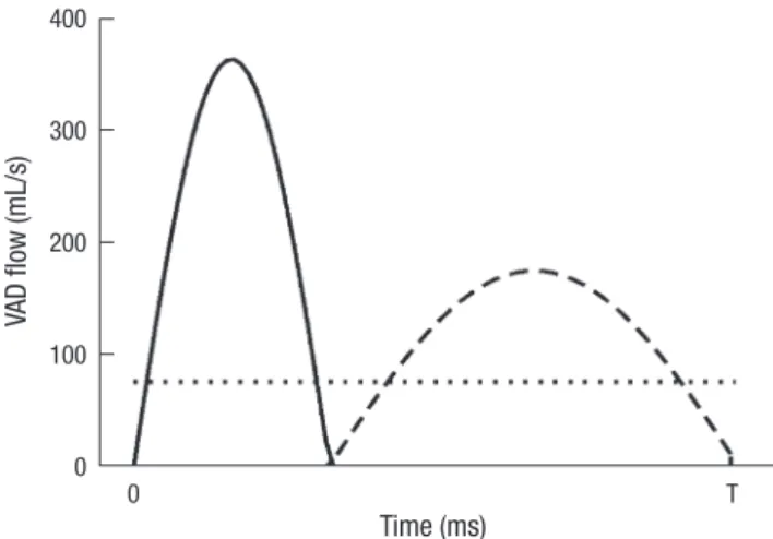
LVAD function was modeled as a flow generator with a mean flow rate of 75 mL/s, which is enough pumping flow for 100% assistance. There are many types of pumping mechanisms for continuous and pulsatile LVADs. To apply a generalized wave-form of continuous and pulsatile LVADs to the model, we used a constant flow rate for continuous LVAD and a harmonic pro-file of flow rate for pulsatile LVAD (Fig. 2). In the case of
pulsa-tile LVAD, the pumping phase was synchronized with the failed ventricle as a counter-pulsation. The governing equations and solving methods of the integrated model, which is based on the windkessel method, have been described in detail (5, 11), and are described briefly in the Appendix section.
Heart failure condition
We applied the left heart failure condition to the model by re-ducing the end-systolic elastance of the left ventricle to 30% of the normal value. This produced hemodynamic results that were consistent with the data reported for systolic HF patients in clin-ical studies (13, 14). Fig. 3A depicts the time-varying elastance of normal and failed left ventricles. Fig. 3B shows the following pressure-volume curves for normal and failed left ventricles. The ejection fraction of the failed ventricle decreased, to 26%. Fig. 3C, D show the following pressure waveforms of the left atrium, left ventricle, and ascending aorta for the normal and HF con-ditions. The mean arterial pressure decreased to 59 mmHg un-der the HF condition.
Simulation protocol
Using the model of the LVAD-implanted cardiovascular system under the HF condition, we performed six independent simu-lations to compare hemodynamic effects with regard to
cannu-VA D flo w (m L/ s) Time (ms) 0 T 400 300 200 100 0
Fig. 2. Flow waveforms of pulsatile and continuous LVADs. LVAD flow waveforms during one cycle (T; 860 milliseconds) in continuous and pulsatile LVADs. Solid line, pulsatile LVAD inflow; dashed line, pulsatile LVAD outflow; dotted line, continuous LVAD flow. El as ta nc e 1 0.5 0 Time (ms) HF Normal 0 T A Pr es su re (m m Hg ) 100 50 0 Volume (mL) HF Normal 0 50 100 150 200 B Pr es su re (m m Hg ) 100 50 0 Time (ms) LV LA Ao 0 T C Pr es su re (m m Hg ) 100 50 0 Time (ms) 0 T D LV LA Ao
Fig. 3. Comparison of cardiovascular characteristics between normal and heart failure patient. Relative value of time-varying elastance of the left ventricle under HF condition compared with normal condition (A) and following hemodynamic responses: pressure and volume curves for normal and HF conditions (B); pressure waveform of the left ventri-cle, left atrium, and aorta for normal (C) and HF condition (D). HF, heart failure; LV, left ventricle; LA, left atrium; Ao, aorta; T, cycle length (860 milliseconds).
lation methods of the LVAD. Three cannulation methods, LV-AA, LA-LV-AA, and AA DA, were simulated under the conditions of continuous and pulsatile LVADs. The pulsatile LVAD pumps blood synchronously with the failed ventricle as counter-pulsa-tion in the model. Each simulacounter-pulsa-tion was performed for 20 s to ob-tain a steady-state response of the cardiovascular system. RESULTS
Fig. 4 illustrates the simulated pressure waveforms in the left ven-tricle and atrium and systemic artery according to three cannu-lation methods, LV-AA, LA-AA and AA-DA, for the LVAD-im-planted HF model operating in continuous flow (A–C) and coun-ter-pulsating flow (D–F) mode. In both modes, the cannulation method, LV-AA, showed the lowest ventricular peak pressure (36 and 12 mmHg in continuous and pulsatile mode, respective-ly), as expected based on previous results (15). As mentioned,
the direct cannulation into the ventricle is not suitable for LVAD support as a bridge to recovery because it results in the injury of the myocardium. The Cannulation method, LA-AA, showed 87 and 76 mmHg left ventricular peak pressure with continuous and pulsatile LVADs, respectively. The in-series cannulation method, AA-DA, showed 75 and 62 mmHg left ventricular peak pressure, respectively. Between the two methods that are not connected directly to the diseased ventricle, the in- series can-nulation method provided 14% and 19% less ventricular pres-sure than the LA-AA method in continuous and pulsatile LVAD modes, respectively.
Fig. 5 presents the simulated pressure-volume curves of the left ventricle according to cannulation methods, LV-AA, LA-AA and AA-DA, for the LVAD-implanted HF model operating in con-tinuous flow (A) or counter-pulsating flow (B) mode. The LV-AA cannulation method showed the lowest end-diastolic ventricu-lar volume among the three methods tested, under both contin-Continuous LVAD support Counter-pulsating LVAD support
Pr es su re (m m Hg ) 100 50 0 A LVAA Time (ms) 0 T LV LA AA B LAAA 0 T C AADA 0 T D LVAA 0 T D LAAA 0 T F AADA 0 T
Fig. 4. Cardiovascular pressure waveforms according to LVAD cannulation type. Simulated pressure waveforms in the left ventricle (LV) and atrium (LA) and ascending aorta (AA) according to cannulation locations, from left ventricle to ascending aorta (LV-AA), from left atrium to ascending aorta (LA-AA), and ascending aorta to descending aorta (AA-DA), for the LVAD-implanted HF model operating in continuous flow (A, B, and C) and counter-pulsating flow (D, E, and F) modes. T, cycle length (860 milliseconds).
Pr es su re (m m Hg ) Pr es su re (m m Hg ) Volume (mL) Volume (mL) LA-AA LV-AA AA-DA 0 50 100 150 200 0 50 100 150 200 100 50 0 100 50 0 A B
Fig. 5. Pressure-volume diagram according to LVAD cannulation type. Simulated pressures-volume curves of the left ventricle according to cannulation locations, from left ven-tricle to ascending aorta (LV-AA), from left atrium to ascending aorta (LA-AA), and ascending aorta to descending aorta (AA-DA), for LVAD-implanted HF model operating in con-tinuous flow (A) and counter-pulsating flow (B) mode.
uous and pulsatile LVAD support (95 mL in continuous LVAD and 99 mL in pulsatile LVAD). The ejection fraction was 31% with continuous LVAD and 50% with pulsatile LVAD support (Table 1). The LA-AA cannulation method showed an ejection fraction of zero and maintained a constant ventricular volume of 138 mL in continuous LVAD and 124 mL in pulsatile LVAD support. The in-series cannulation method showed 168 mL of end-diastolic ventricular volume and 27% ejection fraction under continuous LVAD support, compared with 154 mL and 32% under pulsatile LVAD support.
Under the condition of the in-series cannulation, the total car-diac output, sum of flows out of LV and LVAD, was less than the LVAD outflow. This means that the bulk of blood in the descend-ing aorta from the LVAD outflow ran back to the ascenddescend-ing aor-ta and LVAD inlet. This recirculation occurred only in the in-se-ries cannulation (AA-DA). The recirculated blood volume was 32% of the LVAD outflow with continuous LVAD support and 25% with pulsatile LVAD support (Table 1).
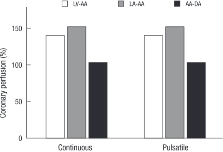
Finally, coronary perfusion was compared among the three cannulation methods under continuous and pulsatile LVAD sup-ports. Fig. 6 shows the relative value of coronary flow to the val-ue calculated under the condition of HF without LVAD support. All LVAD supports improved coronary flow. The LA-AA cannu-lation method demonstrated the most significant improvement of coronary flow under both continuous and pulsatile LVAD sup-port (152% and 162%, respectively). The AA-DA cannulation method showed a slight improvement of coronary perfusion under continuous LVAD support (103%), and better performance under pulsatile LVAD support (123%).
DISCUSSION
Although the LV-AA cannulation is being used for LVAD support, its use has a critical disadvantage for HF patients who need a LVAD support as a bridge to recovery. Left ventricular muscle injury is inevitable during the cannulation. For the purpose of bridge to recovery, it is better not to connect the LVAD directly to the failed ventricle to keep the ventricular muscle intact until
the LVAD is weaned out. For this reason, the LA-AA cannulation instead of LV-AA is used in HF patients for whom cardiac recov-ery is expected with planned weaning of the LVAD in the future. However, it has been reported that LVAD therapy with the LA-AA cannulation method shows poor performance in ventricu-lar unloading and has some problems, including valve stenosis and blood thrombosis due to the small ejection fraction. As an alternative method to overcome the problems of LA-AA cannulation, we theoretically examined the previously de-scribed in-series cannulation method (15). We developed a the-oretical model of a LVAD-implanted cardiovascular system and quantitatively compared the ventricular unloading effect, ejec-tion fracejec-tion, and coronary perfusion during LVAD support ac-cording to three different cannulation methods, LV-AA, LA-AA and AA-DA, under continuous and pulsatile LVAD.
The in-series cannulation method showed better performance in ventricular unloading in terms of pressure (Fig. 4) and vol-ume (Fig. 5) compared with that of LA-AA cannulation under both continuous and pulsatile LVAD supports. In particular, the ventricular unloading effects were more significant in counter-pulsating LVAD support; the advantage of counter-counter-pulsating LVAD on ventricular unloading was also demonstrated theoret-ically in our previous paper (5).
However, with the in-series cannulation method, the bulk of blood in the descending aorta that comes from the LVAD ran back to the ascending aorta due to reduced pressure in the as-cending aorta. Recirculation occurred only in the AA-DA can-nulation method because there was no valve between the LVAD inflow site (ascending aorta) and the LVAD outflow site (descend-ing aorta). The pulsatile LVAD showed less amount of recircula-tion than the continuous LVAD did. This is because the tempo-ral periods of LVAD inflow and outflow under pulsatile condi-tions are totally separate, whereas under continuous condicondi-tions, Table 1. Cardiac responses during pulsatile and continuous LVAD supports: total
car-diac output, ventricular ejection fraction, and recirculation during different types of LVAD supports
No LVAD
LV-AA LA-AA AA-DA
CLVAD PLVAD CLVAD PLVAD CLVAD PLVAD
TCO (mL/s) 51 75 75 75 75 51 56
EF 27% 31% 50% 0 0 27% 32%
Recir. 0 0 0 0 0 32% 25%
CLVAD, continuous left ventricular assist device (LVAD); PLVAD, pulsatile LVAD; LV-AA, cannulation method connected from left ventricle to ascending aorta; LA-AA, cannula-tion method connected from left atrium to ascending aorta; AA-DA, cannulacannula-tion meth-od connected from ascending aorta to descending aorta; TCO, total cardiac output; EF, ventricular ejection fraction; Recir., recirculation.
Co ro na ry p er fu si on (% ) 150 100 50 0 Continuous Pulsatile
LV-AA LA-AA AA-DA
Fig. 6. Coronary perfusion according to LVAD cannulation type. Simulated coronary perfusion according to cannulation locations, from left ventricle to ascending aorta (LV-AA), from left atrium to ascending aorta (LA-AA), and ascending aorta to descend-ing aorta (AA-DA), for LVAD-implanted HF model operatdescend-ing in continuous flow and counter-pulsating flow mode.
they are not (Fig. 2). Thus, it is recommended to choose pulsa-tile LVAD when the AA-DA cannulation method is applied. The LA-AA cannulation method showed an ejection fraction of zero and maintained a constant large volume of the left ventri-cle. In this case, both the mitral and aortic valve remained closed during LVAD support and no blood passed through either valve. This situation would lead to blood thrombosis and valve steno-sis. Even in the case of the LV-AA cannulation method, the aor-tic valve remained closed and no blood passed through the valve because the aortic pressure remained higher than the left ven-tricular pressure throughout the cardiac cycle. Thus, the LV-AA cannulation method also carries the possibility of aortic steno-sis. However, in the case of the AA-DA cannulation method, the mitral and aortic valves opened and closed regularly and posi-tive ejection fraction was provided, so it would not be expected to have such problem as valve stenosis and blood thrombosis. Use of the LA-AA cannulation method improved coronary perfusion most significantly among the three different cannula-tion methods tested; coronary perfusion was lowest in the in-series cannulation method, but it did show an improvement of coronary perfusion compared with that in the HF patient with-out LVAD support. Counter-pulsating LVAD demonstrated bet-ter performance than continuous LVAD, which was also dem-onstrated in our previous study (5). Although this was a simula-tion study, our results may be used as reference data when con-sidering the clinical use of a LVAD to increase ventricular un-loading when expecting cardiac recovery.
To examine different cannulation methods for LVAD support as a bridge to recovery, we theoretically compared cardiovascu-lar response during LVAD supports with LA-AA and AA DA can-nulation methods. The effects of the cancan-nulation method on ven-tricular unloading, ejection fraction of diseased ventricle and coronary perfusion were delineated using a mathematical mod-el of a LVAD-implanted cardiovascular system under HF condi-tions. The LA-AA cannulation method provided more coronary perfusion than the AA-DA cannulation method, though both methods improved the coronary flow rate compared with that under HF without LVAD support. Indeed, the AA-DA cannula-tion method provided better performance in ventricular unload-ing and generated a greater ejection fraction than the LA-AA cannulation method. Each cannulation method had its own ad-vantages and weak points in this study. Thus, these results com-prise basis data for the selection of a specific type of cannulation according to the state of the patient when LVAD implantation is needed as a bridge to recovery. In conclusion, the LA-AA meth-od is more advantageous to the increase of ventricular unload-ing and the AA-DA method is better in the aspect of coronary perfusion.
REFERENCES
1. Kar B, Delgado RM 3rd, Frazier OH, Gregoric ID, Harting MT, Wadia Y, Myers TJ, Moser RD, Freund J. The effect of LVAD aortic outflow-graft
place-ment on hemodynamics and flow: implantation technique and com-puter flow modeling. Tex Heart Inst J 2005; 32: 294-8.
2. Bartoli CR, Giridharan GA, Litwak KN, Sobieski M, Prabhu SD, Slaugh-ter MS, Koenig SC. Hemodynamic responses to continuous versus
pulsa-tile mechanical unloading of the failing left ventricle. ASAIO J 2010; 56: 410-6.
3. Haithcock BE, Morita H, Fanous NH, Suzuki G, Sabbah HN.
Hemody-namic unloading of the failing left ventricle using an arterial-to-arterial extracorporeal flow circuit. Ann Thorac Surg 2004; 77: 158-63.
4. Korakianitis T, Shi Y. Numerical comparison of hemodynamics with
atri-um to aorta and ventricular apex to aorta VAD support. ASAIO J 2007; 53: 537-48.
5. Lim KM, Kim IS, Choi SW, Min BG, Won YS, Kim HY, Shim EB.
Compu-tational analysis of the effect of the type of LVAD flow on coronary perfu-sion and ventricular afterload. J Physiol Sci 2009; 59: 307-16.
6. Timms D, Gregory S, Hsu PL, Thomson B, Pearcy M, McNeil K, Fraser J, Steinseifer U. Atrial versus ventricular cannulation for a rotary
ventricu-lar assist device. Artif Organs 2010; 34: 714-20.
7. Rose AG, Park SJ. Pathology in patients with ventricular assist devices: a
study of 21 autopsies, 24 ventricular apical core biopsies and 24 explant-ed hearts. Cardiovasc Pathol 2005; 14: 19-23.
8. Shi Y, Shi Y, Korakianitis T. Physiological control of an in-series connected
pulsatile VAD: numerical simulation study. Comput Methods Biomech Biomed Engin 2010; 14: 1. doi: 10.1080/10255842.2010.504030.
9. Shim EB, Leem CH, Abe Y, Noma A. A new multi-scale simulation model
of the circulation: from cells to system. Philos Transact A Math Phys Eng Sci 2006; 364: 1483-500.
10. Shim EB, Amano A, Takahata T, Shimayoshi T, Noma A. The cross-bridge
dynamics during ventricular contraction predicted by coupling the car-diac cell model with a circulation model. J Physiol Sci 2007; 57: 275-85.
11. Heldt T, Shim EB, Kamm RD, Mark RG. Computational modeling of
car-diovascular response to orthostatic stress. J Appl Physiol 2002; 92: 1239-54.
12. Schreiner W, Neumann F, Mohl W. The role of intramyocardial pressure
during coronary sinus interventions: a computer model study. IEEE Trans Biomed Eng 1990; 37: 956-67.
13. Monrad ES, Baim DS, Smith HS, Lanoue AS. Milrinone, dobutamine,
and nitroprusside: comparative effects on hemodynamics and myocar-dial energetics in patients with severe congestive heart failure. Circula-tion 1986; 73: III168-74.
14. Grose R, Strain J, Greenberg M, LeJemtel TH. Systemic and coronary
ef-fects of intravenous milrinone and dobutamine in congestive heart fail-ure. J Am Coll Cardiol 1986; 7: 1107-13.
15. Shi Y, Korakianitis T, Bowles C. Numerical simulation of cardiovascular
dynamics with different types of VAD assistance. J Biomech 2007; 40: 2919-33.
AUTHOR SUMMARY
Theoretical Estimation of Cannulation Methods for Left Ventricular Assist Device
Support as a Bridge to Recovery
Ki Moo Lim, Jeong Sang Lee, Jin-Ho Song, Chan-Hyun Youn, Jae-Sung Choi and Eun Bo Shim
Left ventricular assist device (LVAD) support under cannulation connected from the left atrium to the aorta (LA-AA) is used as a bridge to recovery in heart failure (HF) patients. However, it sometimes has serious problems, such as valve stenosis and blood thrombosis. Using mathematical simulation model, we estimated the effect of the in-series cannulation, connected from ascending aorta to descending aorta (DA), on ventricular unloading as an alternative to the LA-AA method. It was predicted that the AA-DA method provided better scores than the LA-AA method.
The time derivative of the eighteen-compartmental volume can be expressed as
dVi
= Qi,in(t)-Qi,out (t) (1)
dt
where V indicates volume, Q is blood flow rate, t indicates time, i means each compartmental index,
in means inflow to i node, and out means outflow from i node.
Each compartmental pressure is calculated as:
Pi(t) = V i(t)-Vi,d
(2)
Ci
where P indicates pressure and Vi,d indicates dead volume in the i compartment.
In the compartment of coronary circulation, pressure is calculated as:
Pi(t) = Pi,IMP(t) + Vi(t)-Vi,d (3)
Ci
IMP indicates intra-myocardial pressure, which is shown below.
Pi,IMP(t) = γnormPLV (t) (4)
where γnorm is the proportionality factor for left ventricular squeezing.
Compliance of the coronary vein is defined in terms of volume-dependent compliance, as in the equation below. This explains the characteristic of increasing vessel stiffness with progressive distension.
CCV(VCV) = χ[1 - σVCV(t)]
-1 · exp [ -σ(V
CV(t) - VCV,d)] (5)
where CV indicates coronary vein, and χ and σ are derived coefficients for the equation.
Flow resistance of left circumflex artery is not constant but has a volume-dependent value as in the equations below.
RLCX1(t) = RLCX when Pbif - pcap > 0 (6)
RLCX + β/(VLCX (t))2 when Pbif - pcap > 0
RLCX2(t) = RLCX + β/(VLCX (t))
2 when P
cap - pven > 0
(7) RLCX when Pcap - pven < 0
A detailed explanation of the governing equations and parameters are presented in Heldt et al. (11) and Schreiner et al. (12).