© 2012 The Korean Society of Pathologists/The Korean Society for Cytopathology pISSN 1738-1843
The primary mucinous cystadenocarcinoma (MCA) of the breast is an extremely rare neoplasm. The most common sites of MCA include the ovary, pancreas and appendix. Only 13 cases of primary MCA of the breast have been reported, since Koenig and Tavassoli reported the first 4 cases of MCA in 1998 (Tables
1, 2).1-10 The breast MCAs showed unique clinico-pathologic
characteristics, distinguishable from ordinary mucinous carci-noma (MC) and signet ring cell carcicarci-noma. Clinically, MCA in the breast is found in postmenopausal women and present as a relatively large, cystic mass with a favorable prognosis. Micro-scopically, the cysts are lined by tall columnar mucinous cells, resembling those of endocervical glands. The MCA tumors demonstrate consistent immunohistochemical staining (IHC) results with negative estrogen receptor (ER), negative proges-terone receptor (PR), positive cytokeratin 7 (CK7), and nega-tive cytokeratin 20 (CK20). The c-erbB2 IHC results are avail-able in five cases.4,5,7-9 They are negative in four and positive in
one with HER-2 gene amplification confirmed by fluorescence
in situ hybridization.9
In the case being reported here, the mass was only 9 mm, though the size before core biopsy was assessed to be 14 mm by ultrasound examination. Other characteristics of this new case share those clinical, cytological, and histopathological findings described in previously published cases (Tables 1, 2). In this pa-per, we present a rare cytologic finding of primary MCA in the breast and the characteristics of mucin (MUC).
CASE REPORT Clinical summary
A 59-year-old postmenopausal woman presented with an ill-defined, hard mass in her left breast for a year. Radiologic evalu-ations demonstrated a hypo-echoic mass (14 mm) with a specu-lated margin in ultrasonography and a focal asymmetric density in the upper medial portion by mammography. Fine needle as-piration followed by needle biopsy was performed with the
di-Primary Mucinous Cystadenocarcinoma of the Breast: Cytologic Finding
and Expression of MUC5 Are Different from Mucinous Carcinoma
Sung Eun Kim · Ji Hye Park SoonWon Hong · Ja Seung Koo Joon Jeong1· Woo-Hee Jung Departments of Pathology and 1Surgery,
Gangnam Sevrance Hsopital, Yonsei University College of Medicine, Seoul, Korea
Mucinous cystadenocarcinoma (MCA) in the breast is a rare neoplasm. There have been 13 cas-es of primary breast MCA reported. The MCA prcas-esents as a large, partially cystic mass in post-menopausal woman with a good prognosis. The microscopic findings resemble those of ovarian, pancreatic, or appendiceal MCA. The aspiration findings showed mucin-containing cell clusters in the background of mucin and necrotic material. The cell clusters had intracytoplasmic mucin displacing atypical nuclei to the periphery. Histologically, the tumor revealed an abundant mucin pool with small floating clusters of mucin-containing tumor cells. There were also small cysts lined by a single layer of tall columnar mucinous cells, resembling those of the uterine endocervix. The cancer cells were positive for mucin (MUC) 5 and negative for MUC2 and MUC6. This mucin pro-file is different from ordinary mucinous carcinoma and may be a unique characteristic of breast MCA.
Key Words: Mucinous cystadenocarcinoma; Breast; MUC5; MUC2 Received: April 10, 2012
Revised: July 2, 2012 Accepted: July 17, 2012 Corresponding Author Woo-Hee Jung, M.D.
Department of Pathology, Gangnam Severance Hospital, Yonsei University College of Medicine, 211 Eonju-ro, Gangnam-gu, Seoul 135-720, Korea Tel: +82-2-2019-3541
Fax: +82-2-3463-2103 E-mail: [email protected]
*Sung Eun Kim’s present address: Department of Pathology, Cha Gangnam Medical Center, Cha University, Seoul, Korea
612 •Kim SE, et al.
agnosis of MC and invasive carcinoma with abundant mucin pool formation, respectively. She underwent partial mastectomy with sentinel lymph node biopsy. The sentinel lymph node was free from metastasis. She has been followed for 3 months with chemotherapy for adjuvant treatment and remains disease-free.
Pathologic findings
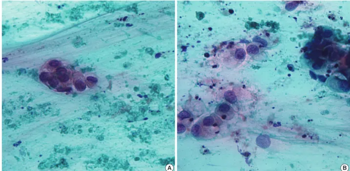
The aspiration showed a few scattered, variably sized, irregu-lar clusters of columnar cells in the greenish blue mucinous back-ground with necrotic debris. The columnar cells contained abun-dant mucin vacuoles in cytoplasm which had displaced their nuclei. The nuclear membrane was irregular and sharply angu-lated and the nuclei revealed a coarse chromatin pattern with prominent nucleoli (Fig. 1).
On gross examination, the cut surface showed an irregular, white to tan, solid and firm mass (9×7 mm), with a glistening appearance. Most MCAs in previous reports have demonstrated grossly cystic cut surfaces, except one case of MCA reported from Koenig and Tavassoli2 in 1998.1,3-10 The case in 1998 was
small in size (8 mm) and had a grossly solid appearance. The present case was 9 mm in maximal diameter and did not con-tain a macroscopic cyst. It is assumed that the small size of the MCA meant that it did not produce enough mucin to fill out and dilate cysts and ductal structures. The microscopic findings revealed a few cysts and ductal carcinoma in situ, adjacent to the invasive carcinoma. The cysts were lined by a single layer of tall columnar mucinous cells with focal areas of micropapillary struc-tures, resembling those of the uterine endocervix. The luminal
Table 1. Clinical feature of mucinous cystadenocarcinoma in the breast
Case Age (yr) Size (mm) TNM Duration Operation Adjuvant
treatment Follow-up
11 79 60 T2N0M0 N/A MRM No Died with other reason 9 yr
22 54 190 T3N2M0 N/A MRM/c LND No Alive with no evidence of disease 24 mo
32 67 23 T2N0M0 N/A MRM/c LND No Alive with no evidence of disease 22 mo
42 49 85 T3N0M0 N/A MRM/c LND Chm+RT Alive with no evidence of disease 11 mo
52 61 8 T1N0M0 N/A Lumpectomy/c LND No N/A Recent
63 74 100 T3N0M0 2 yr MRM/c LND No Alive with no evidence of disease N/A
74 96 20 T2N2M0 5 yr Lumpectomy/c LND No Died with other reason 46 mo
85 65 30 T2N0M0 >3 mo MRM/c LND No Alive with no evidence of disease 8 mo
96 51 40 T2N0M0 N/A Lumpectomy N/A N/A N/A
107 55 25 T2N0M0 N/A Lumpectomy No Alive with no evidence of disease 6 mo
118 52 100 T3N0M0 N/A Excision Tamoxifen Alive with no evidence of disease 24 mo
129 73 45 T2N0M0 Few months MRM/c LND N/A N/A N/A
1310 65 30 T2N0M0 N/A PM/c LND No Alive with no evidence of disease 6 mo
Present case 59 9 T1N0M0 1 yr PM/c LND Chm Alive with no evidence of disease 3 mo TNM, tumor-node-metastasis; N/A, not acquired; MRM, modified radical mastectomy; c LND, with lymph node dissection; Chm+RT, chromatography+radia-tion therapy; PM, partial mastectomy.
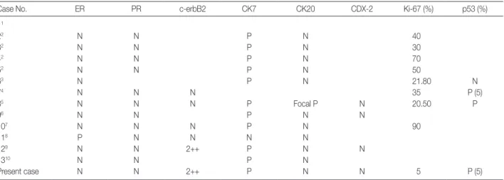
Table 2. Immunohistochemical feature of mucinous cystadenocarcinoma in the breast
Case No. ER PR c-erbB2 CK7 CK20 CDX-2 Ki-67 (%) p53 (%) 11 22 N N P N 40 32 N N P N 30 42 N N P N 70 52 N N P N 50 63 N P N 21.80 N 74 N N N 35 P (5) 85 N N N P Focal P N 20.50 P 96 N N P N N 107 N N N P N 90 118 P N N N N 129 N N 2++ P N N 1310 N N P N Present case N N 2++ P N N 5 P (5)
space contained mucin (Fig. 2). The invasive cancer area con-tained an abundant mucin pool in stroma with floating cell clus-ters. The floating cells contained mucin vacuoles in cytoplasm displacing atypical nuclei to the periphery.
Special staining and IHC on paraffin embedded tissue were performed. Both intracytoplasmic and extracytoplasmic mucin were stained by periodic acid-Schiff with diastase, alcian blue and mucicarcine, representing the acidic and neutral nature of mucin. The tumor cells were positive for CK7 and negative for CK20 and CDX-2. The IHC for MUC proteins was performed. The cancer cells were positive for MUC5 and MUC1 and nega-tive for MUC2 and MUC6. Mucin of the intracytoplasm and stroma revealed positivity for MUC5 and negativity for the oth-er MUC proteins (MUC1, MUC2, and MUC6). The hormone receptors, ER and PR, were negative. The c-erbB2 was 2-posi-tive (Fig. 3), but silver in situ hybridization demonstrated no amplification of the HER-2 gene.
DISCUSSION
In the breasts, mucin-producing tumors are classified as MC,
signet ring cell carcinoma (SRC), or mucocele-like tumors.11
The differences among mucin-containing tumors in the breast
are shown in Table 3.12-14 MC and mucocele-like tumors are
characterized by abundant extracellular mucin with floating neoplastic cells that do not contain intracytoplasmic mucin. The mucocele-like tumor is benign and the floating tumor cells are very rare. MC is specific type of invasive ductal caricinoma. The cytologic features show tight clusters of bland looking cells
with moderate cellularity in the mucinous background.13 SRC
demonstrates mucin only in the cytoplasm. The aspiration re-veals a cellular smear of pleomorphic and plasmocytoid cells with intracytoplasmic mucin in the bloody or necrotic back-ground.14 MCA has similar characteristics to both MC and SRC.
The aspiration smears reveal tight clusters of mucin-containing cells of low to moderate cellularity in the background of muci-nous and necrotic material. The histopathologic findings con-firm both, intracytoplasmic and extracytoplasmic mucin.
Mucin is a high molecular weight glycoprotein that consists of one protein core and O-linked carbohydrate side chains at-tached.15 Epithelial cells produce two types of mucin. One is
se-cretory mucin (MUC2, MUC5, and MUC6) and the other is trans-membrane mucin (MUC1, MUC3, MCU4, MUC12, and MUC17).16 They are expressed differently according to the types
of organs and tumors. For example, MC of the colon shows high
positivity for MUC2.17 SRC of the colon shows MUC2 and
MUC4 positivity.16 Among breast cancers, MC expresses
main-ly MUC2 along with a low level of MUC1, while SRC of the
A B
Fig. 1. Cytologic features of mucinous cystadenocarcinoma. (A) A few scattered, variably sized, irregular clusters of columnar cells with pleo-morphism and anisocytosis in a greenish blue mucinous background and necrotic debris. (B) The cells contain abundant mucin vacuoles in cytoplasm that attenuate and displace their nuclei. The nuclear membrane is irregular and sharply angulated and the nuclei reveal a coarse chromatin pattern with prominent nucleoli.
614 •Kim SE, et al.
breast has a high level of MUC1 expression with a variable level
of MUC2.18 In the breasts, MUC5 is not frequently observed.
MCs of the breast express MUC5 in only small proportions (less
than 5%).19 In our case of MCA reported here, the MUC IHC
demonstrates a high level of expression of MUC5 and no ex-pression of MUC2 or MUC6. This mucin profile is different
from ordinary mucinous carcinoma arising in the breast and other organs. MUC5-positive and MUC2-negative immuno-profiles have not been reported before, in breast carcinoma.
Because of the rarity of mammary MCA, the diagnosis has to be made cautiously. Thus metastasis from distant organs should be initially considered. The usual immunoprofiles to rule out
C A
D B
Fig. 2. Histologic features of mucinous cystadenocarcinoma. (A) The tumor shows an abundant mucin pool in stroma with floating cell clus-ters. (B) The ductal carcinoma in situ is found in the form of small cysts, adjacent to the invasive carcinoma. The cysts are lined by a single layer of tall columnar mucinous cells with focal areas of micropapillary and small tufted structures, resembling those of the uterine endocer-vix. (C) The invasive cells are pleomorphic and contained mucin vacuoles in cytoplasm displacing atypical nuclei to the periphery. (D) Both the intracytoplasmic and extracytoplasmic mucin are stained by alcian blue.
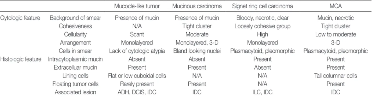
Table 3. Comparison of mucin-producing tumors in the breast
Mucocle-like tumor Mucinous carcinoma Signet ring cell carcinoma MCA Cytologic feature Background of smear Presence of mucin Presence of mucin Bloody, necrotic, clear Mucin, necrotic
Cohesiveness N/A Tight cluster Loosely cohesive group Tight cluster Cellularity Scant Moderate High Low to moderate Arrangement Monolalyered Monolayered, 3-D Monolayered 3-D
Cells in smear Lack of cytologic atypia Bland looking nuclei Plasmacytoid, pleomorphic Plasmacytoid, pleomorphic Histologic feature Intracytoplasmic mucin Absent Absent Present Present
Extracelluar mucin Present Present Absent Present Lining cells Flat or low cuboidal cells N/A N/A Tall columnar cells Floating tumor cells Rarely present Present N/A Present
Associated lesion ADH, DCIS, IDC IDC ILC, IDC IDC
MCA, mucinous cystadenocarcinoma; N/A, not availale; ADH, atypical ductal hyperplasia; DCIS, ductal carcinoma in situ; IDC, invasive ductal carcinoma; ILC, infiltrating lobular carcinoma.
Fig. 3. Immunohistochemical staining of mucinous cystadenocarcinoma. The tumor cells are positive for cytekeratin 7 (A) and negative for cytokeratin 20 (B). The hormone receptors, estrogen receptor and progesterone receptor, are negative and the c-erbB2 is 2-positive (C). The tumor cells are positive for mucin (MUC) 1 (D) along the cytoplasmic membrane and negative for MUC2 (E). Mucin of the intracytoplasm and stroma reveal positivity for MUC5 (F).
A B C
D E F
metastasis from distant MCA are following: CK7 positive, CK20 negative, and CDX-2 negative. These results can help to ex-clude the possibility of metastatic MCA from the ovary, pancre-as, and gastrointestinal tract. Being MUC5 positive and MUC2 negative may be the unique characteristics of MCA in the breast, together with the immunoprofiles of being CK7 positive, CK20
negative, CDX-2 negative, ER negative, and PR negative.
Conflicts of Interest
No potential conflict of interest relevant to this article was reported.
616 •Kim SE, et al. REFERENCES 1. Rosen PP, Scott M. Cystic hypersecretory duct carcinoma of the breast. Am J Surg Pathol 1984; 8: 31-41. 2. Koenig C, Tavassoli FA. Mucinous cystadenocarcinoma of the breast. Am J Surg Pathol 1998; 22: 698-703. 3. Domoto H, Terahata S, Yamazaki T, Sato K, Takeo H, Tamai S. Mu- cinous cystadenocarcinoma of the breast showing sulfomucin pro-duction. Histopathology 2000; 36: 567-9. 4. Honma N, Sakamoto G, Ikenaga M, Kuroiwa K, Younes M, Takubo K. Mucinous cystadenocarcinoma of the breast: a case report and review of the literature. Arch Pathol Lab Med 2003; 127: 1031-3. 5. Chen WY, Chen CS, Chen HC, Hung YJ, Chu JS. Mucinous cystad- enocarcinoma of the breast coexisting with infiltrating ductal carci-noma. Pathol Int 2004; 54: 781-6. 6. Coyne JD, Irion L. Mammary mucinous cystadenocarcinoma. His-topathology 2006; 49: 659-60. 7. Lee SH, Chaung CR. Mucinous metaplasia of breast carcinoma with macrocystic transformation resembling ovarian mucinous cystade-nocarcinoma in a case of synchronous bilateral infiltrating ductal carcinoma. Pathol Int 2008; 58: 601-5. 8. Rakici S, Gönüllü G, Gürsel SB, Yildiz L, Bayrak IK, Yücel I. Muci- nous cystadenocarcinoma of the breast with estrogen receptor ex-pression: a case report and review of the literature. Case Rep Oncol 2009; 2: 210-6. 9. Petersson F, Pang B, Thamboo TP, Putti TC. Mucinous cystadeno- carcinoma of the breast with amplification of the HER2-gene con-firmed by FISH: The first case reported. Hum Pathol 2010; 41: 910-3. 10. Sentani K, Tashiro T, Uraoka N, et al. Primary mammary mucinous cystadenocarcinoma: cytological and histological findings. Diagn Cytopathol 2012; 40: 624-8. 11. Rosen PP. Rosen’s breast pathology. 3rd ed. Philadelphia: Wolters Kluwer/Lippincott Williams & Wilkins, 2009; 515-34. 12. Cheng L, Lee WY, Chang TW. Benign mucocele-like lesion of the breast: how to differentiate from mucinous carcinoma before sur-gery. Cytopathology 2004; 15: 104-8. 13. Laucirica R, Bentz JS, Khalbuss WE, Clayton AC, Souers RJ, Mori- arty AT. Performance characteristics of mucinous (colloid) carcino- ma of the breast in fine-needle aspirates: observations from the Col-lege of American Pathologists Interlaboratory Comparison Program in Nongynecologic Cytopathology. Arch Pathol Lab Med 2011; 135: 1533-8. 14. Kelten C, Akbulut M, Zekioglu O, Kapkac M, Erhan Y, Ozdemir N. Signet ring cells in fine needle aspiration cytology of breast carcino- mas: review of the cytological findings in ten cases identified by his-tology. Cytopathology 2009; 20: 321-7. 15. Lau SK, Weiss LM, Chu PG. Differential expression of MUC1, MUC2, and MUC5AC in carcinomas of various sites: an immunohistoche-mical study. Am J Clin Pathol 2004; 122: 61-9. 16. Nguyen MD, Plasil B, Wen P, Frankel WL. Mucin profiles in signet-ring cell carcinoma. Arch Pathol Lab Med 2006; 130: 799-804. 17. Chu PG, Chung L, Weiss LM, Lau SK. Determining the site of ori-gin of mucinous adenocarcinoma: an immunohistochemical study of 175 cases. Am J Surg Pathol 2011; 35: 1830-6. 18. Hanski C, Hofmeier M, Schmitt-Gräff A, et al. Overexpression or ectopic expression of MUC2 is the common property of mucinous carcinomas of the colon, pancreas, breast, and ovary. J Pathol 1997; 182: 385-91. 19. Matsukita S, Nomoto M, Kitajima S, et al. Expression of mucins (MUC1, MUC2, MUC5AC and MUC6) in mucinous carcinoma of the breast: comparison with invasive ductal carcinoma. Histopa-thology 2003; 42: 26-36.