i
The proinflammatory cytokine effects on neurotrophins
production in bone marrow mesenchymal stem cells
Jang-Kyeun Jung
The Graduate School
Yonsei University
ii
The proinflammatory cytokine effects on neurotrophins
production in bone marrow mesenchymal stem cells
A Doctoral Dissertation
Submitted to the Department of Medicine
and the Graduate School of Yonsei University
in partial fulfillment of the
requirements for the degree of
Doctor of Philosophy
Jang-Kyeun Jung
iii
This certifies that the doctoral dissertation of
Jang-Kyeun Jung is approved.
___________________________ Thesis Supervisor: Prof. Dong-Joon Park
___________________________ Thesis Committee Member: Prof. In-Deok Kong
___________________________ Thesis Committee Member: Prof. Kum-Hwang
___________________________ Thesis Committee Member: Prof. Eung-Ho Choi
___________________________ Thesis Committee Member: Prof. Hyun-Soo Kim
The Graduate School
Yonsei University
iv
Acknowledgement
First of all, I would like to express my heartfelt gratitude to God for precious opportunity to write this thesis. It was entirely by God's grace and divine guidance that I was able to accomplish it.
I also thank my supervisor, professor Dong- Joon Park from the bottom of my heart for his assistance, information and encouragement.
I appreciate professor In Deok Kong at the department of physiology, professor Kum Hwang at the department of neurosurgery, professor Eung Ho Choi at the department of dermatology, professor Hyun Soo Kim at the department of medical oncology for their advice and helpful suggestions.
Finally, I give thanks to my wife, Mee Yon Cho who is deeply in love with me and who supported me in deep and profound ways.
v
Contents
ABSTRACT……….…1
I. Introduction ……….…………3
II. Materials & Methods ……….…...…...8
1. Bone marrow stromal cell culture ……….……….8
2. Experimental groups ……….……….8
3. Analysis of NGF, BDNF mRNA production …………8
4. ELISA for NGF and BDNF protein measurement ……9
5. Protein isolation and Western blot analysis………..…10
6. Si RNA transfection ……….…11
7. Statistical analysis ………11
III. Results ………..13
1. Pro-inflammatory cytokine TNF-α, but not IL-1β,
enhanced NGF secretion from BMSC ……….13
2. Activation of MAPK and NF-kB pathways by TNF-α in
BMSC ………..17
vi
3. MEK-ERK and NF-kB pathway are involved in the
TNF-α–enhanced NGF secretion ………19
4. RSK is involved in the TNF-α–enhanced NGF
production ………23
IV. Discussion ………..…….25
V. Conclusion ………28
References ……….29
vii
List of Figures
Fig. 1. Effects of TNF-α on NGF mRNA expression from BMSC…………..14
Fig. 2. Expression of NGF mRNA (A) and protein (B) in BMSC induced by
TNF-α……….15
Fig. 3. Effects of IL-1β on NGF mRNA expression from BMSC………..16
Fig. 4. Effect of TNF-α on phosphorylation of p38, ERK and NF-kB in
BMSCs………..……….18
Fig. 5. The effects of ERK inhibitor on NGF secretion……….20
Fig. 6. The effects of p38 MAPK inhibitor on NGF secretion………..21
Fig. 7. The effects of NF-kB inhibitor on NGF secretion……….22
viii
List of Table
1
ABSTRACT
The proinflammatory cytokine effects on neurotrophins production
in bone marrow mesenchymal stem cells
Jang-Kyeun Jung
The Graduate School
Yonsei University Department of Medicine
(Directed by professor Dong-Joon Park)
Understanding the mechanisms by which bone marrow stem cells produce neurotrophic factors may represent an important way to optimize their beneficial paracrine and autocrine effects. Components of the damaged nervous microenvironment may stimulate neurotrophic factor production to promote stem cell-mediated repair. We hypothesized that the proimflammatory cytokines (tumor necrosis factor-alpha or Interlukin1beta) may activate human bone marrow mesenchymal stem cells (BMSC) to increase the release of neurotrophic factors (nerve growth factor, brain derived nerve growth factor) and that nuclear factor-kappa B (NF kappa B), mitogen-activated protein kinases(MAPKs) pathway mediates neurotrophic factor production from human BMSC. Furthermore, to modify BMSC ex vivo to capitalize on the positive effects of cytokines, understanding of detail signal pathway is important. To
2
study this, human BMSC were cultured, passaged, divided into four groups (100,000 cells, triplicates) and treated as follows: 1) with vehicle; 2) with stimulant alone (24 h TNF-alpha or 24 h IL-1β); 3) with inhibitor alone [NF kappa B (PDTC), p38MAPK(SB203580), or ERK (PD98059)]; and 4) with stimulant and the various inhibitors. After 24 h incubation, BMSC activation was determined by measuring expression for NGF, BDNF (RT-PCR, ELISA, Western blot). TNF-alpha but not IL-1β significantly increased human BMSC NGF production versus controls. Stem cells exposed to TNF-alpha demonstrated increased activation of NF kappa B, ERK, and p38MAPK. NGF expression was significantly reduced by NF kappa B and ERK inhibition but not p38MAPK inhibition. Inhibitor alone did not activate BMSC NGF expression over controls. Moreover, siRNA for RSK1 but not MSK1 could knock down NGF expression. With these results, TNF-alpha activates human BMSC to increase NGF expression, which depends on an NFkB and MEK/Erk/RSK pathway mechanism.
Key words ; Nerve growth factor, brain derived neurotrophic factor, mesenchymal stem cells, tumor necrosis factor, cell therapy.
3
The proinflammatory cytokine effects on neurotrophins production
in bone marrow mesenchymal stem cells
Jang-Kyeun Jung
The Graduate School
Yonsei University Department of Medicine
(Directed by professor Dong-Joon Park)
Introduction
Stem cell therapy has the potential to be a key component in the field of regenerative medicine (1). Mesenchymal stem cells, in particular, may be a leading candidate for cell-based therapy for the neurological disease. In otolaryngology, treatment of neural disorders, such as anosmia, facial nerve palsy or sensory neural hearing loss, is one of the most difficult problems in modern medicine due to the limitations of neuronal regeneration. Mesenchymal stem cells are a unique subset of stem cells that can be isolated from the bone marrow, adipose tissue, and even umbilical cord blood (2, 3).
Among several kinds of cell-based therapies, bone marrow-derive mesenchymal stem cells or bone marrow stromal cell (BMSC) is a strong therapeutic candidate. BMSCs enriched in
4
immunological and logistical problems associated with embryonic or adult neural stem cell therapies. Furthermore, there is increasing evidence showing that BMSCs survive, selectively migrate to injured areas and provide therapeutic benefits in a variety of neuronal diseases, such as cerebral ischemia (4-6), traumatic brain injury (7), spinal cord injury(8,9) anosmia and facial palsy. These studies suggest the possibility of transplantation therapy using BMSCs for patients with various neural disorders. However, the mechanisms by which BMSCs provide therapeutic benefits remain unclear. BMSCs, including stem and progenitor cells, are multipotent and capable of differentiation into mesodermal derivatives such as bone, cartilage, fatty tissue and even neural cells such as neurons (10, 11). Although the transdifferentiation theory is attractive, it is inconsistent with in vivo data (12). Despite some encouraging results, these studies have shown that adult stem cells typically exhibit low level of engraftment and transdifferentiation within diseased or injured tissue and therefore do not contribute physically to tissue regeneration to a significant extent.
Rodents after middle cerebral artery occlusion (MCAo) obtain therapeutic benefit within days, and very few BMSCs express neural markers (13). Clearly, weeks or months are needed for BMSCs to transdifferentiate into the lost neural cells and appropriately integrate into complex neural connections (14, 15). Alternatively, orthotropic BMSCs naturally secrete a variety of cytokines and growth factors, which mainly support hematopoietic stem cells to differentiate into mature blood cells (16). Interestingly, the pattern and quantity of such functional secretion of BMSCs could be changed in response to their existing microenvironment (17). BMSCs in ischemic conditions increase the synthesis of some cytokines and growth factors (18–20).
These findings suggest that BMSCs may work as ‘small molecular factories’ by secreting cytokines, neurotrophins, growth factors, and other supportive substances at least acutely
5
after stroke which activates the restorative properties on endogenous brain parenchymal cells. Altered neurotrophic and growth factor gene expression may be the initial spark for therapeutic progress, which may produce therapeutic benefits in the ischemic brain.
Neurotrophic factors are target-derived factors, with critical roles in the survival, differentiation, and the maintenance of the function of different neurons both in peripheral and central nervous system (CNS). In mammals, the neurotrophic factor family consists of four members: Nerve growth factor (NGF), Brain derived neurotrophic factor (BDNF), Neurotrophin-3 (NT-3) and Neurotrophin-4/5 (NT-4/5). Neurotrophic factors encode structurally related proteins, which are proteolytically processed and secreted in the extracellular space.
In our previous study, chemically damaged rat olfactory mucosa showed spontaneous recovery within nine weeks. During this period, we found active biological activities in the regenerating olfactory mucosa related to the expression of neurotrophic factor mRNAs. Using extracts of this biologically active olfactory mucosa, we demonstrated increased synthesis of neurotrophic factors in BMSCs under the influence of regenerating olfactory mucosa conditioned medium (in press). Our results support the hypothesis that transplanted stem cells secrete essential neurotrophic factors that promote the regeneration of damaged nerve tissue.
Common condition influencing in ischemic brain and damaged olfactory mucosal microenvironments may be the inflammation. The inflammatory condition of the recipient site may activate the secretion of neurotrophic factors for the transplanted stem cells.
Tumor necrosis factor alpha (TNF-α) is a pro-inflammatory cytokine that is produced by a variety of cell types. The function of TNF-α is ambiguous because the effect of TNF-α is mediated by two receptors with different activation paths (21). The activation of TNF receptor 1 has been reported to decrease BMSC growth factor production (22), generate
6
reactive oxygen species, and induce apoptosis (23). In contrast, TNF receptor 2 cannot transmit apoptotic signals, instead its activation leads to nuclear factor κB (NF-κB) activation, which may be essential for cell survival, proliferation, growth factor production and expression of anti-apoptotic proteins (24-26). Even though the effect of BMSCs on inflammation has been widely studied, the influence of inflammation on BMSCs is still poorly understood. It has been previously reported that TNF-α has a significant effect on human BMSC proliferation and growth factor production in vitro and that these effects are mediated through IκB kinase 2 (IKK-2) and NF-κB pathway activation (26,-27). It is also known that in response to TNF-α, stem cells increase the release of paracrine factors by a p38 mitogen-activated protein kinase and signal transducer and activator of transcription 3 (STAT3)-dependent mechanism (28, 29). In addition, TNF-α-exposure is known to increase the expression of intracellular adhesion molecule-1 (ICAM-1, CD54) in endothelial cells (30). However, the influence of TNF-α to neurotrophic factors secretion in BMSC remains unclear. Interleukin 1 β (IL-1β) is another major proinflammatory cytokine can also considerably up regulate the expression of NGF (31, 32). It is also unclear about its effects on BMSC.
Mitogen-activated protein kinases (MAPKs) are ubiquitous kinases and are involved in signal transduction in eukaryotic organisms. This family of kinases is characterized by their activation by MAPKs through the dual phosphorylation of Thr and Tyr residues in their activation loop. The MAPK family includes extracellular signal-regulated kinases (ERK), which are activated in response to growth factors, via the Ras proto-oncogene. Moreover, c-Jun N-terminal kinase (JNK) and p38 MAPK constitute two other families, collectively known as stress-activated protein kinases (SAPK), because they are induced by UV radiation, heat-shock, oxidative stress. The stimulation of ERK initiates a cascade of activating events, including the phosphorylation of p90 ribosomal S6 protein kinase 1 (RSK1), and its
7
translocation to the nucleus, where RSK1 phosphorylates nuclear substrates (33). Moreover, the phosphorylation of mitogen- and stress-activated protein kinase (MSK), which localized in the nuclei (34), could lead to the phosphorylation and activation several transcription factors like cAMP-response element-binding protein (CREB) and activating transcription factor 1 (ATF1) (35).
We examined the mechanism by which the important proinflammatory mediator, TNF-α increases NGF expression levels. Here we show that NF kB and ERK MAPK are essential for TNF-α induced NGF expression in normal human BMSC. We also show that RSK1 phosphorylation mediates the TNF-α induced NGF production in BMSC.
8
Material and Methods
Bone marrow stromal cells.
Human mesenchymal stem cell was purchased from Pharmicell (Sungnam, Korea). Mesenchymal stem cells were harvested and cultured from normal human bone marrow. Thawing of cells and initiation of culture process were performed based on the manufacturer’s instructions. Human BMSCs were plated in T-225 tissue culture flasks (Corning, Coring, NY) and cultured with mesenchymal stem cell basal medium containing 10% fetal bovine serum (FBS), Mesenchymal Cell Growth Supplement (Camber BioScience), 4 mML-glutamine, and penicillin-streptomycin at 37°C, 5% CO2 and 90% humidity. Medium was changed every 3 days.
Experimental groups.
After cells were 70% confluent, BMSCs were plated in T-25 tissue culture flask (Corning) in a concentration of 0.1 x 10⁶ cells /flask/ml. Cells were divided into the following groups: 1) with vehicle; 2) with stimulant alone [TNF-alpha (1 to 20ng/ml) or IL-1β (1 to 50ng/ml) ]; 3) with inhibitor alone [NF kappa B (PDTC, 0.1 and 1mM), p38MAPK(SB203580, 10 and 20 µM ), or ERK (PD98059, 1 and 10µM)]; and 4) with stimulant and the various inhibitors. Inhibitors were obtained from Calbiochem (San Diego, CA). After 12 to 48 hr incubation, cells and supernatants were harvested for NGF, BDNF assay.
Analysis of NGF, BDNF mRNA production
Cells were grown in T-25 culture flask and incubated for 12–48 h in a fresh medium containing stimuli as indicated. After discarding growth medium, total RNA was isolated
9
from cells using Trizol reagent (Invotrogen, Carlsbad, CA, USA), following the manufacturer’s instructions. Reverse transcription (RT) was performed with SuperScript II reverse transcriptase (Invitrogen, Burlington, ON, Canada), according to the manufacturer’s instructions. One microgram of RNA and 20 pmol/µl primers were preincubated at 70oC for 5 min and transferred to a mixture tube. The reaction volume was 20 µl. cDNA synthesis was performed at 42 oC for 60 min, followed by RT inactivation at 94 oC for 5 min. Thereafter, the RT-generated DNA (2–5 µl) was amplified using AccuPower PCR PreMix (Bioneer). The primers used for cDNA amplification and conditions were as follows (Table 1). RT-PCR products were subsequently separated by electrophoresis on 2% agarose gels containing 0.5 µg/ml ethium bromide and visualized with UV light. All data were normalized to the level of GAPDH mRNA.
ELISA for NGF and BDNF protein measurement
The supernatants were collected, cleared by centrifugation, and kept at -20oC until evaluation by ELISA. For measurement of neurotrophic factors concentrations in cell culture supernatants, 96-well microtiter plates (MaxiSorp; Nunc) were coated with 0.2 µg/well goat anti-human NGF (rabbit polyclonal IgG, epitope mapping at the N-terminus of the mature chain of NGF of human origin, 200µg/ml of PBS with<0.1% sodium azide and 0.1% gelatin, Santacruz, CA, USA) and BDNF (rabbit polyclonal IgG, epitope mapping within an internal region of BDNF of human origin, 200µg/ml of PBS with<0.1% sodium azide and 0.1% gelatin, Santacruz, CA, USA) . Abs in 50 µl of PBS at 4 oC overnight. All further steps were conducted at room temperature. After washing three times with PBS, nonspecific binding sites were blocked by incubation with 150 µl PBS + 1% BSA/ 0.05% Tween 20/well for 2 h. After three washes with PBS, supernatant was added to the 96-well plates, and the captured
10
NGF and BDNF were detected using biotinylated goat anti-human IgG. After 30-min incubation with streptavidin-horseradish peroxidase, substrate solutions (1:1 mixture of H2O2 and tetramethylbenzidine) were added to the wells for 20 min and the reaction was stopped by adding 2 N H2SO4. The plates were read at 450 nm on a microtiter plate reader. ELISA reagents were from R&D Systems. All samples and standards were measured in duplicate. Values are expressed as means ±SE.
Protein isolation and Western blot analysis.
Western blot analysis was performed to measure NGF and the activation of ERK, p38 MAPK and NF-kB. Cells were collected in cold buffer containing 20 mM Tris (pH 7.5), 150 mM NaCl, 1 mM EDTA, 1 mM EGTA, 1% Triton X-100, 2.5 mM sodium pyrophosphate, 1 mM-glycerophosphate, 1 mM Na3VO4, 1 µg/ml leupeptin, 1 mM PMSF, and centrifuged at 12,000 rpm for 10 min. The protein extracts (10 µg/lane) were electrophoresed on a 4–12% Bis-Tris gel (Invitrogen, Carlsbad, CA) and transferred to a nitrocellulose membrane, which was stained by naphthol blue-black to confirm equal protein loading. The membranes
were incubated in 5% dry milk for 1 h and then incubated with the following primary antibodies: NGF antibody, ERK antibody, phospho-ERK (Thr202/Tyr204) antibody,
p38 MAPK antibody, phospho- p38 MAPK (Thr180/Tyr182) antibody, NF-κB p65 antibody and phospho-NF-κB p65 (Ser276) antibody (Cell Signaling Technology, Beverly, MA), followed by incubation with horseradish peroxidase-conjugated goat anti-rabbit IgG secondary antibody and detection using supersignal west pico stable peroxide solution (Pierce, Rockford, IL). The exposure time of the film was adjusted based on the intensity of the signal. Band densities were compared using NIH Image-J software. Equal loading was corrected by β-actin immunoreactivity or of a relative non-phosphorylation antibody.
11
siRNA transfection
RSK1 siRNA and MSK1 siRNA for silencing of BMSC were purchased from Cell Signaling Technology ( Beverly, MA) and Santacruz Inc. (CA, USA) respectively. A double-stranded non-specific siRNA was used as a negative control. The siRNA transfection was conducted with Lipofectamine 2000 reagent (Invitrogen) according to the manufacturer's instructions. Cells plated for 24 hours were transfected with 150 nM of siRNA. At 24 hours after transfection, the cells were treated with TNG-alpha, t hen the expression of NGF was analyzed.
Statistical analysis
Data are presented as means ± SE and were analyzed using one-way ANOVA and Student's
12
Table 1. Sequences of primers used for RT-PCR
Gene Primer sequences(5’ to 3’) length(bp) TM(oC)
NGF Forward : GCC CAC TGG ACT AAA CTT CAG C Reverse : CCG TGG CTG TGG TCT TAT CTC 364 60.0 60.0 BDNF Forward : CGA CGT CCC TGG CTG ACA CTT TT
Reverse : AGT AAG GGC CCG AAC ATA CGA TTG G
450 62.0
62.2
GAPDH Forward : TCC CAT TCT TCC ACC TTT GAT GCT
Reverse : ACC CTG TTG CTG TAG CCA TAT TCA T
102 58.7
13
Results
Pro-inflammatory cytokine TNF-α, but not IL-1β, enhanced NGF secretion from BMSC
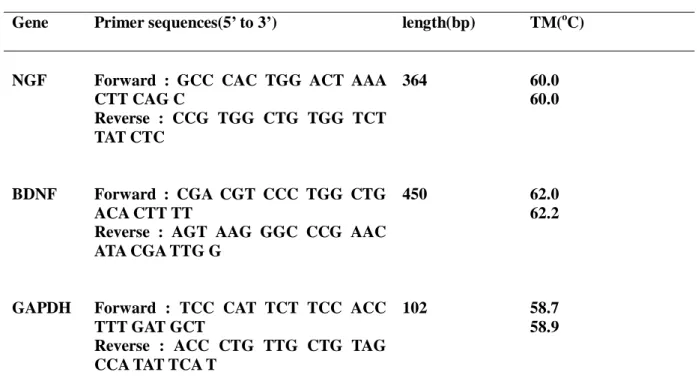
First, we examined whether pro-inflammatory cytokines enhance NGF or BDNF secretion from BMSC. BMSC constitutively secreted a low amount of NGF and BDNF without any stimulation. NGF secretion was at a concetration-dependently increased by TNF-α; the stimulatory effect of TNF-α peaked at 10 ng/ml and maintained up to 20 ng/ml, which is 3-fold the secretion of basal level. However BDNF was not stimulated by TNF-α (Fig. 1).
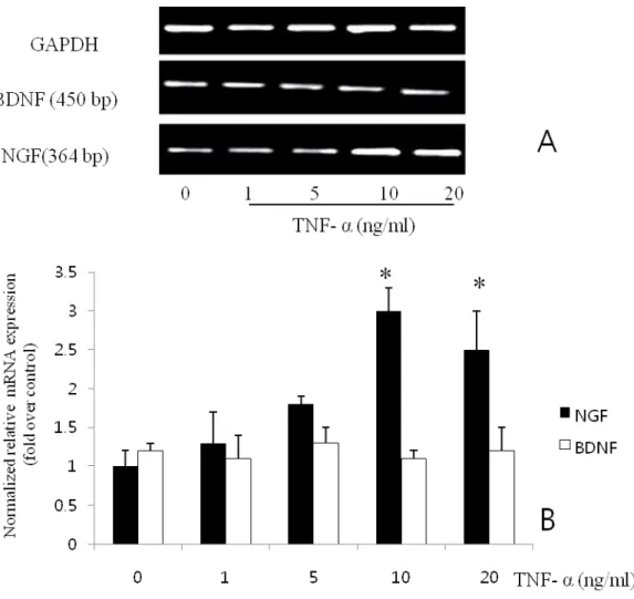
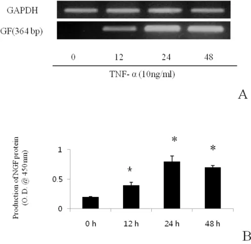
Maximal induction was seen after 24 h of exposure to 10 ng/ml TNF- α. When the level of NGF mRNA in the induced cultures was analyzed by RT-PCR (Fig. 2 A), the increase protein (Fig.2. B) secretion correlated with the accumulation of NGF mRNA. In contrast to TNF-α, IL-1β did not enhance NGF or BDNF secretion (Fig. 3).
14
Fig. 1. Effects of TNF-α on NGF mRNA expression from BMSC. RT-PCR (A) and semi-quantitative analysis of NGF and BDNF mRNA (B). Results are means ± SE; n= 3, *P< 0.05 vs. control.
15
Fig. 2. Expression of NGF mRNA (A) and protein (B) in BMSC induced by TNF-α. The production increased in a time dependent manner. Results are means ± SE; n= 3, *P< 0.05 vs. control.
16
Fig. 3. Effects of IL-1β on NGF mRNA expression from BMSC. RT-PCR (A) and semi-quantitative analysis of NGF and BDNF mRNA (B). There was no statistical significance. Results are means ± SE; n= 3.
17
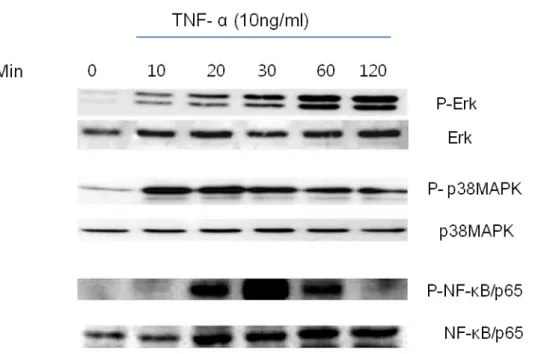
Activation of MAPK and NF-kB pathways by TNF-α in BMSC
TNF-α is known to be a potent stimulus for activation of ERK, p38MAPK, and NF-kB. As shown by the results in Fig. 4, TNF-α activated ERKs phosphorylation at 15 min, and increased sharpely and remained for the following 2 h. TNF markedly activated p38 MAPK phosphorylation and NF-kB at 10min and 20 min respectively. Concentration of 10 ng/ml TNF-α induced increase in ERK, p38MAPK and NF-kB activities.
18
Fig. 4. Effect of TNF-α on phosphorylation of p38, ERK and NF-kB in BMSCs. The cells were treated with TNF-a (10 ng/ml). Cell lysates were analyzed by Western blot. This similar data were obtained in three independent experiments.
19
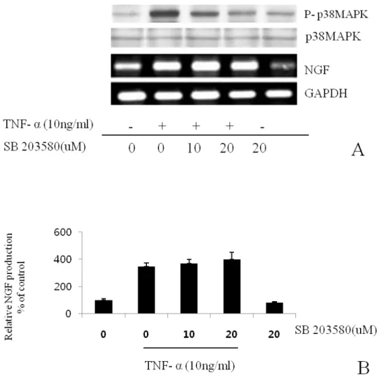
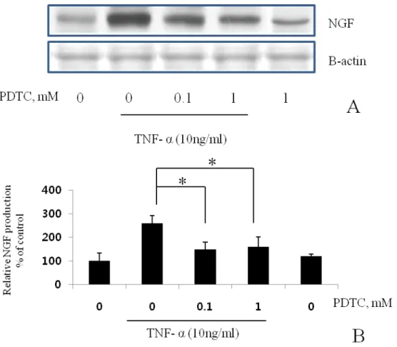
MEK-ERK and NF-kB pathway are involved in the TNF-α–enhanced NGF secretion
To identify the signal transduction pathway of the TNF-α–enhanced NGF secretion, we examined the effect of typical MAPK inhibitors, PD98059, SB203580, and NF-kB inhibitor, PDTC. PD98059, which selectively blocks ERK activity through the inhibition of ERK1/2 phosphorylation by MEK1/2, reduced the TNF-α–enhanced NGF secretion to the basal level at the concentration from 1.0 μM and completely inhibited it at 10 μM (Fig. 5). On the other hand, SB203580, a specific inhibitor of p38 MAPK, did not inhibit the TNF-α–enhanced NGF secretion (Fig. 6). PDTC, a specific inhibitor of NF-kB, inhibited NGF secretion (Fig. 7).
20
Fig. 5. The effects of ERK inhibitor on NGF secretion. BMSCs were pre-incubated with the indicated concentrations of PD98059 for 15 min and then cultured with TNF-α (10 ng/ml) for 24 h in the presence of the inhibitor. Inhibition of the phosphorylation was analyzed by Western blot. NGF mRNA was analyzed by the RT-PCR (A). Semi- quantification of Western blot result for NGF production by NIH image software (B). Values are each the mean ± S.E.M. of triplicate cultures of three independent experiments. *P<0.05 vs. control cultures without inhibitor.
21
Fig. 6. The effects of p38 MAPK inhibitor on NGF secretion. BMSCs were pre-incubated with the indicated concentrations of SB203580 for 15 min and then cultured with TNF-α (10 ng/ml) for 24 h in the presence of the inhibitor. Inhibition of the phosphorylation was analyzed by Western blot. NGF mRNA was analyzed by the RT-PCR (A). Semi-quantification of Western blot result for NGF production by NIH image software (B). There were no statistical significances. Values are each the mean ± S.E.M. of triplicate cultures of three independent experiments.
22
Fig. 7. The effects of NF-kB inhibitor on NGF secretion. BMSCs were pre-incubated with the indicated concentrations of PDTC for 15 min and then cultured with TNF-α (10 ng/ml) for 24 h in the presence of the inhibitor. Inhibition of the phosphorylation was analyzed by Western blot. NGF mRNA was analyzed by the RT-PCR (A). Semi-quantification of Western blot result for NGF production by NIH image software (B). Values are each the mean ± S.E.M. of triplicate cultures of three independent experiments. *P<0.05 vs. control cultures without inhibitor.
23
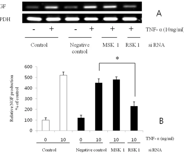
RSK is involved in the TNF-α–enhanced NGF production
It is well known that RSK and MSK are involved in the downstream portion of the MEK-ERK pathway. We thus examined whether RSK1 or MSK1 are also involved in the TNF-α– induced enhancement of NGF secretion. RSK1 siRNA inhibited the TNF-α–enhanced NGF production, but not the basal level (Fig. 8). However, MSK1 siRNA did not affect the NGF production . These results indicate that RSK1 is involved in the signaling pathway of the TNF-α–enhanced NGF secretion..
24
Fig. 8. Inhibition of NGF production by siRNA transfection. TNF-a stimulated NGF mRNA production was inhibited by RSK1 siRNA transfection. However, MSK1 si RNA transfection did not affect (A). Semi-quantification of Western blot result for NGF production by NIH image software (B). Values are each the mean ± S.E.M. of triplicate cultures of three independent experiments. *P<0.05 vs. control.
25
Discussion
While stem cell therapy has the potential to be a key component of regeneration therapies for neurologic disorder, the efficacy is not so high. If we can make neurotrophic factors secreting bone marrow stromal cells, the stem cell therapy for injured nervous system would be more effective. Recently some pioneer studies to make a neurotrophins producing stem cells are tried. Major portion of those studies includes neurotrophins encoding gene transfections(36). However, so far, the gene transfer technique is not safe and problem of toxicity of the vector is not overcome.
Cytokines likely affect the function of BMSCs and represent one possible explanation for the variable results seen in the literature. Therefore, increasing our understanding of the cytokines that affect BMSC function and the methods available to clinicians to modify BMSCs ex vivo to capitalize on the positive effects of cytokines may lead to future therapeutic gains.
The results of present study represent the first demonstration that: 1) TNF- α stimulates BMSC production of NGF 2) NF kB is involved in production of NGF in BMSCs 3) MEK/ERK/RSK pathway is also involved in production of NGF. Stem cells transplanted into injured nerve tissue express several paracrine signaling factors, including cytokines, chemokines, and growth factors, which are involved in stem cell-mediated repair. A critically important part of this process may be their chronically inflammed tissue. Although the particular local signaling molecules contributing to this regenerating effect remain to be defined, the list most likely includes neurotrophic factors in neuronal lesion. NGF is a strong promoter of neurogenesis, has been shown to be intimately involved with nerve cell proliferation and may be a more potent neurogenic factor than BDNF. The results of present study demonstrate that BMSC in cell culture exposed to specific stimuli such as TNF-α
26
significantly increase release of NFG but not BDNF. Crisostomo et al demonstrated that TNF- α stimulates VEGF, FGF, HGF, IGF-1 production in BMSC (37). This increase in growth factors may improve regional nerve regeneration as well as promote autocrine self survival (38). Increased perfusion due to the stem cell angiogenic growth factor production has also been associated with improved end organ function (39). Furthermore, VEGF over expressing bone marrow stem cells demonstrate greater protection of injured tissue than controls (40). Thus NGF as well as other growth factors may be important paracrine signaling molecules in stem cell-mediated angiogenesis, protection, and nerve survival. Further understanding of the mechanisms by which these paracrine growth factors are released may enable us to maximize stem cell paracrine effects when transplanted into injured tissue. It remains unknown by what mechanisms injury stimuli induce BMSCs to release growth factors.
NF kB is an important rapid acting transcription factor found in all cell types and is involved in cellular responses to stimuli such as stress, cytokines, free radicals, ultraviolet irradiation, and bacterial or viral antigens. In stem cells, recent data suggest that NF kB plays a role in proliferation, migration, and differentiation (41). NGF has been implicated in as an important upstream component of the NF kB proliferation process in neural and hepatic stem cells (42). NF kB inhibition (PDTC) significantly decreased the production of NGF. The results of present study shed further light on the role of NF kB in stem cell-mediated protection. Interestingly, umbilical cord blood, which has a higher proliferation rate than adult stem cells, was recently found to have higher expression of NF kB compared with that of adults (43). Thus pharmacological activation of NF kB to enhance growth factor production and proliferation may be a strategy to improve their paracrine effects before cell transplant; however, this remains to be determined.
27
Wang et al have previously shown that the increase in BMSCs production of VEGF, HGF, and IGF-1 was associated with the increase of p38 MAPK activation exposure to TNF-α (44). p38 MAPK inhibitor administration also resulted in a decreased release of growth factors in hMSCs response to TNF-α. MAPKs are critically involved in regulatory signaling pathways that ultimately leads to inflammation (45-47). Activation of p38 MAPK and JNK is a critical step in the generation of deleterious inflammatory cytokines after tissue injury, whereas ERK activation has been found to improve functional recovery after ischemia (48-50). However, the results of present study demonstrate that although TNF-α exposure increases ERK and p38 activation, inhibition of p38 MAPK does not significantly affect stem cell production of NGF. Thus it appears that TNF-α induced stem cell production of NGF depends on ERK activation but does not depend on increased p38 MAPK phosphorylation.
To know further downstream pathway, we used the siRNA specific for RSK1 and MSK1, the results showed that RSK1 knockdown NGF production. Besides MSK1, the RSK1 was another downstream kinase for ERK1/2.
28
Conclusions
Taken together, TNF-alpha activates human BMSC to increase NGF expression, which depends on an NF kB and MEK/Erk/RSK pathway mechanism. This study highlights the effect of TNF-a as an important local factor, on BMSC release of NGF. The enhancing of NGF production in BMSC may be an alternative strategy to stem cell therapy for the treatment of neurologic disorder.
29
References
1. Jolicoeur EM, Granger CB, Fakunding JL, et al. Bringing cardiovascular cell-based therapy to clinical application: perspectives based on a National Heart, Lung, and Blood Institute Cell Therapy Working Group meeting. Am Heart J 2007;153:732– 42.
2. Kumar S, Chanda D, Ponnazhagan S. Therapeutic potential of genetically modified mesenchymal stem cells. Gene Ther 2008;15:711–5.
3. Kestendjieva S, Kyurkchiev D, Tsvetkova G, et al. Characterization of mesenchymal stem cells isolated from the human umbilical cord. Cell Biol Int 2008;32:724 –32.
4. Zhao LR, Duan WM, Reyes M et al. Human bone marrow stem cells exhibit neural phenotypes and ameliorate neurological deficits after grafting into the ischemic brain of rats. Exp Neurol 2002; 174: 11–20.
5. Shen LH, Li Y, Chen J et al. Therapeutic benefit of bone marrow stromal cells administered 1 month after stroke. J Cereb Blood Flow Metab 2007; 27: 6–13.
6. Chen J, Li Y, Wang L et al. Therapeutic benefit of intravenous administration of bone marrow stromal cells after cerebral ischemia in rats. Stroke 2001; 32:1005–1011.
7. Mahmood A, Lu D, Qu C et al. Long-term recovery after bone marrow stromal cell treatment of traumatic brain injury in rats. J Neurosurg 2006; 104: 272–277.
30
8. Chopp M, Zhang XH, Li Y et al. Spinal cord injury in rat: treatment with bone marrow stromal cell transplantation. Neuroreport 2000; 11: 3001–3005.
9. Himes BT, Neuhuber B, Coleman C et al. Recovery of function following grafting of human bone marrowderived stromal cells into the injured spinal cord. Neurorehabil Neural Repair 2006; 20: 278–296.
10. Jiang Y, Jahagirdar BN, Reinhardt RL et al. Pluripotency of mesenchymal stem cells derived from adult marrow. Nature 2002; 418: 41–49.
11. Pittenger MF, Mackay AM, Beck SC et al. Multilineage potential of adult human mesenchymal stem cells. Science 1999; 284: 143–147.
12. Castro RF, Jackson KA, Goodell MA et al. Failure of bone marrow cells to transdifferentiate into neural cells in vivo. Science 2002; 297: 1299.
13. Li Y, Chen J, Chopp M. Adult bone marrow transplantation after stroke in adult rats. Cell Transplant 2001;10: 31–40.
14. Brazelton TR, Rossi FM, Keshet GI et al. From marrow to brain: expression of neuronal phenotypes in adult mice. Science 2000; 290: 1775–1779.
15. Mezey E, Chandross KJ, Harta G et al. Turning blood into brain: cells bearing neuronal antigens generated in vivo from bone marrow. Science 2000; 290: 1779–1782.
31
16. Friedenstein AJ, Chailakhyan RK, Latsinik NV et al. Stromal cells responsible for transferring the microenvironment of the hemopoietic tissues. Cloning in vitro and retransplantation in vivo. Transplantation 1974; 17:331–340.
17. Caplan AI, Dennis JE. Mesenchymal stem cells as trophic mediators. J Cell Biochem 2006; 98: 1076–1084.
18. Li Y, Chen J, Chen XG et al. Human marrow stromal cell therapy for stroke in rat: neurotrophins and functional recovery. Neurology 2002; 59: 514–523.
19. Chen X, Li Y,Wang L et al. Ischemic rat brain extracts induce human marrow stromal cell growth factor production.Neuropathology 2002; 22: 275–279.
20. Tokumine J, Kakinohana O, Cizkova D et al. Changes in spinal GDNF, BDNF, and NT-3 expression after transient spinal cord ischemia in the rat. J Neurosci Res 2003; 74: 552–561.
21. Wallach D, Boldin MP, Goltsev YV, Tumor necrosis factor receptor and Fas signalling mechanisms, Ann. Rev. Immunol. 1999; 17: 331–367.
22. Markel TA, Crisostomo PR, Wang M, Herring CM, Meldrum DR, Activation of individual tumor necrosis factor receptors differentially affects stem cell growth factor and cytokine production, Am. J. Physiol. Gastrointest. Liver Physiol. 2007; 293: G657–G662.
32
by receptor compartmentalization, Nat. Rev. Mol. Cell Biol. 2008; 9: 655–662.
24. Marchetti L, Klein M, Schlett K, Pfizenmaier K, Eisel UL, TNF mediated neuroprotection against glutamate induced excitotoxicity is enhanced by NMDA receptor activation: essential
role of a TNF receptor 2 mediated, PI3 kinase dependent NF-kB pathway, J. Biol. Chem. 2004; 279: 32869–32881.
25. Crisostomo PR, Wang Y, Markel TA, Wang M, Lahm T, Meldrum DR, Human mesenchymal stem cells stimulated by TNF-alpha, LPS, or hypoxia produce growth factors by an NF kappa B- but not JNK-dependent mechanism, Am. J. Physiol. Cell Physiol. 2008; 294: C675–C682.
26. Ait-AD, Turquier V, Tanguy Y, Thouënnon E, Ghzili H, Mounien L, Derambure C, Jégou S, Salier JP, Vaudry H, Eiden LE, Anouar Y, Tumor necrosis factor (TNF)-alpha persistently activates nuclear factor-kappaB signaling through the type 2 TNF receptor in chromaffin cells: implications for long-term regulation of neuropeptide gene expression in inflammation, Endocrinology 2008; 149: 2840–2852.
27. Böcker W, Docheva D, Prall DC, Egea V, Pappou E, Rossmann O, Popov C, Mutschler W, Ries C, Schieker M, IKK-2 is required for TNF-alpha-induced invasion and proliferation of human mesenchymal stem cells, J. Mol. Med. 2008; 86:1183–1192.
33
cells from bone marrow or adipose tissue produce VEGF, HGF, and IGF-I in response to TNF by a p38 MAPK-dependent mechanism, Am. J. Physiol. Regul. Integr. Comp. Physiol. 2006;291: R880–R884.
29. Wang M, Zhang W, Crisostomo P, Markel T, Meldrum KK, Fu XY, Meldrum DR, STAT3 mediates bone marrow mesenchymal stem cell VEGF production, J. Mol. Cell. Cardiol. 2007; 42: 1009–1015.
30. Pober JS, Lapierre LA, Stolpen AH, Brock TA, Springer TA, Fiers W, Bevilacqua MP, Mendrick DL, Gimbrone MA Jr., Activation of cultured human endothelial cells by recombinant lymphotoxin: comparison with tumor necrosis factor and interleukin 1 species, J. Immunol. 1987; 138: 3319–3324.
31. Purmessur D, Freemont AJ, Hoyland JA, Expression and regulation of neurotrophins in the nondegenerate and degenerate human intervertebral disc. Arthritis Res Ther. 2008:10(4)R99.
32. Manni L, Lundeberg T, Fiorito S, Bonini S, Vigneti E, Aloe L, Nerve growth factor release by human synovial fibroblasts prior to and following exposure to tumour necrosis factor-alpha, interleukin-1 beta and cholecystokinin-8: the possible role of NGF in the inflammatory response. Clin Exp Rheumatol.2003:21:617-624.
33. Richards SA, Dreisbach VC, Murphy LO, Blenis J. Characterization of regulatory events associated with membrane targeting of p90 ribosomal S6 kinase 1. Mol. Cell Biol. 2001;21:
34
7470–7480
34. Deak, M., Clifton, A. D., Lucocq, L. M., and Alessi, D. R. Mitogen- and stress-activated protein kinase-1 (MSK1) is directly activated by MAPK and SAPK2/p38, and may mediate activation of CREB. EMBO J. 1998; 17:4426–4441
35. Van Seuningen I, Pigny P, Perrais M, Porchet N, Aubert JP. Transcriptional regulation of the 11p15 mucin genes. Towards new biological tools in human therapy, in inflammatory diseases and cancer? Front. Biosci 2001;6: 1216–1234
36. Leng S, He J, Fan W, Cheng S, Long D, He H. Bone mesenchymal stem cells for gene transfer of NGF to the adult rat brain: Rescue the NGFR p75 positive neurons from fimbria– fornix lesion-induced degeneration. Neuroscience Letters 2008; 448: 282–287
37. Crisostomo PR, Wang Y, Markel TA,Wang M, Lahm T, Meldrum DR, Human mesenchymal stem cells stimulated by TNF-a, LPS, or hypoxia produce growth factors by an NFkB- but not JNK-dependent mechanism Am J Physiol Cell Physiol 294: C675–C682, 2008.
38. Houchen CW, George RJ, Sturmoski MA, Cohn SM. FGF-2 enhances intestinal stem cell survival and its expression is induced after radiation injury. Am J Physiol Gastrointest Liver Physiol 1999; 276: G249–G258
39. Kinnaird T, Stabile E, Burnett MS, Shou M, Lee CW, Barr S, Fuchs S, Epstein SE. Local delivery of marrow-derived stromal cells augments collateral perfusion through paracrine
35
mechanisms. Circulation 2004; 109: 1543–1549.
40. Wang Y, Haider HK, Ahmad N, Xu M, Ge R, Ashraf M. Combining pharmacological mobilization with intramyocardial delivery of bone marrow cells over-expressing VEGF is more effective for cardiac repair. J Mol Cell Cardiol 2006; 40: 736–745.
41. Widera D, Mikenberg I, Kaltschmidt B, Kaltschmidt C. Potential role of NF-kappaB in adult neural stem cells: the underrated steersman? Int J Dev Neurosci 2006;24: 91–102.
42. Yao P, Zhan Y, Xu W, Li C, Yue P, Xu C, Hu D, Qu CK, Yang X. Hepatocyte growth factor-induced proliferation of hepatic stem-like cells depends on activation of NF-kappaB. J Hepatol 2004; 40: 391–398.
43. Panepucci RA, Calado RT, Rocha V, Proto-Siqueira R, Silva WA Jr, Zago MA. Higher expression of transcription targets and components of the nuclear factor-kappaB pathway is a distinctive feature of umbilical cord blood CD34 precursors. Stem Cells 2007;25: 189–196.
44. Wang M, Crisostomo PR, Herring C, Meldrum KK, Meldrum DR. Human progenitor cells from bone marrow or adipose tissue produce VEGF, HGF, and IGF-I in response to TNF by a p38 MAPK-dependent mechanism. Am J Physiol Regul Integr Comp Physiol 2006; 291: R880–R884,
45. Clerk A, Michael A, Sugden PH. Stimulation of the p38 mitogenactivated protein kinase pathway in neonatal rat ventricular myocytes by the G protein-coupled receptor agonists,
36
endothelin-1 and phenylephrine: a role in cardiac myocyte hypertrophy? J Cell Biol 1998; 142: 523–535.
46. Meldrum KK, Meldrum DR, Hile KL, Yerkes EB, Ayala A, Cain MP, Rink RC, Casale AJ, Kaefer MA. p38 MAPK mediates renal tubular cell TNF-a production and TNF-a-dependent apoptosis during simulated ischemia. Am J Physiol Cell Physiol 2001;281: C563–C570.
47. Song GY, Chung CS, Chaudry IH, Ayala A. MAPK p38 antagonism as a novel method of inhibiting lymphoid immune suppression in polymicrobial sepsis. Am J Physiol Cell Physiol 2001; 281: C662–C669.
48. Jarrar D, Wang P, Song GY, Cioffi WG, Bland KI, Chaudry IH. Inhibition of tyrosine kinase signaling after trauma-hemorrhage: a novel approach for improving organ function and decreasing susceptibility to subsequent sepsis. Ann Surg 2000; 231: 399–407.
49. Khan TA, Bianchi C, Ruel M, Voisine P, Sellke FW. Mitogen-activated protein kinase pathways and cardiac surgery. J Thorac Cardiovasc Surg, 2004; 127: 806–811.
50. Schwacha MG, Chaudry IH, Alexander M. Regulation of macrophage IL-10 production postinjury via beta2 integrin signaling and the P38 MAP kinase pathway. Shock 2003;20: 529–535.
37
Abstract in Korean (국문 요약)
전염증성 싸이토카인이 중간엽줄기세포에서 뉴로트로핀 생성에
미치는 영향
정 장 균
연세대학교 대학원 의학과
지도교수 박동준
목적 ; 병변에 이식된 줄기세포가 새로운 세포로 분화되어 조직을 재생 한다기 보단, 조직의 치유나 재생에 유익한 물질을 줄기세포가 분비 함으로서 회복을 유도한다는 설이 최근에 학자들 사이에서 공감을 받고 있다. 대부분의 창상이나 병변에는 염증반응이 수반된다. 이에 공통적으로 기능을 하고 있는 물질이 TNF-a 나 IL-1B 같은 전염증성 싸이토카인 (proinflammatory cytokine) 이다. 이에 본 연구는 이러한 전염증성 싸이토카인이 과연 줄기세포를 자극해 신경재생에 유익한 물질인 neurotorphic factor (neurotrophin) 를 생산할수 있는지 알아보고 과연 그렇다면 이의 signal transduction pathway 를 추정하여 효과적인 신경재생 세포치료에 도움을 주고자 하였다.방법 ; 사람 중간엽 줄기세포를 구입하여 계대 배양 후 TNF-a 나 IL-1B 를 농도별, 시간별로 자극하여 nerve growth factor(NGF), brain derived neurotrophic factor(BDNF)의 생성을 RT-PCR, Western blot, ELISA 로 분석하였다. 또한 이
38
싸이토카인에 의한 Erk, p38MAPK, NFkB 의 phosphorylation 을 측정하고 각각의 억제제 및 si RNA 를 사용하여 신호전달체계를 추측하였다.
결과 ; TNF-a 는 사람 중간엽 줄기세포에서 농도에 비례하여 NGF 의 생성을 자극 하였으나 IL-1B 는 별 영향을 주지 않았다. 또한 TNF-a 는 Erk, p38MAPK, NFkB 를 모두 활성화 시켰으나, Erk, NFkB 억제제에 의해서만 NGF 의 생성이 감소 되었다. Erk 의 하위 전달체계인 RSK1 siRNA 에 의해 NGF 의 생성이 감소 되었으나, MSK1 siRNA 에는 영향을 받지 않았다. 결론 ; TNF-a 는 사람 중간엽 줄기세포를 자극하여 NGF 의 생성을 촉진 시키며, 이는 NFkB, MEK/Erk/RSK pathway 에 의한 것임을 알 수 있었으며 향후 연구를 통해 정확한 transcription factor 를 확인하면 효과적인 신경재생 세포치료에 응용 할 수 있을 것이라 생각된다. 핵심단어 ; 신경성장 인자, 뇌유래 신경인자, 중배엽성줄기세포, 세포치료