J Korean Neurol Assoc / Volume 22 / June, 2004
Received July 31, 2003 Accepted December 10, 2003 *Address for correspondence
Young-Chul Choi, M.D., Ph.D.
Department of Neurology, Yonsei University College of Medicine, Yongdong Severance Hospital
146-92 Dogok-dong Gangnam-gu, Seoul, 135-720, Korea Tel:+82-2-3497-3323 Fax:+82-2-3462-5904 E-mail:[email protected]
근위축성측삭경화증 환자에서 경부 척수 자기공명영상을 통한
경부 척추증의 평가
연세대학교 의과대학 신경과학교실 나상준 김원주 이진구 이경열 최영철Evaluation of Cervical Spondylosis by Cervical MRI in Patients with
Amyotrophic Lateral Sclerosis
Sang-Jun Na, M.D., Won-Joo Kim, M.D., Jin-Goo Lee, M.D., Kyung-Yul Lee, M.D., Young-Chul Choi, M.D., Ph.D.
Department of Neurology, Yonsei University College of Medicine, Yongdong Severance Hospital, Seoul, Korea
Background: Amyotrophic lateral sclerosis (ALS) is a progressive degenerative disease involving upper and lower
motor neurons, which leads to respiratory paralysis and death. Although ALS has been well known to be combined with cervical spondylosis, its frequency has not been established. The purpose of this study is to investigate the frequency of the cervical spondylosis in patients with ALS. Methods: We evaluated the frequency and degree of spinal stenosis in ALS patients using cervical MRI. The study groups were composed of 30 ALS patients and 30 healthy controls. Using the T2-weighted cervical MRI, we measured the anteroposterior diameters of the cord and canal in the sagittal and axial views at the narrowest and C3-4 levels, and calculated the ratios of cord/canal (CCr). We also measured the areas of the cord and canal in the axial views at the narrowest and C3-4 levels. Results: Of the 30 patients, 20 (66.7%) had cervical spondylosis. On sagittal and axial view, the average CCr at the narrowest / C3-4 level were 71.2±10.4 / 67.8±9.6% and 73.1±9.3 / 71.3±8.2% in ALS, and 59.2±4.4 / 56.7±5.9% and 62.5±5.6 / 60.4±6.4% in controls. The area of spinal cord at the narrowest / C3-4 level was 69.1±9.8 / 70.3±8.7 mm2 in ALS, and 78.4±11.3 / 78.9±10.8 mm2 in controls.
The area of canal at the narrowest / C3-4 level was 149.7±12.5 / 151.5±7.8 mm2 in ALS, and 172.5±12.5 / 173.8±11.6
mm2 in controls. Conclusions: Our results showed the high frequency of cervical spondylosis and spinal cord atrophy in ALS.
J Korean Neurol Assoc 22(3):213~218, 2004
Key Words: Amyotrophic lateral sclerosis, Cervical spondylosis, Cord / canal ratio (CCr)
서 론 근위축성측삭경화증(amyotrophic lateral sclerosis;
ALS)은 상부운동신경원과 하부운동신경원의 퇴행을 보 이는 진행성 신경 질환으로 서서히 진행하는 근력저하 로 결국에는 호흡 마비를 일으켜 사망하는 질환이다. 따 라서 ALS 진단에 있어서 치료가 가능한 질환을 감별하 는 것이 무엇보다도 중요하다. 감별 질환으로 경부 척추 증(cervical spondylosis)에 의해 이차적으로 생기는 경 추증성 척수증(cervical spondylotic myelopathy; CSM) 이 있다. 경부 척추증은 경추의 퇴행성 변화를 기술하는 용어로 사용되며 경추의 관절, 추간판, 인대, 결합 조직
Figure 1. The measurements of diameter and area in spinal cord and canal. Sagittal CCr (the ratio of cord/canal) measured by spinal
canal diameter (a) and spinal cord diameter (b) (A). Axial CCr (the ratio of cord/canal) measured by spinal canal diameter (a) and spinal cord diameter (b) (B). Area of spinal canal (a) and spinal cord (b) (C).
의 퇴행성 변화로 척추관이 좁아지고 척수 압박을 일 으켜 ALS와 비슷한 증상을 일으킬 수 있다.1 경부 척 추증의 원인으로 후방 종측인대 골화(ossification of posterior longitudinal ligament; OPLL), 골 돌기체 (osteophyte), 추간판 탈출(herniated cervical disc; HCD), 황색인대 비대(ligamentum flavum hypertrophy) 등이 있으며, 이러한 척수관 협착을 경부 척수 자기공명 영상으로 확인할 수 있다. 그러나 무증상이지만 경부 척 추증이 있는 경우는 일반인의 약 35.4~39.0%에서 관찰 된다는 보고가 있다.2-5 또한 척추 전산화단층촬영에서 추간판 탈출이 보이는 경우의 1/3 이상은 증상이 없다는 보고도 있다.6 임상적으로 ALS로 진단된 환자에서 경부 척추증이 동반되는 경우를 관찰할 수 있어 CSM과 ALS 를 감별하는 데 어려움이 있다. CSM과 ALS는 치료 및 예후가 서로 다르기 때문에 이 두 질병을 감별하는 것은 매우 중요하지만 현재까지 ALS 환자에서 경부 척추증 의 빈도가 보고된 바는 없다. 본 연구에서는 경부 척수 자기공명영상을 이용하여 ALS 환자에서 경부 척추증의 빈도와 그 원인에 대해 알아 보고 경추 협착증의 정도를 대조군과 비교하여 분석하고자 한다. 대상과 방법 2000년 4월 1일부터 2003년 3월 30일까지 영동세브란 스병원에 내원하여 임상 양상, 신경학적검사 및 근전도 검사 등에 의해 ALS로 진단받은 환자 30명을 대상으로 하였으며 이를 Benjamin Rix Brooks 등7이 제시한 Revised El Escorial criteria의 진단 기준과 비교하여 볼 때 모두 definite ALS로 진단되었다. 대조군은 환자군과 연령, 성비를 고려한 30명으로 건강 검진 시 경부 척수 자기공명영상촬영한 사람 중 설문지 등을 통해 말초신 경병이나 척수병증 등의 특이 질환이 없는 건강한 사람 을 대상으로 하였다. ALS 환자에서 경부 척추증을 진 단하기 위해서 경부 척수 자기공명영상(SIEMENS MAGNETOM vision 1.5 T)을 촬영하였고 본원 신경방 사선과 전문의의 판독을 통하여 그 빈도와 원인을 분석 하였다. 본 연구에 참여한 두 명의 신경과 의사는 경부 척수 자기공명영상의 가장 좁아진 부위와 경부 척추증 이 드문 C3-4 부위의 시상 관점(sagittal view)과 축상 관 점(axial view)에서 척수관(spinal canal)의 직경과 척수 (spinal cord)의 직경을 컴퓨터 프로그램(GE pathspeed web)을 이용하여 측정하였다(Fig. 1). 측정치의 실험자 간 신뢰도(inter-observer reliability)는 통계적으로 유 의하였으며(r=0.88, p<0.05 by Spearmann correlation co-efficiency) 연구자들이 측정한 값의 평균값을 사용하 였다. 척수 직경/척수관 직경의 비율(CCr)은 척수의 직 경에서 척수관의 직경을 나눈 후 100을 곱한 값으로 정 의하였다[CCr (%)=diameter of cord / diameter of canal ×100]. 또한 그 지점에서의 척수 면적과 척수관 면적을 동일한 컴퓨터 프로그램을 이용하여 구하였다. ALS 환 자의 CCr, 척수 면적, 척수관 면적 등을 대조군과 비교하 기 위하여 Mann-Whitney U test를 통해 분석하였다. 환 자군에서 상지 근력을 수기로 측정하여 MRC (medical research council) grading에 따라 motor grade III, IV를 경도(mild), motor grade II를 중등도(moderate), motor grade I을 중증도(severe)로 분류하였으며 CCr의 연관 성을 비교하기 위하여 Kruskal Wallis test를 통해 분석 하였다. 결 과 본 연구의 대상이 되었던 환자군은 남자가 19명, 여자 가 11명이었고 평균 연령은 52±11.2세였으며 대조군은 남자가 19명, 여자가 11명으로 평균 연령은 49±8.7세였 다. 환자군의 평균 유병 기간은 2년 6개월이었고 환자군 과 대조군 사이의 연령과 성비를 비교해 보았을 때 통계
Finding of cervical MR imaging Number of ALS (%) Number of controls (%) Spondylosis Cervical stenosis HCD OPLL 20 (66.7%) 14 (70%) 4 (20%) 2 (10%) 11 (36.7%) 5 (45.6%) 4 (36.3%) 2 (18.1%) Normal 10 (33.3%) 19 (66.3%) Total 30 (100%) 30 (100%)
HCD; herniated cervical disc, OPLL; ossification of posterior longitudinal ligament
Table 1. Cervical MRI findings in patients with ALS and
controls
Case Sex/Age Cervical spondylosis Pyramidal signs Amyotrophy Weakness 1 2 3 4 5 6 7 8 9 10 11 12 13 14 15 16 17 18 19 20 21 22 23 24 25 26 27 28 29 30 M/21 M/27 M/65 F/49 M/67 M/60 M/63 M/33 F/73 M/33 M/23 M/61 M/73 M/60 F/60 M/64 M/55 M/62 F/54 F/58 F/61 M/58 F/62 M/42 M/34 F/54 F/56 F/52 M/48 F/46 None None HCD, C5-6
Spinal stenosis, C5-6, mild Spinal stenosis, C5-6, moderate Spinal stenosis, C5-7, mild Spinal stenosis, C5-7, mild None
Spinal stenosis, C5-7, mild HCD, C4-6, mild None
Spinal stenosis, C5-6, mild Spinal stenosis, C5-6, mild Spinal stenosis, C6-7, mild Spinal stenosis, C5-7, severe HCD, C4-5, mild
None
Spinal stenosis, C4-6, mild Spinal stenosis, C5-6, mild OPLL, C4-5, mild None
Spinal stenosis, C6-7, mild None
Spinal stenosis, C7-8, mild HCD, C4-5, mild None
Spinal stenosis, C6-7, mild None None OPLL, C5-7, mild 4 limbs L limbs None 4 limbs Lower limbs 4 limbs Lower limbs 4 limbs Lower limbs None None L limbs Lower limbs Lower limbs L limbs 4 limbs 4 limbs 4 limbs None Lower limbs 4 limbs 4 limbs Lower limbs 4 limbs None 4 limbs 4 limbs 4 limbs Lower limbs Lower limbs Both hands R upper limb R upper limb Upper limbs Both hands Upper limbs 4 limbs Both hands Both hands Both hands R upper limb L hand 4 limbs 4 limbs Both hands L hand Both hands Both hands Both hands R upper limb Both hands Upper limbs Upper limbs 4 limbs Both hands Upper limbs 4 limbs Both hands R upper limb Both hands 4 limbs R upper limb
R upper limb, bulbar muscles 4 limbs
Lower limbs, L upper limb 4 limbs
4 limbs 4 limbs Upper limbs
4 limbs, bulbar muscles R upper limb
4 limbs
4 limbs, bulbar muscles 4 limbs
4 limbs 4 limbs
4 limbs, bulbar muscles 4 limbs
Upper limbs Upper limbs
4 limbs, bulbar muscles 4 limbs
4 limbs 4 limbs Upper limbs
Upper limbs, bulbar muscles 4 limbs
Upper limbs Upper limbs 4 limbs HCD; herniated cervical disc, OPLL; ossification of posterior longitudinal ligament, L; left, R; right.
Table 2. Clinical findings in 30 patients with ALS
학적으로 유의한 차이는 없었다. 전체 30명 환자 중에서 경부 척수 자기공명영상이 정상인 경우는 10명(33.3%) 이었으며 경부 척추증이 확인된 경우는 20명(66.7%)이 었다. 경부 척추증이 있는 20명 중 경부 척추관 협착은 14명(70%), 경추 추간판탈출증은 4명(20%), 후방 종측 인대 골화는 2명(10%)이었다(Table 1). 대조군에서 경 부 척추 자기공명영상으로 경부 척추증이 확인된 경우 는 11명(36.7%)이었다. 경부 척추증이 있었던 ALS 환자 에서 신경학적 소견과 경부 척추 자기공명영상을 비교 해 보았을 때, 경부 척추증은 환자가 보였던 ALS 증상 과는 관련이 적었고 경부 척추증의 정도도 심하지 않았 다(Table 2). 시상 관점에서 척수관 직경에 대한 척수 직 경의 비(CCr)로 경부 척추증의 정도를 비교한 결과, 가 장 좁아진 부위에서 환자군의 시상 관점에서 평균 척수 관 직경은 11.7±2.3 mm, 평균 척수 직경은 7.9±1.2 mm 였고, 평균 CCr은 71.2±10.4%였으며, C3-4 부위에서의 평균 척수관 직경은 12.1±1.8 mm, 평균 척수 직경은 8.2 ±2.3 mm였고, 평균 CCr은 67.8±9.6%였다. 대조군에서 는 시상 관점의 가장 좁아진 부위에서의 평균 척수관 직 경은 14.7±3.2 mm, 평균 척수 직경은 8.5±1.3 mm였고,
ALS (n=30) Controls (n=30) p-value* Sagittal / Axial CCr (%) Narrowest level C3-4 level 71.2±10.4 / 73.1±9.3 67.8±9.6 / 71.3±8.2 59.2±4.4 / 62.5±5.6 56.7±5.9 / 60.4±6.4 < 0.05 < 0.05 Cord diameter (mm) Narrowest level C3-4 level 7.9±1.2 / 8.2±4.1 8.2±2.3 / 8.7±3.2 8.5±1.3 / 9.1±8.7 8.9±2.3 / 10.4±2.6 < 0.05 < 0.05 Canal diameter (mm) Narrowest level C3-4 level 11.7±2.3 / 10.9±6.7 12.1±1.8 / 12.2±2.4 14.7±3.2 / 16.0±1.4 15.7±4.1 / 17.2±2.1 < 0.05 < 0.05 CCr; the ratio of cord/canal, *by Mann-Whitney U test
Table 3. Degree of cervical spinal stenosis in patients with ALS and controls in sagittal/axial view at the narrowest and C3-4 level
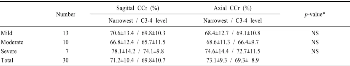
Number Sagittal CCr (%) Axial CCr (%) p-value*
Narrowest / C3-4 level Narrowest / C3-4 level Mild Moderate Severe Total 13 10 7 30 70.6±13.4 / 69.8±10.3 66.8±12.4 / 65.7±11.5 78.1±14.2 / 74.1±9.8 71.2±10.4 / 69.8±10.7 68.4±12.7 / 69.1±10.8 68.6±11.3 / 66.4±9.7 74.6±14.4 / 72.7±11.5 73.1±9.3 / 69.3± 8.9 NS NS NS NS; not-significant, CCr; the ratio of cord/canal, *by kruskal wallis test.
Table 5. Relationship between upper limb weakness and CCr in patients with ALS at the narrowest / C3-4 levels
ALS (n=30) Controls (n=30) p-value* Cord area (mm2) Narrowest level C3-4 level 69.1±9.8 70.3±8.7 78.4±11.3 78.9±10.8 < 0.05 < 0.05 Canal area (mm2) Narrowest level C3-4 level 149.7±12.5 151.5±7.8 172.5±12.5 173.8±11.6 < 0.05 < 0.05 *by Mann-Whitney U test.
Table 4. Area of spinal cord and spinal canal in patients with
ALS and controls in axial view at the narrowest and C3-4 level
평균 CCr은 59.2±4.4%였고, C3-4 부위에서는 평균 척 수관 직경은 15.7±4.1 mm, 평균 척수 직경은 8.9±2.3 mm였고, 평균 CCr은 56.7±5.9%였다. 시상 관점에서 환 자군의 평균 CCr은 대조군의 평균 CCr보다 통계학적으 로 유의하게 높았다(Table 3). 또한 CCr이 70% 이상인 경우는 전체 ALS 환자(30명)에서 16명으로 53.3%였고 대조군에서는 보이지 않았다. 축상 관점에서 경부 척추 증의 정도를 비교해 보면, 가장 좁아진 부위에서 환자군 에서는 평균 척수관 직경은 10.9±6.7 mm, 평균 척수 직 경은 8.2±4.1 mm였고, CCr은 73.1±9.3%였고, C3-4 부 위에서는 평균 척수관 직경은 12.2±2.4 mm, 평균 척수 직경은 8.7±3.2 mm였고, 평균 CCr은 71.3±8.2%였다. 대조군에서는 축상 관점의 가장 좁아진 부위에서 평균 척수관 직경은 16.0±1.4 mm, 평균 척수 직경은 9.1±8.7 mm였고 평균 CCr은 62.5±5.6%였고, C3-4 부위에서는 평균 척수관 직경은 17.2±2.1 mm, 평균 척수 직경은 10.4±2.6 mm였고, 평균 CCr은 60.4±6.4%였다(Table 3). 축상 관점에서 환자군의 평균 CCr은 대조군의 평 균 CCr보다 통계학적으로 유의하게 높았다. 또한 CCr 이 70% 이상인 경우는 전체 ALS 환자(30명)에서 17명 으로 56.7%였고 대조군에서는 보이지 않았다. 환자군에 서 평균 척수 직경과 평균 척수관 직경은 대조군보다 통 계적으로 유의하게 감소되었으며 대조군과 비교해 볼 때 척수관 직경의 감소가 척수 직경의 감소보다 더 심했 다(Table 3). 환자군에서 축상 관점의 가장 좁아진 부위 에서 평균 척수 면적은 69.1±9.8 mm2, 평균 척수관 면 적은 149.7±12.5 mm2였고, C3-4 부위에서 평균 척수 면 적은 70.3±8.7 mm2, 평균 척수관 면적은 151.5±7.8 mm2였다. 대조군에서는 가장 좁아진 부위에서 평균 척 수 면적은 78.4±11.3 mm2, 평균 척수관 면적은 172.5± 12.5 mm2였고, C3-4 부위에서 평균 척수 면적은 78.9± 10.8 mm2, 평균 척수관 면적은 173.8±11.6 mm2였다. 환 자군에서 평균 척수 면적과 평균 척수관 면적은 대조군 보다 통계적으로 유의하게 감소되었으며 대조군과 비교 해 볼 때 척수관 면적의 감소가 척수 면적의 감소보다 더 심했다(Table 4). 이번 연구에서 상지 근력저하와 CCr의 상관 관계를 살펴볼 때, 상지 근력저하가 경도인
경우는 13명으로 시상 관점의 가장 좁아진 부위와 C3-4 부위에서 CCr은 각각 70.6±13.4%와 69.8±10.3%, 중등 도인 경우는 10명으로 66.8±12.4%와 65.7±11.5%, 중증 도인 경우는 7명으로 78.1±14.2%와 74.1±9.8%였다. 축 상 관점의 가장 좁아진 부위와 C3-4부위에서 각각 상지 근력저하가 경도인 경우는 68.4±12.7%와 69.1±10.8%, 중등도인 경우는 68.6±11.3%와 66.4±9.7%, 중증도인 경우는 74.6±14.4%와 72.7±11.5%였다. 상지 근력저하 정도와 CCr과의 관계는 통계학적으로 유의하지 않았다 (Table 5). 고 찰 ALS는 상하지 근력저하를 보이는 퇴행성 질환으로 뇌간의 운동 신경원 및 척수 전각세포가 소실되고 피질 척수로의 퇴행과 운동 피질의 소실이 와서 상부 및 하부 운동신경원 징후가 나타난다.8 소실된 척수 전각세포는 섬유성 성상세포로 대치되며 살아있는 신경세포는 작아 지고 감소하여 지갈소(lipofuscin)로 채워지는데 이러한 일련의 과정을 통해서 척수 위축이 온다고 한다.9 경부 척추증은 경추부 추간판의 변화로부터 시작되며,21 CSM 은 경부 척추증으로부터 이차적으로 흔히 발생한다.22 CSM은 경추의 변성에 의한 신경 압박으로 일어나는 경 추부 척수의 기능장애를 말한다. 즉 나이가 증가함에 따 라 추간판의 변성으로 추간판 간격이 감소되고 후관절 낭 및 인대의 이완으로 불안정성이 생기고 이에 대한 조 직반응으로 추간판 변연부의 골극 형성과 황색인대 비 후 등이 경추부 척수를 압박하여 증상이 나타나게 된다. 가장 흔한 초기 증상은 손의 근력약화, 부자연스러운 손 놀림과 감각이상, 하지의 근력약화로 인한 보행장애이 며, 수개월에 걸쳐 서서히 나타난다.10,16,23-24 경부 척추증 은 골 돌기체가 척수관을 좁게하고 척수를 압박해 생긴 다. 그리고 척수병증의 정도는 골 돌기체의 위치와 척수 관의 좁아짐 정도에 비례한다. 또한 척수에서 탈수초화 도 나타나는데 척수 측주에서 흔히 나타난다.11 경부 척 추증에서의 외측 피질 척수로 손상으로 과다반사, 경직, clonus, 바빈스키 징후와 같은 상부운동신경원 징후가 나타난다. 후주(dorsal column)는 보통 잘 손상되지 않 으며 전각세포의 소실로 하부운동신경원 징후도 나타난 다.12 일반적으로 상부운동신경원 징후는 ALS에서도 나 타나지만 CSM에서 보다 자주 관찰되고13 감각이상의 증 상은 대부분의 ALS 환자에서는 보이지 않고 대부분의 경우에 CSM은 감각이상 증상과 징후가 자주 관찰된 다.14-16 Crandall 등의 연구에서는 CSM 환자 55명 중 20 명은 감각이상보다는 근력저하 증상을 호소한 것을 보 고한 바 있다.15-16 또한 ALS에서도 주관적이고 미약하지 만 감각이상이 질병 초기에 일시적으로 나타날 수 있 다.13 따라서 근전도검사상 상지뿐만 아니라 혀나 하지의 운동신경원을 침범한 소견이 보인다면 ALS를 진단하는 데 유용하지만17,18 그렇지 않은 경우는 ALS 진단에 있 어 CSM과의 감별에 어려움이 있다. 이와 같이 ALS 와 CSM은 임상적으로 비슷한 양상을 보일 수 있어 상 부운동신경원 징후와 하부운동신경원 징후가 모두 나타 날 수 있다. 따라서 상부운동신경원 징후와 하부운동신 경원 징후가 모두 나타나는 경우에는 ALS와 CSM을 정 확하게 진단하는 것은 매우 어렵다.15,16 특히 초기에 두 질병을 감별하는 것은 더 어렵지만 이 시기에 정확하게 두 질병을 감별하는 것은 무엇보다도 중요하다. 왜냐하 면 초기에 적절한 수술적 중재는 CSM의 진행을 멈추게 하고 만족할 만한 회복이 가능하게 하기 때문이다. 반면 에 무분별한 수술은 신경근 기능을 빠르게 악화시키고 심지어 ALS 환자를 죽음으로 이르게 하기도 한다.19 Norris 등에 의하면 ALS 환자가 초기에 CSM으로 진 단 받아 불필요한 수술적 중재를 받은 경우가 약 15% 에 이른다고 한다.13 또한 Mulder에 의하면 ALS로 진 단된 46명 중에 16명이 척수에 병변이 있었다고 하며 46명중 12명(26%)이 심한 경부 척추증이 있었고 수술적 감압으로 호전되었다고 한다.20 본 연구에서 확인할 수 있었던 점은 첫째로 ALS 환자에서 경부 척추증이 동반 되는 경우가 66.7%였고 대조군에서는 36.7%였으며 이러 한 사실은 ALS 환자에서 경부 척추증이 빈도가 높다는 것을 객관적으로 반영하나 그 병태 생리에 대해서는 아 직도 의문이다. 둘째로 ALS 환자에서 대조군에 비해 척 수 위축과 척추관 협착이 의미 있게 관찰되었다. 그리고 척수 위축에 의한 평균 척수 면적의 감소보다는 경부 척 추증에 의한 척수관 면적의 감소 정도가 더 심하였다. 이 러한 사실들을 종합해 볼 때 ALS에서 경부 척추 자기공 명영상의 정량적인 분석을 통해서 척수 위축이 온다는 사실과 경부 척추증이 많이 동반된다는 사실을 객관적 으로 확인할 수 있었다. 셋째로 ALS 환자에서 상지의 근력저하 정도와 CCr과의 상관 관계는 없었다. 이는 상 지 근력저하가 경부 척추증에 의한 이차적인 것이라기 보다는 ALS 자체에 의한 것이라는 것을 의미한다. 결론 적으로 본 연구에서 임상적으로 증상이 있는 ALS 환자 는 일반인에 비해 척수 위축을 보이고 경부 척추증이 많 이 동반되며, 경부 척추 협착증의 정도도 심하다는 사실 을 확인할 수 있었고 이러한 결과에 대한 원인을 밝히기 위해 앞으로 더 많은 연구가 필요할 것으로 생각한다. REFERENCES
1. Young WF. Cervical spondylotic myelopathy: a common cause of spinal cord dysfunction in older persons. Am Fam
Physician 2000;62:1064-1070.
2. MCRae DL. Asymptomatic inadvertible disc protrusions. Acta
Radiol 1956;46:9-27.
3. Hitselberger WE, Witten RM. Abnormal myelograms in asymptomatic patients. J Neurosurg 1968;28:204-206. 4. Holt EP Jr. The question of lumbar discography. J Bone
Joint Surg Am 1968;5:720-726.
5. Wiesel SW, Tsourmas N, Feffer HL, Citrin CM, Patronas N. A study of computer-assisted tomography 1. The incidence of positive CAT scans in an asymptomatic group of patients.
Spine 1984;9:549-551.
6. Armstrong P, Wastie ML. Diagnostic imaging. 1st ed. London: Blackwell Scientific publications, 1989;336. 7. Brooks BR, Miller RG, Swash M, Munsat TL;World
Federation of Neurology Research Group on Motor Neuron Diseases. El Escorial revisited: revised criteria for the diagnosis of amyotrophic lateral sclerosis. Amyotroph Lateral
Scler Other Motor Neuron Disord 2000;1:293-299.
8. Tandan R, Bradley WG. Amyotrophic lateral sclerosis: Part 1. clinical feature,pathology, and ethical issues in management.
Ann Neurol 1985;18:271-280.
9. Victor M, Ropper AH. Adams and Victor's Principle of NEUROLOGY. 7th ed. International:McGraw-Hill, 2001; 1156.
10. Bohlman HH. Cervical spondylosis and myelopathy. Instr
Course Lect 1995;44:81-97.
11. Hoff JT, Wilson CB. The pathophysiology of cervical spondylotic radiculopathy and myelopathy. Clin Neurosurg 1977;24:474-487.
12. Stark RJ, Kennard C, Swash M. Hand wasting in spondylotic high cord compression:an electromyographic syudy. Ann
Neurol 1981;9:58-62.
13. Norris FH Jr, Denys EH, Ü KS. Differential diagnosis of
adult motor neuron disease. The diagnosis and treatment of amyotrophic lateral sclerosis. Mulder DW(ed). Boston:
Houghton Mufflin, 1980;53-78.
14. Brain WR, Northfield D, Wilkinson M. The neurological
manifestations of cervical spondylosis. Brain 1952;75:187- 225.
15. Crandall PH, Batzdorf U. Cervical spondylotic myelopathy. J
Neurosurg 1966;25:57-66.
16. Gregorius FK, Estrin T, Crandall PH. Cervical spondylotic radiculopathy and myelopathy. A long term follow-up study.
Arch Neurol 1976;33:618-625.
17. Gao XX, Tang XF. Relationship between the clinical manifestation and electromyographic findings in motor neuron disease. Zhonghua Shen Jing Jing Shen Ka Za Zhi 1991;24:98-100.
18. Lee KS, Kelly DL Jr. Amyotrophic lateral sclerosis and cervical spondylotic myelopathy in a patient with a posterior fossa arachnoid cyst:diagnostic dilemma. South Med J 1987; 80:1580-1583.
19. Zhao DL, Zhang WM, Li GD. Cervical Spondylosis. 1st ed. Shangahi: scientific and Technological Literature Press,1988; 158-160.
20. Mulder DW(ed). The diagnosis and treatment of amyotrophic
lateral sclerosis. Boston: Houghton Mufflin, 1980;1-397.
21. Lunsford LD, Bissonette DJ, Zorub DS. Anterior surgery for cervical disc disease. Part 2: Treatment of cervical spondylotic myelopathy in 32 cases. J Neurosurg 1980;53: 12-19.
22. Wilberger JE Jr, Chedid MK. Acute cervical spondylytic myelopathy. Neurosurgery 1988;22:142-146.
23. Clark CR. Cervical spondylotic myelopathy: History and physical findings. Spine 1988;13:847-849.
24. Lipson SJ. Cervical disc disease: Pathogenesis and natural history. Seminars in spine 1989;1:69-93.